

Chronic fluoride toxicity: dental fluorosis
- PMID: 21701193
- PMCID: PMC3433161
- DOI: 10.1159/000327028
Chronic fluoride toxicity: dental fluorosis
Abstract
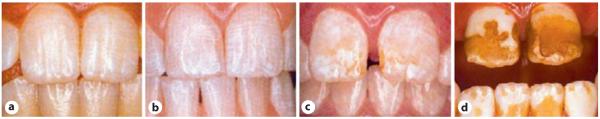
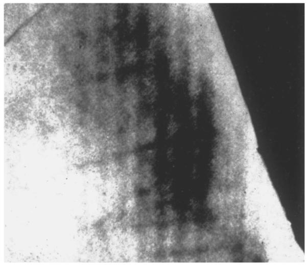
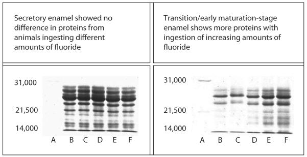
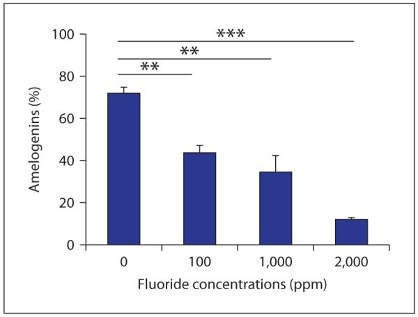
Dental fluorosis occurs as a result of excess fluoride ingestion during tooth formation. Enamel fluorosis and primary dentin fluorosis can only occur when teeth are forming, and therefore fluoride exposure (as it relates to dental fluorosis) occurs during childhood. In the permanent dentition, this would begin with the lower incisors, which complete mineralization at approximately 2-3 years of age, and end after mineralization of the third molars. The white opaque appearance of fluorosed enamel is caused by a hypomineralized enamel subsurface. With more severe dental fluorosis, pitting and a loss of the enamel surface occurs, leading to secondary staining (appearing as a brown color). Many of the changes caused by fluoride are related to cell/matrix interactions as the teeth are forming. At the early maturation stage, the relative quantity of amelogenin protein is increased in fluorosed enamel in a dose-related manner. This appears to result from a delay in the removal of amelogenins as the enamel matures. In vitro, when fluoride is incorporated into the mineral, more protein binds to the forming mineral, and protein removal by proteinases is delayed. This suggests that altered protein/mineral interactions are in part responsible for retention of amelogenins and the resultant hypomineralization that occurs in fluorosed enamel. Fluoride also appears to enhance mineral precipitation in forming teeth, resulting in hypermineralized bands of enamel, which are then followed by hypomineralized bands. Enhanced mineral precipitation with local increases in matrix acidity may affect maturation stage ameloblast modulation, potentially explaining the dose-related decrease in cycles of ameloblast modulation from ruffle-ended to smooth-ended cells that occur with fluoride exposure in rodents. Specific cellular effects of fluoride have been implicated, but more research is needed to determine which of these changes are relevant to the formation of fluorosed teeth. As further studies are done, we will better understand the mechanisms responsible for dental fluorosis.
Copyright © 2011 S. Karger AG, Basel.
Figures
References
-
- Aoba T, Moreno EC, Tanabe T, Fukae M. Effects of fluoride on matrix proteins and their properties in rat secretory enamel. J Dent Res. 1990;69:1248–1250. - PubMed
-
- Richards A. Nature and mechanisms of dental fluorosis in animals. J Dent Res. 1990;69(spec No):701–705. discussion 721. - PubMed
-
- Giambro NJ, Prostak K, DenBesten PK. Characterization of fluorosed human enamel by color reflectance, ultrastructure, and elemental composition. Caries Res. 1995;29:251–257. - PubMed
-
- Angmar-Mansson B, Ericsson Y, Ekberg O. Plasma fluoride and enamel fluorosis. Calcif Tissue Res. 1976;22:77–84. - PubMed
-
- Angmar-Mansson B, Whitford GM. Enamel fluorosis related to plasma F levels in the rat. Caries Res. 1984;18:25–32. - PubMed
Publication types
MeSH terms
Substances
Grants and funding
LinkOut - more resources
Full Text Sources
Research Materials
