. 2011 Oct;36(4):381-9.
doi: 10.1097/SHK.0b013e31822793c4.

Epidermal growth factor improves survival and prevents intestinal injury in a murine model of pseudomonas aeruginosa pneumonia

Affiliations

Epidermal growth factor improves survival and prevents intestinal injury in a murine model of pseudomonas aeruginosa pneumonia

Jessica A Dominguez et al. Shock. 2011 Oct.

Abstract

Mortality from pneumonia is mediated, in part, through extrapulmonary causes. Epidermal growth factor (EGF) has broad cytoprotective effects, including potent restorative properties in the injured intestine. The purpose of this study was to determine the efficacy of EGF treatment following Pseudomonas aeruginosa pneumonia. FVB/N mice underwent intratracheal injection of either P. aeruginosa or saline and were then randomized to receive either systemic EGF or vehicle beginning immediately or 24 h after the onset of pneumonia. Systemic EGF decreased 7-day mortality from 65% to 10% when initiated immediately after the onset of pneumonia and to 27% when initiated 24 h after the onset of pneumonia. Even though injury in pneumonia is initiated in the lungs, the survival advantage conferred by EGF was not associated with improvements in pulmonary pathology. In contrast, EGF prevented intestinal injury by reversing pneumonia-induced increases in intestinal epithelial apoptosis and decreases in intestinal proliferation and villus length. Systemic cytokines and kidney and liver function were unaffected by EGF therapy, although EGF decreased pneumonia-induced splenocyte apoptosis. To determine whether the intestine was sufficient to account for extrapulmonary effects induced by EGF, a separate set of experiments was done using transgenic mice with enterocyte-specific overexpression of EGF (IFABP-EGF [intestinal fatty acid-binding protein linked to mouse EGF] mice), which were compared with wild-type mice subjected to pneumonia. IFABP-EGF mice had improved survival compared with wild-type mice following pneumonia (50% vs. 28%, respectively, P < 0.05) and were protected from pneumonia-induced intestinal injury. Thus, EGF may be a potential adjunctive therapy for pneumonia, mediated in part by its effects on the intestine.


Figures

Figure 1
Figure 1. Effect of EGF treatment on mortality in P. aeruginosa pneumonia
Mice were subjected to pneumonia with or without EGF treatment (150 µg/kg/day i.p., twice daily for 7 days) that was initiated immediately after surgery (0 hours) or 24 hours later. Sham mice received intratracheal injections of saline. All mice were given antibiotics and were followed for survival for 7 days. Mice given EGF immediately after the onset of pneumonia exhibited significantly improved survival compared to untreated mice (p<0.001). Even if EGF treatment was delayed for 24 hours after the onset of infection, there was still a significant survival advantage (p<0.05). All sham mice survived.
Figure 2
Figure 2. Effect of EGF treatment on pulmonary pathology
The severity of pneumonia was significantly increased following intratracheal injection of P. aeruginosa regardless of EGF treatment (A). Representative H&E-stained micrographs of the lung are shown (B). Magnification 400×. n=6–10/group.
Figure 3
Figure 3. Effect of EGF treatment on pulmonary neutrophil infiltration, MIP-2 levels, apoptosis, and bacterial clearance
Myeloperoxidase (MPO) activity was evaluated as an index of neutrophil infiltration and degranulation in lung tissue (A) and in BAL fluid (B). Mice subjected to P. aeruginosa pneumonia exhibited elevated MPO activity in both lung tissue and BAL fluid compared to shams. EGF treatment after the onset of sepsis did not significantly alter neutrophil activation. MIP-2 levels in BAL fluid (C) were elevated in mice subjected to P. aeruginosa pneumonia compared to sham but there were no statistically significant differences in MIP-2 levels following EGF treatment. Apoptosis in lung tissue was evaluated by active caspase-3 staining (D) in 5 random high powered fields (RHPF). Mice subjected to P. aeruginosa pneumonia exhibited increased lung apoptosis regardless of EGF treatment. Mice with pneumonia had increased levels of bacteria in the BAL fluid (E), and EGF treatment significantly improved clearance of infection from the lung compared to untreated mice with pneumonia. n=4–10/group.
Figure 4
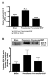
Figure 4. Effect of EGF treatment on intestinal EGF-R expression
Relative mRNA levels were quantified in jejunal tissue via qRT-PCR (A). Gene expression of intestinal EGF-R was increased in mice with pneumonia and decreased when systemic EGF was given after the onset of pneumonia. Intestinal lysates were probed for protein expression of EGF-R (B). Mice with pneumonia exhibited a trend for increased expression of EGF-R, while EGF-treated mice with pneumonia had decreased EGF-R expression to sham levels. Representative blots for EGF-R are depicted; β-actin was used as a control for equal protein loading in each lane. n=4–5/group.
Figure 5
Figure 5. Effect of EGF treatment on intestinal integrity
Mice subjected to P. aeruginosa pneumonia had significantly shorter villi than sham mice (A). Mice with pneumonia treated with EGF had villus lengths similar to those seen in sham animals. Intestinal epithelial apoptosis was evaluated by active caspase-3 staining (C,D) and H&E staining (E,F) in 100 crypts. Representative micrographs are shown for both methods. Magnification 400×. Mice subjected to P. aeruginosa pneumonia exhibited increased intestinal apoptosis by both methods. Mice with pneumonia treated with EGF had similar levels of intestinal apoptosis as sham mice. S-phase cells were also quantified in 100 crypts. Mice subjected to P. aeruginosa pneumonia had significantly decreased intestinal proliferation compared to sham mice (B). Mice with pneumonia treated with EGF had significantly increased intestinal proliferation compared to untreated mice; however, EGF treatment was not able to fully restore the proliferative response to levels seen in sham animals. n=11–22/group.
Figure 6
Figure 6. Effect of EGF treatment of EGF on splenocyte apoptosis
Splenocyte apoptosis was evaluated by active caspase-3 staining (A) and H&E staining (B) in 5 random high powered fields (RHPF). Mice subjected to P. aeruginosa pneumonia exhibited significantly increased splenocyte apoptosis compared to sham mice. EGF treatment resulted in significantly decreased apoptosis compared to untreated mice with pneumonia; however, levels of apoptosis were still increased in these mice compared to shams. n=5/group.
Figure 7
Figure 7. Effect of enterocyte-specific overexpression of EGF on mortality, intestinal epithelial apoptosis and proliferation
WT and IFABP-EGF mice were subjected to pneumonia, given antibiotics, and followed for survival for 7 days (A). Mice that overexpress EGF exclusively in enterocytes had improved survival compared to WT mice following pneumonia. WT and IFABP-EGF mice were subjected to pneumonia or saline and were euthanized 24 hours later. Intestinal epithelial apoptosis was evaluated by active caspase-3 staining (B) and H&E staining (C) in 100 crypts. Proliferation was evaluated by quantifying S-phase cells in 100 crypts (D). IFABP-EGF mice subjected to P. aeruginosa pneumonia exhibited decreased intestinal apoptosis and increased proliferation to levels seen in sham mice. n=7/group.
Figure 8
Figure 8. Working model for the protective effects of EGF in pneumonia
Dissemination of P. aeruginosa from the lungs leads to bacteremia and a systemic inflammatory response. Proinflammatory mediators can induce apoptosis in the intestinal epithelium and immune cells in the spleen. In the intestine, increased apoptosis, decreased proliferation, and villus atrophy contribute to intestinal barrier dysfunction, thereby perpetuating systemic inflammation. We hypothesize that EGF enhances pulmonary bacterial clearance by inducing secretion of cytokines and chemokines to the lung. However, these mice still become bacteremic and exhibit similar levels of systemic inflammation. The direct actions of EGF on the intestinal epithelium prevent intestinal barrier dysfunction, thereby dampening systemic inflammation and improved survival. Systemic EGF also prevents splenocyte apoptosis which plays a role in mediating the improved survival seen with systemic EGF compared with intestine-specific EGF.

Similar articles

Cited by

References

    1. Heron M, Hoyert DL, Murphy SL, Xu J, Kochanek KD, Tejada-Vera B. Deaths: final data for 2006. Natl Vital Stat Rep 17. 2009;57(14):1–134. - PubMed
    1. American College of Chest Physicians/Society of Critical Care Medicine Consensus Conference: definitions for sepsis and organ failure and guidelines for the use of innovative therapies in sepsis. Crit Care Med. 1992;20(6):864–874. - PubMed
    1. Clark JA, Coopersmith CM. Intestinal crosstalk: a new paradigm for understanding the gut as the "motor" of critical illness. Shock. 2007;28(4):384–393. - PMC - PubMed
    1. Hassoun HT, Kone BC, Mercer DW, Moody FG, Weisbrodt NW, Moore FA. Post-injury multiple organ failure: the role of the gut. Shock. 2001;15(1):1–10. - PubMed
    1. Coopersmith CM, Stromberg PE, Dunne WM, Davis CG, Amiot DM, Buchman TG, Karl IE, Hotchkiss RS. Inhibition of intestinal epithelial apoptosis and survival in a murine model of pneumonia-induced sepsis. JAMA. 2002;287(13):1716–1721. - PubMed

Publication types