. 2011 Nov;25(11):1739-50.
doi: 10.1038/leu.2011.159. Epub 2011 Jun 24.

p53 independent epigenetic-differentiation treatment in xenotransplant models of acute myeloid leukemia

Affiliations

p53 independent epigenetic-differentiation treatment in xenotransplant models of acute myeloid leukemia

K P Ng et al. Leukemia. 2011 Nov.

Abstract

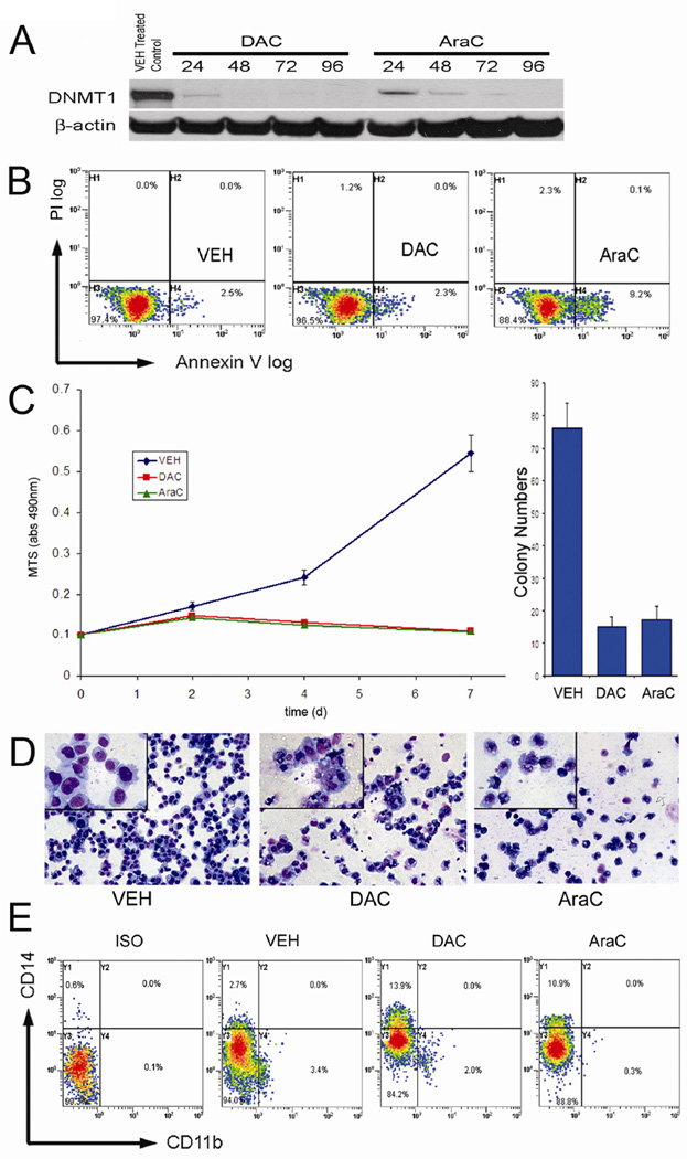
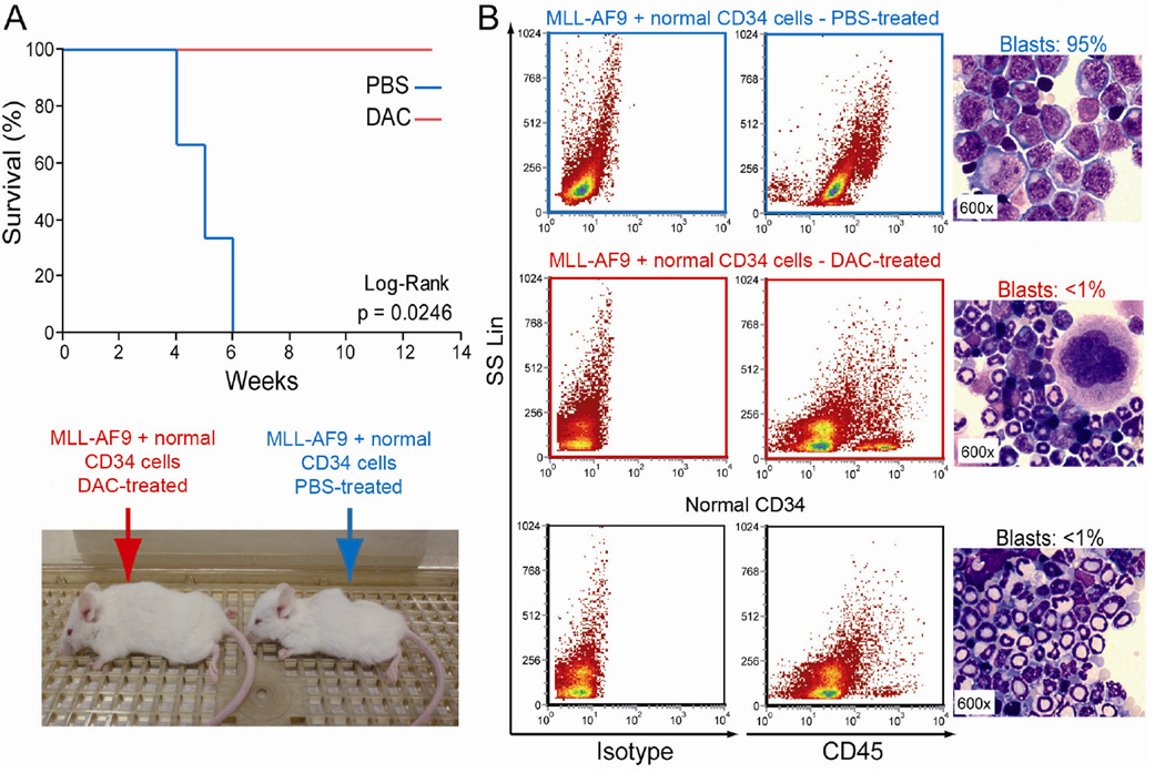
Suppression of apoptosis by TP53 mutation contributes to resistance of acute myeloid leukemia (AML) to conventional cytotoxic treatment. Using differentiation to induce irreversible cell cycle exit in AML cells could be a p53-independent treatment alternative, however, this possibility requires evaluation. In vitro and in vivo regimens of the deoxycytidine analogue decitabine that deplete the chromatin-modifying enzyme DNA methyl-transferase 1 without phosphorylating p53 or inducing early apoptosis were determined. These decitabine regimens but not equimolar DNA-damaging cytarabine upregulated the key late differentiation factors CCAAT enhancer-binding protein ɛ and p27/cyclin dependent kinase inhibitor 1B (CDKN1B), induced cellular differentiation and terminated AML cell cycle, even in cytarabine-resistant p53- and p16/CDKN2A-null AML cells. Leukemia initiation by xenotransplanted AML cells was abrogated but normal hematopoietic stem cell engraftment was preserved. In vivo, the low toxicity allowed frequent drug administration to increase exposure, an important consideration for S phase specific decitabine therapy. In xenotransplant models of p53-null and relapsed/refractory AML, the non-cytotoxic regimen significantly extended survival compared with conventional cytotoxic cytarabine. Modifying in vivo dose and schedule to emphasize this pathway of decitabine action can bypass a mechanism of resistance to standard therapy.


Figures

Figure 1
Figure 1. Non-cytotoxic concentrations of decitabine (DAC) induce differentiation and terminate proliferation of p53 wild-type MLL-AF9 cells
A) DNMT1 protein levels in DAC and cytarabine (AraC) treated cells. DNMT1 measured by Western blot at different time points. DAC or AraC 0.5 µM added at 0 and 72h. B) DAC 0.5 µM did not cause significant early apoptosis. Annexin staining measured by flow-cytometry 24 hours after DAC treatment. Positive control AraC treatment. C) DAC and AraC treatment were anti-proliferative. Cell quantity estimated by MTT assay at day 2, 4 and 7. DAC or AraC 0.5 µM added to cells on Day 1 and 4. Cells were plated into methylcellulose (2000 viable cells per ml) on day 5, to assay for stem/progenitor activity. D) DAC induced morphologic changes of monocytic differentiation (decreased nuclear-cytoplasmic ratio, increased cell size, granulation and vacuolization of the cytoplasm). AraC treated cells were small and disrupted, suggesting apoptosis and necrosis. Day 4 Giemsa stained cytospins. E) DAC and AraC treatment increased expression of CD14. CD14 (monocyte marker) and CD11b (granulocytic marker) measured by flow-cytometry on day 4.
Figure 2
Figure 2. Decitabine (DAC), but not an identical regimen of cytarabine (AraC), terminated proliferation of p53-null MLL-AF9 cells (THP1 cells (
A) DAC 0.5 µM depleted DNMT1. DNMT1 measured by Western blot. B) Neither DAC nor AraC 0.5 µM caused early apoptosis. Annexin staining measured by flow-cytometry 24 hours after DAC or AraC treatment. C) DAC, but not AraC treatment, was anti-proliferative. Cell quantity estimated by MTT assay at day 1, 3, 6 and 8. DAC or AraC 0.5 µM added to cells on Day 1 and 4. Cells were plated into methylcellulose (2000 viable cells per ml) on day 5, to assay for stem/progenitor activity. D) DAC induced morphologic changes of myeloid differentiation (decreased nuclear-cytoplasmic ratio, increased cell size, granulation and vacuolization of the cytoplasm). Arrows = mitotic cells. Day 4 Giemsa stained cytospins. E) DAC, but not AraC, increased expression of CD11b, and to a lesser extent CD14. CD11b and CD14 measured by flow-cytometry on day 4.
Figure 3
Figure 3. Decitabine (DAC) and cytarabine (AraC) differentially regulate apoptosis and differentiation proteins in p53 wild-type and p53-null MLL-AF9 cells
Key events associated with apoptosis and differentiation mediated cell cycle exit were examined in p53 wild-type and p53-null MLL-AF9 cells treated with DAC or AraC 0.5 µM on day 1 and 4. A) Western blots of p53 wild-type MLL-AF9 cells treated with DAC or AraC. Numbers = hours after initiation of treatment. Red boxes = protein upregulated by DAC but not by AraC. Graphs depict results of densitometry analysis. B) Western blots of p53-null MLL-AF9 cells (THP1 cells) treated with DAC or AraC.
Figure 4
Figure 4. Low concentration decitabine inhibits AML leukemia initiating cells (LIC) but spared normal HSC
Engraftment in an immuno-compromised murine host is a functional assay for both normal HSC and LIC (47;48). Normal CD34+ HSC and MLL-AF9 leukemia cells were treated in vitro with the identical decitabine regimen (0.5 µM on day 1, 0.2 µM on day 2, 0.5 µM on day 5, 0.2 µM on day 6) or mock treated with PBS. Cells harvested on day 7 were combined (3 × 10⁵ each MLL-AF9 + normal cells), then transplanted by tail-vein injection into sub-lethally irradiated NOD/SCID recipient mice (n=6). Additional controls: mice transplanted with PBS-treated normal CD34+ cells alone (4 × 10⁶ cells/mouse). A) Significant survival difference between groups. Surviving mice did not demonstrate distress but were sacrificed for analysis at week 13. B) >90% AML cell engraftment in bone marrow of mice receiving PBS-treated cells; ~80% normal, multi-lineage, human hematopoietic cell engraftment in mice receiving decitabine treated cells. Additional engraftment data and controls in figure S1. Cytospin preparations of cells flushed from bone marrow were stained with Giemsa. Measurement of blast percentage in cytospins was blinded to treatment status.
Figure 5
Figure 5. Better survival with non-cytotoxic decitabine (DAC) than with cytotoxic cytarabine (AraC) in a murine xeno-transplantation model of p53-null human AML
A) Kaplan-Meier plot of survival distribution function. X-axis: days after xeno-transplant of AML cells. Non-irradiated NSG mice were transplanted with 3×10⁶ THP1 cells (p53-null MLL-AF9 AML cells) by tail vein injection. Starting day 5 after transplantation, mice were treated with vehicle (PBS, n=5), AraC 75 mg/kg/day IP for five days (n=5) (to model conventional chemotherapy (51)), or DAC 0.2 mg/kg SC 3X/week for 2 weeks then 1X/week (n=5). B) Bioluminescent imaging to show anatomic localization of engrafted THP1 cells. Images from dorsal perspective obtained ten minutes after IP administration of luciferin substrate, on day 28 after transplant of 3 × 10⁶ THP1 cells transfected to express luciferase. PBS, AraC and DAC treatment started on day 3. Red=most intense, blue = least intense bioluminescence/engraftment. C) Liver focus of disease in AraC and DAC treated mice confirmed at sacrifice. * = p <0.05 compared with PBS treated control. ** = p <0.005 compared with PBS treated control. D) Spleens of DAC treated mice were decreased in size compared to vehicle treated mice, but not to a statistically significant extent (organs harvested at different time-points per the Kaplan-Meier plot).
Figure 6
Figure 6. Better survival with non-cytotoxic decitabine (DAC) than cytotoxic cytarabine (AraC) in a murine xeno-transplantation model of refractory/relapsed human AML
Fresh AML cells from a patient with relapsed, chemotherapy treatment refractory AML were transplanted by tail-vein injection (1×10⁶ cells/mouse) into NSG mice. These AML cells contained multiple chromosome abnormalities including t(8;18)(q22:q23) and t(11;13)(q21:q12). Starting day 5 after transplantation, mice were treated with vehicle (control) (n=7), AraC 75 mg/kg/day IP for five days (n=7), or DAC 0.2 mg/kg SC 3X/week for 2 weeks then 1X/week (n=7). A) Better survival in DAC treated mice. Mice were sacrificed for signs of distress. B) Spleens of decitabine treated mice were significantly decreased in size compared to AraC or vehicle treated mice. ** = p <0.005 compared to PBS treated control. Normal = non-transplanted, non-treated mouse. Spleens were harvested at later time-points in DAC treated mice as per the Kaplan-Meier plot. C) Bone marrow replacement by human leukemia cells. Human CD45 staining used to identify human cells in bone marrow after sacrifice at different time-points per the Kaplan-Meier plot.

References

    1. Vazquez A, Bond EE, Levine AJ, Bond GL. The genetics of the p53 pathway, apoptosis and cancer therapy. Nat Rev Drug Discov. 2008;7(12):979–987. - PubMed
    1. Suarez L, Vidriales MB, Garcia-Larana J, Sanz G, Moreno MJ, Lopez A, Barrena S, Martinez R, Tormo M, Palomera L, Lavilla E, Lopez-Berges MC, de Santiago M, de Equiza ME, Miguel JF, Orfao A. CD34+ cells from acute myeloid leukemia, myelodysplastic syndromes, and normal bone marrow display different apoptosis and drug resistance-associated phenotypes. Clin Cancer Res. 2004;10(22):7599–7606. - PubMed
    1. Yin B, Kogan SC, Dickins RA, Lowe SW, Largaespada DA. Trp53 loss during in vitro selection contributes to acquired Ara-C resistance in acute myeloid leukemia. Exp Hematol. 2006;34(5):631–641. - PubMed
    1. Wattel E, Preudhomme C, Hecquet B, Vanrumbeke M, Quesnel B, Dervite I, Morel P, Fenaux P. p53 mutations are associated with resistance to chemotherapy and short survival in hematologic malignancies. Blood. 1994;84(9):3148–3157. - PubMed
    1. Toledo F, Wahl GM. Regulating the p53 pathway: in vitro hypotheses, in vivo veritas. Nat Rev Cancer. 2006;6(12):909–923. - PubMed

Publication types

MeSH terms