. 2012 Jan;19(1):162-9.
doi: 10.1038/cdd.2011.88. Epub 2011 Jun 24.

Autophagy promotes survival of retinal ganglion cells after optic nerve axotomy in mice

Affiliations

Autophagy promotes survival of retinal ganglion cells after optic nerve axotomy in mice

N Rodríguez-Muela et al. Cell Death Differ. 2012 Jan.

Abstract

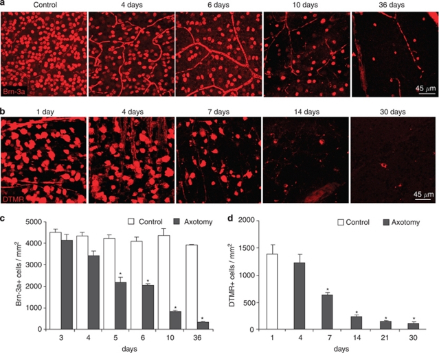
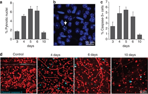
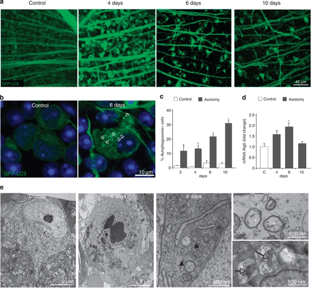
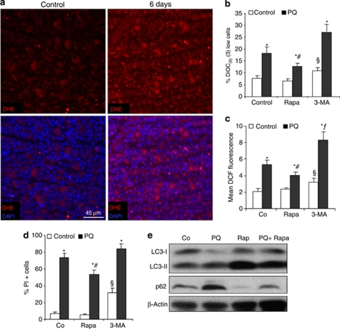
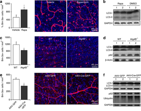
Autophagy is an essential recycling pathway implicated in neurodegeneration either as a pro-survival or a pro-death mechanism. Its role after axonal injury is still uncertain. Axotomy of the optic nerve is a classical model of neurodegeneration. It induces retinal ganglion cell death, a process also occurring in glaucoma and other optic neuropathies. We analyzed autophagy induction and cell survival following optic nerve transection (ONT) in mice. Our results demonstrate activation of autophagy shortly after axotomy with autophagosome formation, upregulation of the autophagy regulator Atg5 and apoptotic death of 50% of the retinal ganglion cells (RGCs) after 5 days. Genetic downregulation of autophagy using knockout mice for Atg4B (another regulator of autophagy) or with specific deletion of Atg5 in retinal ganglion cells, using the Atg5(flox/flox) mice reduces cell survival after ONT, whereas pharmacological induction of autophagy in vivo increases the number of surviving cells. In conclusion, our data support that autophagy has a cytoprotective role in RGCs after traumatic injury and may provide a new therapeutic strategy to ameliorate retinal diseases.


Figures

Figure 1
Figure 1
Retinal ganglion cell loss after ONT. Optic nerves from adult mice were unilaterally sectioned and the animals were killed at different time points. Right (control) and left (axotomy) retinas were then isolated, flat-mounted and fixed. (a and c) Immunostaining and quantification of the number of Brn-3a-positive cells in control and axotomized retinas. (bd) DTMR retrograde labeling of RGCs and its quantification from one day (control) up to 30 days of axotomy
Figure 2
Figure 2
Kinetics of cell death after ONT. (a) Quantification of the percentage of pyknotic nuclei stained with DAPI related to the total number of cells in the ganglion cell layer (GCL). (b) Pyknotic nuclei stained with DAPI (arrow) in the GCL for 6 days after axotomy. (c) Quantification of the percentage of activated caspase-3-positive cells and (d) immunostaining against Brn-3a and cleaved caspase-3 in control and axotomized retinas at different time points after axotomy
Figure 3
Figure 3
Autophagy is activated in RGCs shortly after ONT. (a) GFP-LC3 fluorescence of the GCL in flat-mounted retinas from control and axotomized mice. (b) Magnification of RGCs in a control retina and 6 days after ONT, a 0.5 μm confocal plane is shown. Cells from control retinas display a diffuse and homogeneous green fluorescence in the cytosol, whereas the axotomized RGCs in the right panel showt visible GFP-LC3 dots in the cytosol identified as autophagosomes and pointed with arrowheads. (c) Quantification of the percentage of cells with GFP-LC3 positive APs in the cytosol versus total number of cells in the GCL. (d) Analysis of Atg5 mRNA levels by qPCR in control and axotomized retinas at different time points after the injury. (e) Electron microscopy analysis of representative RGCs from control and 6 days-injured retinas. Panels on the right show a magnification of a degenerating RGC after the lesion. Note the swollen endoplasmic reticulum (black asterisk), damaged mitochondria (white asterisks), autophagosomes (black arrow head) and mitochondria inside autophagosomes (empty arrows)
Figure 4
Figure 4
ROS production in vivo and in vitro and autophagy modulation in vitro. (a) In vivo ROS staining in the GCL of control and 6 days-axotomized retinas after injecting the animals with DHE for 18 h. (bd) FACS analysis of RGC-5 cells treated with paraquat (PQ) alone or combined with rapamycin or 3-MA to determine (b) mitochondrial membrane potential with DiOC(6)(3) at 24 h, (c) ROS production with DCF at 48 h after treatment and (d) cell viability with PI staining at 48 h and. *P<0.05 Control versus PQ, #P<0.05 PQ versus Rapa+PQ, §P<0.05 Control versus 3-MA, fP<0.05 PQ versus PQ+3-MA. (e) Western blot analysis of RGC-5 cells treated with rapamycin and subjected to PQ toxicity during 24 h
Figure 5
Figure 5
Autophagy protects after ONT in vivo. (a) Representative image showing Brn-3a immunostaining of vehicle and rapamycin-treated animals and quantification of the percentage of surviving RGCs 10 days after axotomy. (b) LC3 immunoblot of retinas from mice injected with vehicle or rapamycin at 10 days after the injury. (c) Image of RGCs stained with Brn-3a from WT and Atg4B−/− mice at 10 days after axotomy and quantification of the percentage of RGCs in the GCL. (d) Immunoblot analysis of untreated WT and Atg4B−/− retinas. (e) Representative image of the GCL stained for Brn-3a and its quantification at 10 days after ONT in Atg5flox/flox mice injected with AAV-GFP or AAV-Cre-GFP vectors. (f) Immunoblot analysis of uninjured retinas from mice injected with AAV-GFP or AAV-Cre-GFP

References

    1. Isenmann S, Kretz A, Cellerino A. Molecular determinants of retinal ganglion cell development, survival, and regeneration. Prog Retin Eye Res. 2003;22:483–543. - PubMed
    1. Villegas-Perez MP, Vidalsanz M, Rasminsky M, Bray GM, Aguayo AJ. Rapid and protracted phases of retinal ganglion-cell loss follow axotomy in the optic-nerve of adult-rats. J Neurobiol. 1993;24:23–36. - PubMed
    1. Tezel G. Oxidative stress in glaucomatous neurodegeneration: mechanisms and consequences. Prog Retin Eye Res. 2006;25:490–513. - PMC - PubMed
    1. Yang Z, Klionsky DJ. Mammalian autophagy: core molecular machinery and signaling regulation. Curr Opin Cell Biol. 2010;22:124–131. - PMC - PubMed
    1. Boya P, Gonzalez-Polo RA, Casares N, Perfettini J, Dessen P, Larochette N, et al. Inhibition of macroautophagy triggers apoptosis. Mol Cell Biol. 2005;25:1025–1040. - PMC - PubMed

Publication types

MeSH terms