. 2011 Aug;18(8):598-608.
doi: 10.1038/cgt.2011.30. Epub 2011 Jun 24.

Armed and targeted measles virus for chemovirotherapy of pancreatic cancer

Affiliations

Armed and targeted measles virus for chemovirotherapy of pancreatic cancer

S Bossow et al. Cancer Gene Ther. 2011 Aug.

Abstract

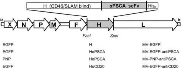
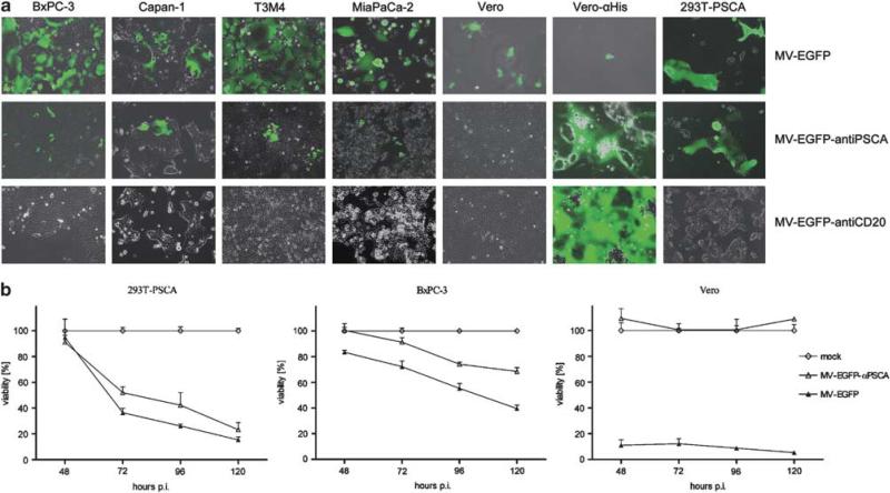
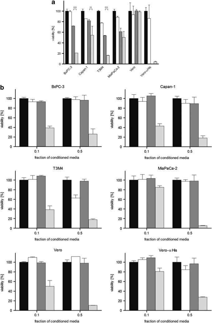
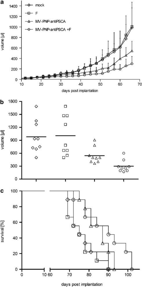
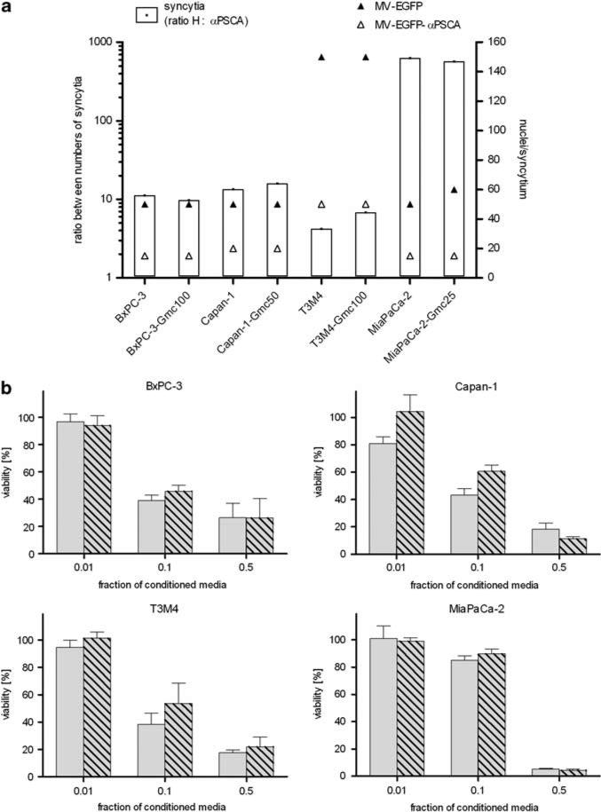
No curative therapy is currently available for locally advanced or metastatic pancreatic cancer. Therefore, new therapeutic approaches must be considered. Measles virus (MV) vaccine strains have shown promising oncolytic activity against a variety of tumor entities. For specific therapy of pancreatic cancer, we generated a fully retargeted MV that enters cells exclusively through the prostate stem cell antigen (PSCA). Besides a high-membrane frequency on prostate cancer cells, this antigen is expressed on pancreatic adenocarcinoma, but not on non-neoplastic tissue. PSCA expression levels differ within heterogeneous tumor bulks and between human pancreatic cell lines, and we could show specific infection of pancreatic adenocarcinoma cell lines with both high- and low-level PSCA expression. Furthermore, we generated a fully retargeted and armed MV-PNP-anti-PSCA to express the prodrug convertase purine nucleoside phosphorylase (PNP). PNP, which activates the prodrug fludarabine effectively, enhanced the oncolytic efficacy of the virus on infected and bystander cells. Beneficial therapeutic effects were shown in a pancreatic cancer xenograft model. Moreover, in the treatment of gemcitabine-resistant pancreatic adenocarcinoma cells, no cross-resistance to both MV oncolysis and activated prodrug was detected.


Figures

Figure 1
Figure 1
Schematic representation of the recombinant measles virus (MV) genomes. Vectors harbor either unmodified hemagglutinin (H) or fully retargeted H against prostate stem cell antigen (PSCA) and CD20 (listed at the bottom, middle), respectively, in combination with the transgene X = enhanced green fluorescent protein (EGFP) or purine nucleoside phosphorylase (PNP) (listed at the bottom, left). Corresponding names of the viruses are listed at the bottom right side.
Figure 2
Figure 2
Susceptibility of pancreatic adenocarcinoma cell lines with high- and low-level prostate stem cell antigen (PSCA) expression to measles virus (MV) infection. (a) Four pancreatic adenocarcinoma cell lines, Vero and Vero-αHis cells were infected with the respective viruses (indicated on the right of each row) at a multiplicity of infection (MOI) of 1. Photographs were taken at 48 h post-infection (p.i.) using a fluorescence microscope (×100 magnification). Merged pictures of enhanced green fluorescent protein (EGFP) fluorescence and phase contrast are shown. (b) In total, 104 293T-PSCA, BxPC-3 and Vero cells each were infected at an MOI of 1 in quadruplicates and cell viability was determined at the indicated time points. Means and standard deviations are shown (white diamond, mock-treated cells defining 100% viability; white triangle, MV-EGFP-anti-PSCA; black triangle, MV-EGFP-anti-PSCA).
Figure 3
Figure 3
Cytotoxic effect of prodrug convertase. (a) In total, 104 cells each were infected with measles virus (MV)-purine nucleoside phosphorylase (PNP)-anti-PSCA (multiplicity of infection (MOI) of 1) for 24 h and incubated with 5 μm fludarabine for 72 h, followed by a viability assay. Means and standard deviations from two experiments are shown (black, mock-treated cells defining 100% viability; white, fludarabine only; dark gray, MV-PNP-anti-PSCA; light gray, MV-PNP-anti-PSCA + fludarabine). (b) Cytotoxic efficacy of activated fludarabine in conditioned media. Vero-αHis cells were mock treated, treated with fludarabine only, infected with MV-PNP-anti-PSCA only (MOI = 0.1) or infected with MV-PNP-anti-PSCA and incubated with 5 μm fludarabine 36 h post-infection (p.i.) for 12 h. Different fractions (0.1 and 0.5) of conditioned and heat-inactivated media were added to a fresh layer of test cells. Viability was determined after 72 h by XTT (2,3-bis-(2-methoxy-4-nitro-5-sulfophenyl)-2H-tetrazolium-5-carboxanilide) assay. Means and standard deviations from three experiments are shown (black, mock-treated cells defining 100% viability; white, fludarabine only; dark gray, MV-PNP-anti-PSCA; light gray, MV-PNP-anti-PSCA + fludarabine).
Figure 4
Figure 4
Oncolytic effects of the measles virus (MV)-purine nucleoside phosphorylase (PNP)-anti-PSCA/fludarabine system in non-obese diabetic/severe combined immunodeficient (NOD/SCID) mice after intratumoral (i.t.) administration. BxPC-3 cells (6.6 × 10⁶) were implanted into the right flank of mice (9 per group) and treatment started at a tumor volume of ca. 50 μl. Mice were injected i.t. on 5 consecutive days (days 19–23) with a dose of 7.2 × 10⁵ cell infectious unit (ciu) of MV-PNP-anti-PSCA each. Fludarabine was given intraperitoneally (i.p.) (250 mg/kg per dose) 3 days after the last MV application on 3 consecutive days (diamond, mock-treated; square, treated with fludarabine only; triangle, MV-PNP-anti-PSCA only; circle, MV-PNP-anti-PSCA + fludarabine). (a) Tumor volume measurements starting on day 3 after subcutaneous implantation (error bars are indicating standard deviation of the mean). (b) Distribution of tumor volumes at day 66 after implantation. Each dot represents one mouse with 9 mice per group. (c) Kaplan–Meier survival curve documenting the effects of oncolytic MV-PNP-anti-PSCA and prodrug, respectively. The defined end point was >1500 μl of tumor volume.
Figure 5
Figure 5
Susceptibility of gemcitabine-resistant pancreatic adenocarcinoma cells to measles virus (MV) infection and cytotoxic efficacy of activated fludarabine. (a) Naive or gemcitabine-resistant pancreatic adenocarcinoma cells were infected with MV-enhanced green fluorescent protein (EGFP) or MV-EGFP-anti-PSCA at a multiplicity of infection (MOI) of 1 and syncytia formation was analyzed 72 h post-infection (p.i.) (means of 3 wells per cell line). Differential viral permissiveness is expressed as the ratio between numbers of syncytia per well induced by the virus variants with unmodified H vs retargeted anti-PSCA (left y axis, white bars) and sizes of the respective syncytia are indicated as nuclei per syncytium (right y axis; black triangle: MV-EGFP; white triangle, MV-EGFP-anti-PSCA). (b) Cytotoxic efficacy of activated fludarabine. Different fractions (0.01, 0.1 and 0.5) of conditioned media (identical preparation procedure as described in legend to Figure 3) were added to a fresh layer of test cells. Cell viability was determined after 72 h by XTT (2,3-bis-(2-methoxy-4-nitro-5-sulfophenyl)-2H-tetrazolium-5-carboxanilide) assay. Means and standard deviations of samples normalized to mock-treated cells from three experiments are shown (light gray bars, naive cells; light gray-shaded bars, gemcitabine-resistant cells).

References

    1. Jemal A, Siegel R, Ward E, Hao Y, Xu J, Murray T, et al. Cancer statistics, 2008. CA Cancer J Clin. 2008;58:71–96. - PubMed
    1. Hidalgo M. Pancreatic cancer. N Engl J Med. 2010;362:1605–1617. - PubMed
    1. Alberts SR, Gores GJ, Kim GP, Roberts LR, Kendrick ML, Rosen CB, et al. Treatment options for hepatobiliary and pancreatic cancer. Mayo Clin Proc. 2007;82:628–637. - PubMed
    1. Moore MJ, Goldstein D, Hamm J, Figer A, Hecht JR, Gallinger S, et al. Erlotinib plus gemcitabine compared with gemcitabine alone in patients with advanced pancreatic cancer: a phase III trial of the National Cancer Institute of Canada Clinical Trials Group. J Clin Oncol. 2007;25:1960–1966. - PubMed
    1. Vaha-Koskela MJ, Heikkila JE, Hinkkanen AE. Oncolytic viruses in cancer therapy. Cancer Lett. 2007;254:178–216. - PMC - PubMed

Publication types

MeSH terms