. 2011;6(6):e21249.
doi: 10.1371/journal.pone.0021249. Epub 2011 Jun 20.

MicroRNA-145 regulates human corneal epithelial differentiation

Affiliations

MicroRNA-145 regulates human corneal epithelial differentiation

Sharon Ka-Wai Lee et al. PLoS One. 2011.

Abstract

Background: Epigenetic factors, such as microRNAs, are important regulators in the self-renewal and differentiation of stem cells and progenies. Here we investigated the microRNAs expressed in human limbal-peripheral corneal (LPC) epithelia containing corneal epithelial progenitor cells (CEPCs) and early transit amplifying cells, and their role in corneal epithelium.

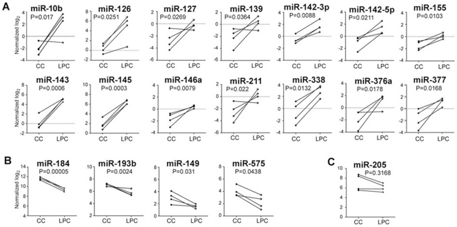
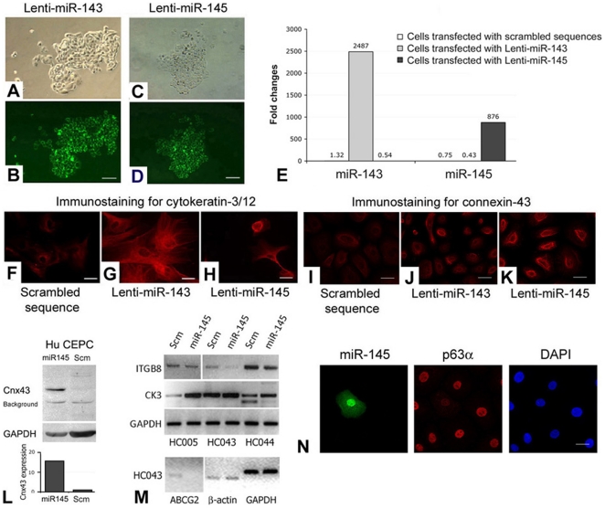
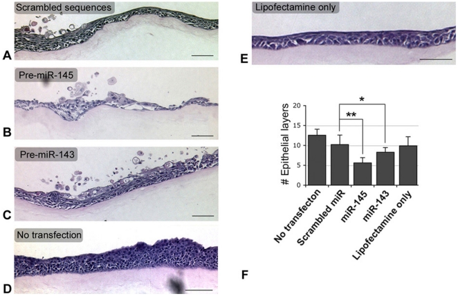
Methodology/principal findings: Human LPC epithelia was extracted for small RNAs or dissociated for CEPC culture. By Agilent Human microRNA Microarray V2 platform and GeneSpring GX11.0 analysis, we found differential expression of 18 microRNAs against central corneal (CC) epithelia, which were devoid of CEPCs. Among them, miR-184 was up-regulated in CC epithelia, similar to reported finding. Cluster miR-143/145 was expressed strongly in LPC but weakly in CC epithelia (P = 0.0004, Mann-Whitney U-test). This was validated by quantitative polymerase chain reaction (qPCR). Locked nucleic acid-based in situ hybridization on corneal rim cryosections showed miR-143/145 presence localized to the parabasal cells of limbal epithelium but negligible in basal and superficial epithelia. With holoclone forming ability, CEPCs transfected with lentiviral plasmid containing mature miR-145 sequence gave rise to defective epithelium in organotypic culture and had increased cytokeratin-3/12 and connexin-43 expressions and decreased ABCG2 and p63 compared with cells transfected with scrambled sequences. Global gene expression was analyzed using Agilent Whole Human Genome Oligo Microarray and GeneSpring GX11.0. With a 5-fold difference compared to cells with scrambled sequences, miR-145 up-regulated 324 genes (containing genes for immune response) and down-regulated 277 genes (containing genes for epithelial development and stem cell maintenance). As validated by qPCR and luciferase reporter assay, our results showed miR-145 suppressed integrin β8 (ITGB8) expression in both human corneal epithelial cells and primary CEPCs.

Conclusion/significance: We found expression of miR-143/145 cluster in human corneal epithelium. Our results also showed that miR-145 regulated the corneal epithelium formation and maintenance of epithelial integrity, via ITGB8 targeting.


Conflict of interest statement

Competing Interests: The authors have declared that no competing interests exist.

Figures

Figure 1
Figure 1. MicroRNA microarray analysis of human LPC and CC epithelial samples.
(A) MicroRNAs expressed with >2 folds higher in LPC compared to CC epithelia. (B) MicroRNAs expressed higher by >2 folds in CC compared to LPC epithelia. (C) miR-205 expression in both epithelia. Data were log(2) transformed and normalized to 75th percentile of signal intensity for standardization. Statistical significance was calculated by unpaired Student's t test.
Figure 2
Figure 2. Validation of miR-143 and miR-145 expression in limbal epithelium.
By qPCR analysis, (A) miR-143 and (B) miR-145 was up-regulated (compared to U6 expression) in LPC epithelia, when compared to CC (P<0.001, Mann Whitney U-test). Red lines indicated mean CT value. In situ hybridization showed (C) miR-143 and (E) miR-145 in limbal epithelium, compared to (G) scrambled sequences and (H) U6. At higher magnification, (D) miR-143 and (F) miR-145 were present in parabasal layers. Scale bars: 150 µm (C, E, G, H); 40 µm (D, F).
Figure 3
Figure 3. Transfection analysis of miR-143 and miR-145.
(A–D) Human P2 CEPCs transfected with (A and B) Lenti-miR-143 and (C and D) Lenti-miR-145. (A, C) Phase-contrast images; (B, D) live GFP imaging. (E) Overexpression levels of miR-143 and 145 in transfected CEPCs by qPCR analysis. Amplification signals from cells with scrambled sequences (Scm) were indicated. Immunofluorescence of (F–H) cytokeratin-3/12 and (I–K) connexin-43 in CEPCs transfected with (F, I) scrambled sequence, (G, J) Lenti-miR-143 and (H, K). (L) Western blotting and densitometry analysis of connexin-43 (Cnx43) and GAPDH in CEPCs transfected with Lenti-miR-145 or scrambled sequences. (M) RT-PCR result of integrin β8 (ITGB8), cytokeratin-3 (CK3), ABCG2, β-actin and GAPDH in different primary CEPCs (at P2) transfected with Lenti-miR-145 or scrambled sequences. (N) Immunofluorescence of miR-145 (revealed by GFP), p63α and nuclear DAPI stain in P2 CEPCs after Lenti-miR-145 transfection. Scale bars: (A–D) 100 µm; (F–K, N) 10 µm.
Figure 4
Figure 4. Corneal epithelial organotypic assay.
Representative hematoxylin-eosin stained pictures from serial sections of cell-denuded AM composite showing the thickest epithelium and the most epithelial layers. (A) CEPCs transfected with scrambled sequences, (B) CEPCs transfected with pre-miR-145, (C) CEPCs transfected with pre-miR-143 and (D) CEPCs with Lipofectamine 2000 and (E) non-transfected CEPCs. Scale bars: 100 µm. (F) Epithelium forming efficiency determined by the number of epithelial layers in 15 sites along the composite. * P = 0.042; ** P = 0.0002 (paired Student's t-test).
Figure 5
Figure 5. Target gene identification of miR-145 in human corneal epithelium.
(A) Gene expression analysis by qPCR showing that ITGB8 was significantly down-regulated (P = 0.00024, paired Student's t-test) and IFNB1 was induced after miR-145 transfection (P<0.005). Wnt7A, Klf4, SOCS7 and FBN3 showed no changes. The dots represented ΔCT values (CT of transfected cells subtracted with CT of control cells). Horizontal lines indicated mean CT values. Scm: scrambled sequences. (B) Sequences of two miR-145 binding sites located in human ITGB8 3′UTR. Yellow shaded regions represent the conserved complementary nucleotides of miR-145 seed sequence in different species. The first 28–34th nucleotide region is only conserved in primates whereas 4421–4427th region is conserved in primates and rodents. (C) HeLa cells co-transfected with wildtype (WT) pCHECK-ITGB8_3′UTR and pre-miR-145 showed lower luciferase reporter activities when compared with cells transfected with scrambled sequences (n = 5) (red labels). Disruption of binding site 4421–4427th region resulted in higher luciferase activity (blue labels) whereas mutation at 28–34th region had no influence and the reduced luciferase activity levels (green labels) were similar as WT. * P<0.005, as compared to scrambled control (one-way ANOVA). (D) Immunofluorescence of ITGB8 in human limbal epithelium. Positive immunoreactivity was observed scattered in basal layer and continuous in superficial layers. No observable expression was noted in the parabasal layers. (E) Immunoperoxidase staining of ITGB8 in organotypic generated epithelia. Distinct ITGB8 expression was observed in epithelial layers generated from CEPCs transfected with scrambled sequences whereas reduced expression was found in epithelia from pre-miR-145-transfected CEPCs. Scale bars (D, E): 50 µm.

References

    1. Pellegrini G, Golisano O, Paterna P, Lambiase A, Bonini S, et al. Location and clonal analysis of stem cells and their differentiated progeny in the human ocular surface. J Cell Biol. 1999;145:769–782. - PMC - PubMed
    1. Cotsarelis G, Cheng SZ, Dong G, Sun TT, Lavker RM. Existence of slow-cycling limbal epithelial basal cells that can be preferentially stimulated to proliferate: implications on epithelial stem cells. Cell. 1989;57:201–209. - PubMed
    1. Schermer A, Galvin S, Sun TT. Differentiation-related expression of a major 64K corneal keratin in vivo and in culture suggests limbal location of corneal epithelial stem cells. J Cell Biol. 1986;103:49–62. - PMC - PubMed
    1. Lekhanont K, Choubtum L, Chuck RS, Sangiampornpanit T, Chuckpaiwong V, et al. A serum- and feeder-free technique of culturing human corneal epithelial stem cells on amniotic membrane. Mol Vis. 2009;15:1294–1302. - PMC - PubMed
    1. Lehrer MS, Sun TT, Lavker RM. Strategies of epithelial repair: modulation of stem cell and transit amplifying cell proliferation. J Cell Sci. 1998;111:2867–2875. - PubMed

Publication types