. 2011 Aug;29(4):128-39.
doi: 10.3109/08977194.2011.593178. Epub 2011 Jun 27.

Divergent activities of osteogenic BMP2, and tenogenic BMP12 and BMP13 independent of receptor binding affinities

Affiliations

Divergent activities of osteogenic BMP2, and tenogenic BMP12 and BMP13 independent of receptor binding affinities

Stephen P Berasi et al. Growth Factors. 2011 Aug.

Abstract

Ectopic expression of recombinant human bone morphogenetic protein 2 (rhBMP2) induces osteogenesis, while ectopic expression of rhBMP12 and rhBMP13 induces the formation of tendon-like tissue. Despite their different in vivo activities, all three ligands bound to the type I bone morphogenic protein receptors (BMPRs), activin receptor-like kinase (ALK)-3 and ALK6, and to the type II BMPRs, activin receptor type-2A, activin receptor type-2B, and BMPR2, with similar affinities. Treatment of C3H10T1/2 cells with rhBMP2 activated SMAD signaling and induced expression of osteoblast markers including osteocalcin mRNA (Ocn). In contrast, treatment with rhBMP12 or rhBMP13 resulted in a dose-dependent induction of a tendon-specific gene (Thbs4) expression with no detectable activation of SMAD 1, 5, and 8. Differential regulation of Thbs4 and Ocn has potential utility as an in vitro biomarker for induction of tenogenic signaling. Such an assay also permits the ability to distinguish between the activities of different BMPs and may prove useful in studies on the molecular mechanisms of BMP tenogenic activity.


Figures

Figure 1
Figure 1
Characterization of purified rhBMP2, rhBMP12, and rhBMP13. (a) The purified proteins were collected and analyzed under reducing (RED) and nonreducing (NR) conditions using 12% polyacrylamide slab gels. (b) SDS-PAGE analysis of rhBMP12 purified from E. coli or CHO cells reduced with b-mercaptoethanol. (c) CD spectroscopy of BMP12 purified from E. coli or CHO cells.
Figure 2
Figure 2
Histology and X-ray analysis of BMP-treated implants after 14 days. Saffranin O stain of representative sections of implants containing (a) 25 µg of rhBMP2, (b) 25 µg of rhBMP12, or (c) 25 µg of rhBMP13. Faxitron X-ray images of representative implants containing (d) 25 µg of rhBMP2, (e) 25 µg of rhBMP12, or (f) 25 µg of rhBMP13. The implants containing rhBMP2 consists of mostly bone, whereas the implants containing rhBMP12 or rhBMP13 consists of only small little bone spicules (arrows).
Figure 3
Figure 3
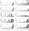
Expression of bone, tendon, and cartilage markers by BMPs in C3H10T1/2 cells. mRNA levels were determined by Taqman for the bone markers (a) Ocn (osteocalcin), (b) Runx2 (runt-related transcription factor 2, (c) Alpl (ALP), and (d) Sp7 (osterix), the tendon markers (e) Thbs4 (thrombospondin 4) and (f) Tnmd, (tenomodulin) and the cartilage markers (g) Sox9 (SRY-box containing gene 9), (h) Col2a1 (collagen, type II, alpha 1), (i) Acan (Aggrecan 1) and (j) Col11a1 (collagen, type 11, alpha 1).
Figure 4
Figure 4
BMP activity in C3H10T1/2 cells. (a) C3H10T1/2 cells infected with BRE-Luc construct were treated with increasing concentration of rhBMP2, rhBMP12, or rhBMP13. (b) ALP activity in C3H10T1/2 cells following 2-day incubation with 0, 1, 10, or 100nM of rhBMP2, rhBMP12, or rhBMP13.
Figure 5
Figure 5
Induction of SMAD signaling by BMPs in C3H10T1/2 cells. Cell lysates from rhBMP2, rhBMP12, or rhBMP13 treated C3H10T1/2 cells were analyzed by western blot with antibodies to phosphorylated SMAD 1/5/8 and to β-actin or tubulin. Cells were treated with (a)10 nM BMPs for 0 to 30 min, (b) 10 nM BMPs for 0 to 120 min or (c) treated with 100 nM BMPs for 0 to 3 h.
Figure 6
Figure 6
Kinase dead ALK3 blocks Thbs4 induction. (a) mRNA levels for Thbs4 were determined by Taqman after treatment with kinase dead ALK3 and rhBMP1 2. Error bars represent SD (N = 3). (b) anti-HA western of protein extract from the infected cells to detect expression of the kinase dead ALK3.

Similar articles

Cited by

  • Pathophysiology and Emerging Molecular Therapeutic Targets in Heterotopic Ossification.
    Felix-Ilemhenbhio F, Pickering GAE, Kiss-Toth E, Wilkinson JM. Felix-Ilemhenbhio F, et al. Int J Mol Sci. 2022 Jun 23;23(13):6983. doi: 10.3390/ijms23136983. Int J Mol Sci. 2022. PMID: 35805978 Free PMC article. Review.
  • Effects of BMSC-Derived EVs on Bone Metabolism.
    Zhou X, Cao H, Guo J, Yuan Y, Ni G. Zhou X, et al. Pharmaceutics. 2022 May 8;14(5):1012. doi: 10.3390/pharmaceutics14051012. Pharmaceutics. 2022. PMID: 35631601 Free PMC article. Review.
  • Polymorphisms near TBX5 and GDF7 are associated with increased risk for Barrett's esophagus.
    Palles C, Chegwidden L, Li X, Findlay JM, Farnham G, Castro Giner F, Peppelenbosch MP, Kovac M, Adams CL, Prenen H, Briggs S, Harrison R, Sanders S, MacDonald D, Haigh C, Tucker A, Love S, Nanji M, deCaestecker J, Ferry D, Rathbone B, Hapeshi J, Barr H, Moayyedi P, Watson P, Zietek B, Maroo N, Gay L, Underwood T, Boulter L, McMurtry H, Monk D, Patel P, Ragunath K, Al Dulaimi D, Murray I, Koss K, Veitch A, Trudgill N, Nwokolo C, Rembacken B, Atherfold P, Green E, Ang Y, Kuipers EJ, Chow W, Paterson S, Kadri S, Beales I, Grimley C, Mullins P, Beckett C, Farrant M, Dixon A, Kelly S, Johnson M, Wajed S, Dhar A, Sawyer E, Roylance R, Onstad L, Gammon MD, Corley DA, Shaheen NJ, Bird NC, Hardie LJ, Reid BJ, Ye W, Liu G, Romero Y, Bernstein L, Wu AH, Casson AG, Fitzgerald R, Whiteman DC, Risch HA, Levine DM, Vaughan TL, Verhaar AP, van den Brande J, Toxopeus EL, Spaander MC, Wijnhoven BP, van der Laan LJ, Krishnadath K, Wijmenga C, Trynka G, McManus R, Reynolds JV, O'Sullivan J, MacMathuna P, McGarrigle SA, Kelleher D, Vermeire S, Cleynen I, Bisschops R, Tomlinson I, Jankowski J. Palles C, et al. Gastroenterology. 2015 Feb;148(2):367-78. doi: 10.1053/j.gastro.2014.10.041. Epub 2014 Nov 5. Gastroenterology. 2015. PMID: 25447851 Free PMC article.
  • Growth Differentiation Factor 7 Prevents Sepsis-Induced Acute Lung Injury in Mice.
    Dong P, Zhang Y, Liu N, Yang JY, Wang HM, Geng Q. Dong P, et al. Evid Based Complement Alternat Med. 2022 Dec 22;2022:3676444. doi: 10.1155/2022/3676444. eCollection 2022. Evid Based Complement Alternat Med. 2022. Retraction in: Evid Based Complement Alternat Med. 2023 Jul 12;2023:9795462. doi: 10.1155/2023/9795462. PMID: 36588594 Free PMC article. Retracted.
  • MB109 as bioactive human bone morphogenetic protein-9 refolded and purified from E. coli inclusion bodies.
    Kuo MM, Nguyen PH, Jeon YH, Kim S, Yoon SM, Choe S. Kuo MM, et al. Microb Cell Fact. 2014 Feb 24;13(1):29. doi: 10.1186/1475-2859-13-29. Microb Cell Fact. 2014. PMID: 24559319 Free PMC article.

References

    1. Amthor H, Christ B, Weil M, Patel K. The importance of timing differentiation during limb muscle development. Curr Biol. 1998;8:642–652. - PubMed
    1. Aspenberg P, Forslund C. Enhanced tendon healing with GDF 5 and 6. Acta Orthop Scand. 1999;70:51–54. - PubMed
    1. Bolt P, Clerk AN, Luu HH, Kang Q, Kummer JL, Deng ZL, Olson K, Primus F, Montag AG, He TC, Haydon RC, Toolan BC. BMP-14 gene therapy increases tendon tensile strength in a rat model of Achilles tendon injury. J Bone Joint Surg Am. 2007;89:1315–1320. - PubMed
    1. Chang SC, Hoang B, Thomas JT, Vukicevic S, Luyten FP, Ryba NJ, Kozak CA, Reddi AH, Moos M., Jr Cartilage-derived morphogenetic proteins. New members of the transforming growth factor-beta superfamily predominantly expressed in long bones during human embryonic development. J Biol Chem. 1994;269:28227–28234. - PubMed
    1. Cook SD, Rueger DC. Osteogenic protein-1: Biology and applications. Clin Orthop Relat Res. 1996:29–38. - PubMed

Publication types

MeSH terms

LinkOut - more resources