. 2011 Aug;44(4):301-10.
doi: 10.1111/j.1365-2184.2011.00765.x.

RNAi-mediated inhibition of the desmosomal cadherin (desmoglein 3) impairs epithelial cell proliferation

Affiliations

RNAi-mediated inhibition of the desmosomal cadherin (desmoglein 3) impairs epithelial cell proliferation

T Mannan et al. Cell Prolif. 2011 Aug.

Abstract

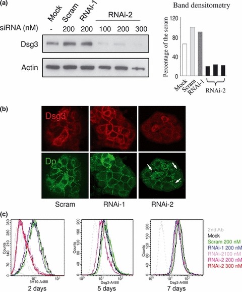
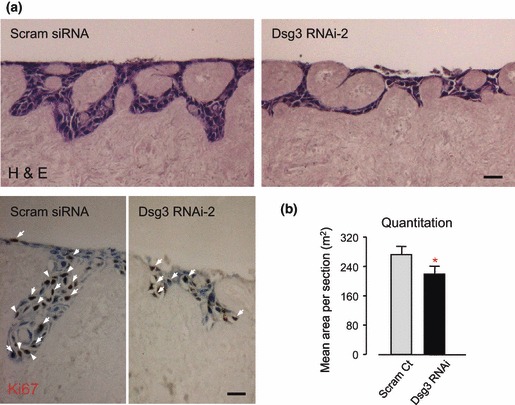
Objectives: Desmoglein 3 (Dsg3) is a desmosomal adhesion protein expressed in basal and immediate suprabasal layers of skin. Importance of Dsg3 in cell-cell adhesion and maintenance of tissue integrity is illustrated by findings of keratinocyte dissociation in the autoimmune disease, pemphigus vulgaris, where autoantibodies target Dsg3 on keratinocyte surfaces and cause Dsg3 depletion from desmosomes. However, recognition of possible participation of involvement of Dsg3 in cell proliferation remains controversial. Currently, available evidence suggests that Dsg3 may have both anti- and pro-proliferative roles in keratinocytes. The aim of this study was to use RNA interference (RNAi) strategy to investigate effects of silencing Dsg3 in cell-cell adhesion and cell proliferation in two cell lines, HaCaT and MDCK.

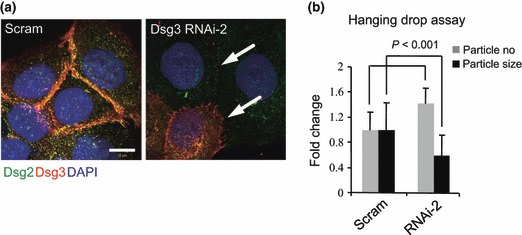
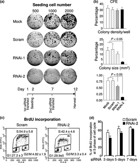
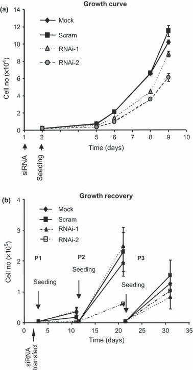
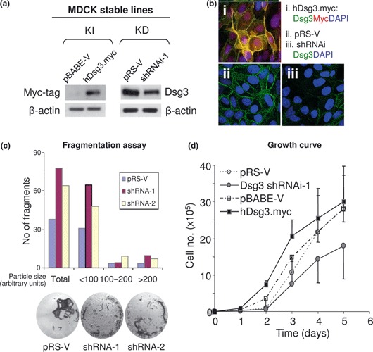
Materials and methods: Cells were transfected with siRNA, and knockdown of Dsg3 was assessed by western blotting, fluorescence-activated cell sorting and confocal microscopy. Cell-cell adhesion was analysed using the hanging drop/fragmentation assay, and cell proliferation by colony forming efficiency, BrdU incorporation, cell counts and organotypic culture.

Results: Silencing Dsg3 caused defects in cell-cell adhesion and concomitant reduction in cell proliferation in both HaCaT and MDCK cells.

Conclusion: These findings suggest that Dsg3 depletion by RNAi reduces cell proliferation, which is likely to be secondary to a defect in cell-cell adhesion, an essential function required for cell differentiation and morphogenesis.


Figures

Figure 1
Figure 1
 Dsg3 knockdown in HaCaTs. (a) Western blotting of Dsg3 in HaCaT cells transfected either with or without scrambled (Scram), RNAi‐1 and RNAi‐2 siRNAs at different concentrations, for 2 days; loading control –β‐actin. Densitometry of Dsg3 blot is shown on the right. Note that Dsg3 levels in RNAi‐2 treated cells were ∼20% of that of scrambled control regardless of different siRNA concentrations. (b) Confocal microscope images of HaCaT cells dual labelled for Dsg3 (AHP319) and Dp (115F) following transfection of scrambled siRNA, RNAi‐1 and RNAi‐2 for 3 days. Disruption of desmosomal junctions was seen in RNAi‐2‐treated cells for Dp staining (arrows). (c) FACS analysis of Dsg3 expression, labelled with mouse Dsg3 Ab 5H10, in HaCaTs transfected similarly (a) for a period of up to 7 days. Again, significant knockdown of Dsg3 was seen after 2 days regardless of siRNA concentration for RNAi‐2. RNAi‐1 showed very subtle effect on Dsg3 expression (blue line). After 7 days, expression of Dsg3 in RNAi treated cells recovered to almost that of normal control levels.
Figure 2
Figure 2
 Dsg3 silencing in HaCaTs caused reduction in Dsg2 and disruption of desmosome junctions. (a) Confocal images of HaCaT cells transfected with either scrambled siRNA (Scram) or Dsg3 RNAi‐2 and dual labelled for Dsg2 (green, with 33‐3D Ab) and Dsg3 (red, with 5H10 Ab) counterstained with DAPI. Note significant reduction in and disruption of desmosomal junctions (indicated by arrows) was evident in cells with both Dsg3 and Dsg2 depletion. Bar = 10 μm. (b) Hanging drop assay indicated significantly increased particle number and reduced particle size in cells with Dsg3 depletion. Data (mean ± SD) pooled from two independent experiments (sample size = 12).
Figure 3
Figure 3
 Dsg3 silencing in HaCaTs resulted in restricted colony growth, but no changes in CFE. (a) Colony‐forming assay of HaCaT cells transfected with either scrambled (Scram) or Dsg3 siRNAs. Cells seeded at 500, 1000 and 2000 cells/well in six‐well plates 1 day after first siRNA transfection. After 7 days, cells were subjected to second siRNA transfection and colonies were fixed and stained after 12 days. (b) Quantification of colonies from six wells of two representative experiments (mean ± SEM). (c) BrdU incorporation assay indicated that cells treated with RNAi‐2 had reduced cell entry into S phase (54.9 ± 5.8 in control versus 42.4 ± 4.6 in RNAi‐2). (d) Time course analysis of BrdU incorporation following Dsg3 knockdown by RNAi. Decreased BrdU incorporation was seen in RNAi‐2‐treated cells after 3–5 days after siRNA transfection compared to scrambled control cells.
Figure 4
Figure 4
 Transiently suppressed cell growth after Dsg3 knockdown. (a) Growth curve of HaCaT cells after transfection with or without scrambled (Scram), RNAi‐1 and RNAi‐2 siRNAs. Note that cells transiently transfected with Dsg3 siRNAs exhibited lower growth rate compared to controls, particularly, RNAi‐2‐treated cells. (b) Cell growth recovery over three passages after transient siRNA transfection in HaCaTs. Cells were seeded at the same density the following day after transfection and allowed to grow for 10 days (P1). Then, all cells were harvested and cell number for each population was determined by direct cell counting before seeding an aliquot of cells for culture again (P2). The same procedure was repeated for P3. Note that population growth rate of cells with Dsg3 knockdown appeared to be recovering gradually over time at P2 and P3.
Figure 5
Figure 5
 Cells with Dsg3 silencing displayed poor quality of skin regeneration. (a) One week organotypic culture of cells with or without Dsg3 knockdown (n > 3). Top: H&E staining; Bottom: Ki67 staining, positive cells marked with white arrows. Bars, 50 μm. (b) Quantification of epithelial areas in organotypic culture sections (n = 12). Note that cells treated with RNAi produced significantly lower cellular mass (*P < 0.05) and less Ki67 positive cells.
Figure 6
Figure 6
 Modulation of Dsg3 expression affected cell proliferation in MDCK cells. (a) Western blotting of cell lysates extracted from cells with either overexpression (knock‐in: KI) or knockdown (KD) of Dsg3. (b) Confocal microscopy of MDCK cells with ectopic Dsg3 expression [myc‐tag in (i)] or with Dsg3 knockdown (iii). Images in (ii) were vector control cells. (c) Dispase fragmentation assay of MDCK cells with either vector or Dsg3 shRNAi transduction. Cells were grown to confluence before being treated with 2.4 units/ml dispase for about 30 min to detach epithelial sheets. These sheets were subjected to mechanical stress by pipetting five times with 1 ml tips. Epithelial fragments were quantified by ImageJ and increased fragments (up to 2‐fold) were seen in cells with Dsg3 silencing by shRNAi‐1 or shRNAi‐2, respectively. Data are averages of duplicates in each group. (d) Growth curve of matched MDCK cells with up‐ or down‐regulation of Dsg3. Cells with overexpression had higher proliferation, but those with Dsg3 knockdown exhibited lower growth rate compared to matched control cells.

References

    1. Bass‐Zubek AE, Godsel LM, Delmar M, Green KJ (2009) Plakophilins: multifunctional scaffolds for adhesion and signaling. Curr. Opin. Cell Biol. 21, 708–716. - PMC - PubMed
    1. Garrod D, Kimura TE (2008) Hyper‐adhesion: a new concept in cell‐cell adhesion. Biochem. Soc. Trans. 36, 195–201. - PubMed
    1. Sharma P, Mao X, Payne AS (2007) Beyond steric hindrance: the role of adhesion signaling pathways in the pathogenesis of pemphigus. J. Dermatol. Sci. 48, 1–14. - PubMed
    1. Garrod D, Chidgey M (2008) Desmosome structure, composition and function. Biochim. Biophys. Acta 1778, 572–587. - PubMed
    1. Allen E, Yu QC, Fuchs E (1996) Mice expressing a mutant desmosomal cadherin exhibit abnormalities in desmosomes, proliferation, and epidermal differentiation. J. Cell Biol. 133, 1367–1382. - PMC - PubMed

Publication types