. 2011 Jun 25:9:97.
doi: 10.1186/1479-5876-9-97.

Protein kinase CK2α is overexpressed in colorectal cancer and modulates cell proliferation and invasion via regulating EMT-related genes

Affiliations

Protein kinase CK2α is overexpressed in colorectal cancer and modulates cell proliferation and invasion via regulating EMT-related genes

Jinjin Zou et al. J Transl Med. .

Abstract

Background: Protein kinase CK2 is a highly conserved, ubiquitous protein serine/threonine kinase that phosphorylates many substrates and has a global role in numerous biological and pathological processes. Overexpression of the protein kinase CK2α subunit (CK2α) has been associated with the malignant transformation of several tissues, with not nearly as much focus on the role of CK2α in colorectal cancer (CRC). The aims of this study are to investigate the function and regulatory mechanism of CK2α in CRC development.

Methods: Expression levels of CK2α were analyzed in 144 patients (104 with CRC and 40 with colorectal adenoma) by immunohistochemistry. Proliferation, senescence, motility and invasion assays as well as immunofluorescence staining and western blots were performed to assess the effect of CK2α in CRC.

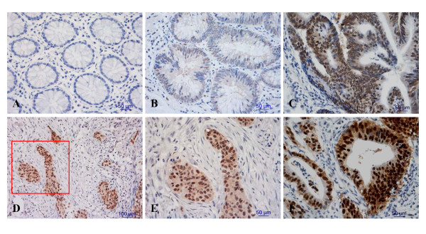
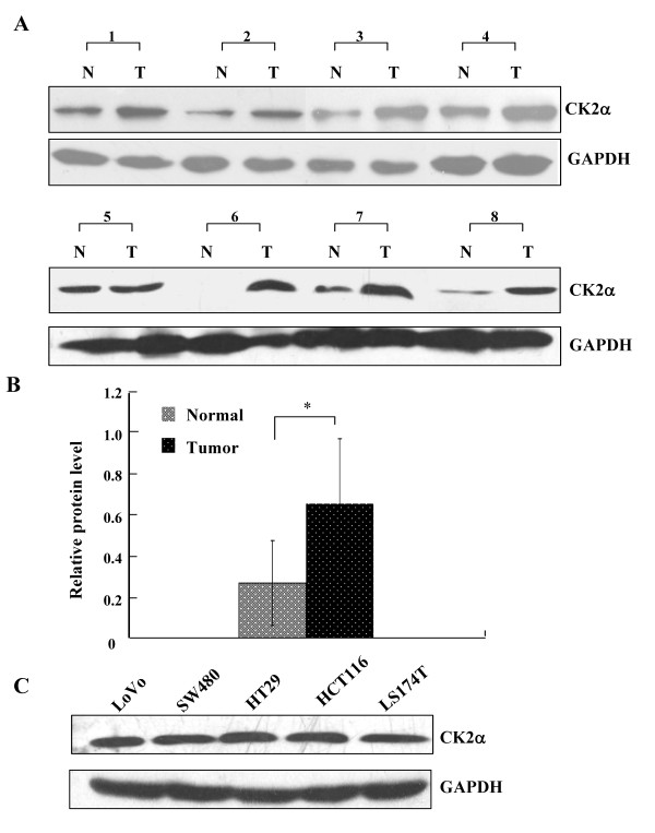
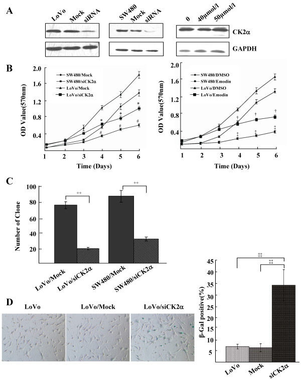
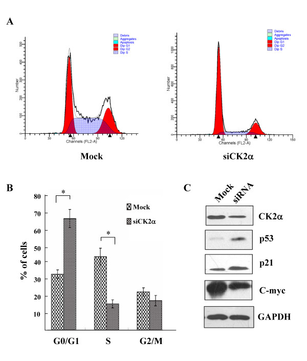
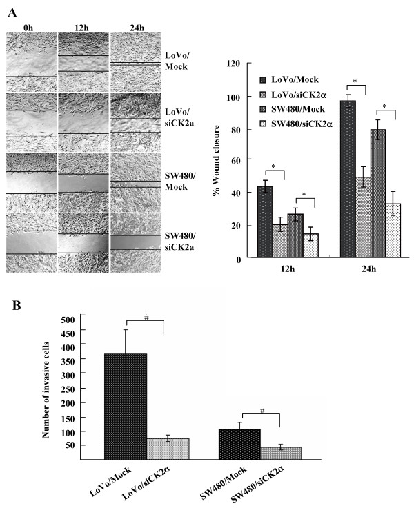
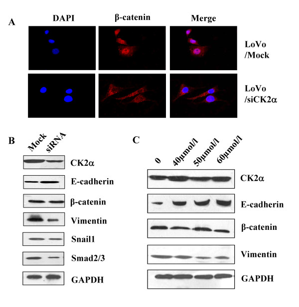
Results: The immunohistochemical expression of nuclear CK2α was stronger in tumor tissues than in adenomas and normal colorectal tissues. Suppression of CK2α by small-interfering RNA or the CK2α activity inhibitor emodin inhibited proliferation of CRC cells, caused G0/G1 phase arrest, induced cell senescence, elevated the expression of p53/p21 and decreased the expression of C-myc. We also found that knockdown of CK2α suppressed cell motility and invasion. Significantly, CK2α inhibition resulted in β-catenin transactivation, decreased the expression levels of vimentin and the transcription factors snail1 and smad2/3, and increased the expression of E-cadherin, suggesting that CK2α regulates the epithelial-mesenchymal transition (EMT) process in cancer cells.

Conclusions: Our results indicate that CK2α plays an essential role in the development of CRC, and inhibition of CK2α may serve as a promising therapeutic strategy for human CRC.


Figures

Figure 1
Figure 1
Immunohistochemical detection of CK2α expression in colorectal cancers, adenomas and adjacent normal colorectal mucosa. Staining was (A) negative in normal colorectal epithelium cells, (B, C) weak to moderate in the nuclei of colorectal adenoma cells, (D, E, F) and strong in the nuclei of colorectal cancer cells. (E is a close-up of the inset in D [framed in red]). Original magnification: × 200 (D), × 400 (A, B, C, E, F).
Figure 2
Figure 2
CK2α protein expression in CRC tissues and cell lines. (A) Western blot analysis of CK2α expression in eight pairs of CRC tissues and adjacent, normal colorectal mucosa tissues. N: normal colorectal mucosa tissue; T: tumor tissue. (B) Quantitative analysis of CK2α protein expression in eight pairs of CRC tissues and adjacent normal colorectal mucosa tissues. Columns, mean CK2α protein level after normalizing the data to GAPDH expression; bars, SD. *P < 0.01. (C) Western blot was used to detect CK2α expression in five CRC cell lines. GAPDH expression was used as a loading control.
Figure 3
Figure 3
Knockdown of CK2α inhibited cell proliferation and promoted cell senescence of CRC cell lines. (A) Western blot analysis of CK2α protein in lysates of cells transfected with a specific CK2α siRNA or treated with emodin. GAPDH expression was used as a loading control. (B) MTT assay of the proliferating cells transfected with a CK2α-specific siRNA or a nonspecific siRNA and treated with emodin. Points, mean of three independent experiments; bars, SD. *P < 0.01 versus LoVo/Mock; #P < 0.01 versus SW480/Mock; †P < 0.01 versus LoVo/DMSO; ‡P < 0.01 versus SW480/DMSO. (C) The number of colonies formed from cells transfected with CK2α siRNA. Colonies were stained with crystal violet and counted. Columns, mean of three independent experiments; bars, SD. ++P < 0.01. (D) The number of SA-β-gal-positive cells (green) 48 h after transfection with CK2α siRNA. Cells were stained with SA-β-gal staining solution. Columns, mean of three independent experiments; bars, SD. ‡‡P < 0.01.
Figure 4
Figure 4
CK2α inhibition induced G0/G1 phase arrest. (A) LoVo cells were transfected with CK2α-specific siRNA or nonspecific siRNA, stained with propidium iodide (PI), and monitored by flow cytometry to determine the cell cycle phase distribution. (B) Comparison of the percentage of cells in each phase of the cell cycle between LoVo cells transfected with CK2α-specific siRNA and nonspecific siRNA. Columns, mean of three independent experiments; bars, SD. *P < 0.01. (C) CK2α, p53, p21, C-myc and GAPDH expression in cells transfected with CK2α-specific siRNA was detected by western blot analysis.
Figure 5
Figure 5
Knockdown of CK2α inhibited cell migration and invasion of CRC cell lines. (A) Monolayers of cells transfected with CK2α-specific siRNAs were wounded by scraping, and wound closure was followed at 0, 12, and 24 h. The distance of the wound was measured. Columns, mean of three independent experiments; bars, SD. *P < 0.01. (B) After transfection with CK2α-specific siRNAs for 18 h, cells that migrated through the filters were counted in five randomly selected fields. Columns, mean of three independent experiments; bars, SD. #P < 0.05.
Figure 6
Figure 6
Inhibition of CK2α reversed the nuclear translocation of β-catenin and altered EMT-related genes expression. Reversal of EGF-induced nuclear translocation of β-catenin occurred in LoVo cells transfected with CK2α-specific siRNA (A), treated with EGF (100 ng/ml) for 2 h, and stained for immunofluorescence with β-catenin antibody (red) and DAPI (blue). (B) Western blot was used to detect the expression levels of CK2α, E-cadherin, β-catenin, vimentin and the transcription factors snail1 and smad2/3 in cells transfected with CK2α-specific siRNA. (C) One week later, in LoVo cells treated with emodin (40 μmol/l, 50 μmol/l and 60 μmol/l), the expressions of E-cadherin, β-catenin and vimentin were detected by western blot analysis. GAPDH expression was used as a loading control.

Similar articles

Cited by

References

    1. Jemal A, Siegel R, Ward E, Hao Y, Xu J, Murray T, Thun MJ. Cancer statistics, 2008. CA Cancer J Clin. 2008;58:71–96. doi: 10.3322/CA.2007.0010. - DOI - PubMed
    1. Sung JJ, Lau JY, Goh KL, Leung WK. Increasing incidence of colorectal cancer in Asia: implications for screening. Lancet Oncol. 2005;6:871–876. doi: 10.1016/S1470-2045(05)70422-8. - DOI - PubMed
    1. Pino MS, Chung DC. The chromosomal instability pathway in colon cancer. Gastroenterology. pp. 2059–2072. - DOI - PMC - PubMed
    1. Boland CR, Goel A. Microsatellite instability in colorectal cancer. Gastroenterology. pp. 2073–2087. e2073. - DOI - PMC - PubMed
    1. Hadziavdic V, Pavlovic-Calic N, Eminovic I. Microsatellite instability and loss of heterozygosity of tumor suppressor genes in Bosnian patients with sporadic colorectal cancer. Bosn J Basic Med Sci. 2008;8:313–321. - PMC - PubMed

Publication types

MeSH terms