. 2011 Dec;55(6):1256-62.
doi: 10.1016/j.jhep.2011.03.014. Epub 2011 Apr 13.

Hepatocyte γ-catenin compensates for conditionally deleted β-catenin at adherens junctions

Affiliations

Hepatocyte γ-catenin compensates for conditionally deleted β-catenin at adherens junctions

Emily Diane Wickline et al. J Hepatol. 2011 Dec.

Abstract

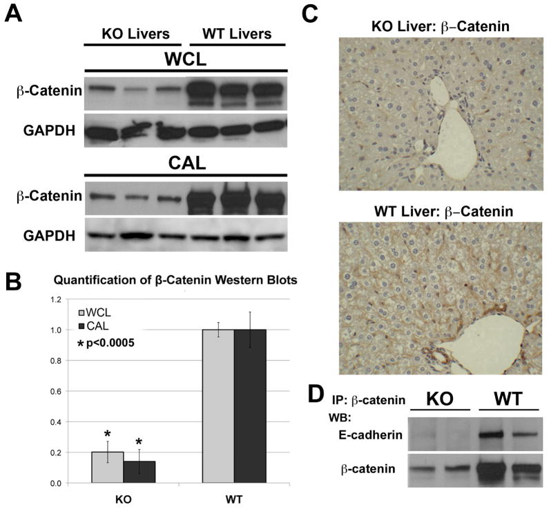
Background & aims: Wnt/β-catenin signaling is important in liver physiology. Moreover, β-catenin is also pivotal in adherens junctions (AJ). Here, we investigate hepatocyte-specific β-catenin conditional null mice (KO) for any alterations in AJ and related tight junctions (TJ).

Methods: Using gene array, PCR, Western blot, immunohistochemistry, immunofluorescence, and co-immunoprecipitation, we compare and contrast the composition of AJ and TJ in KO and littermate wild-type (WT) control livers.

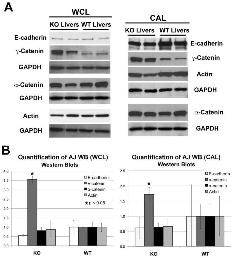
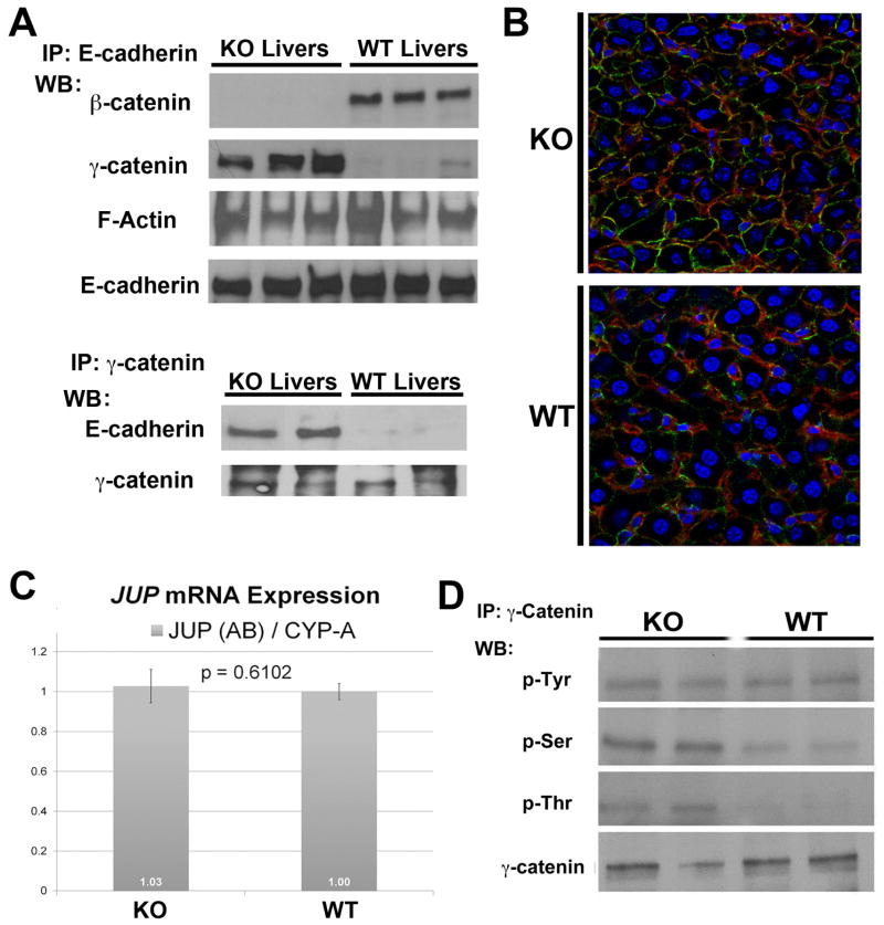
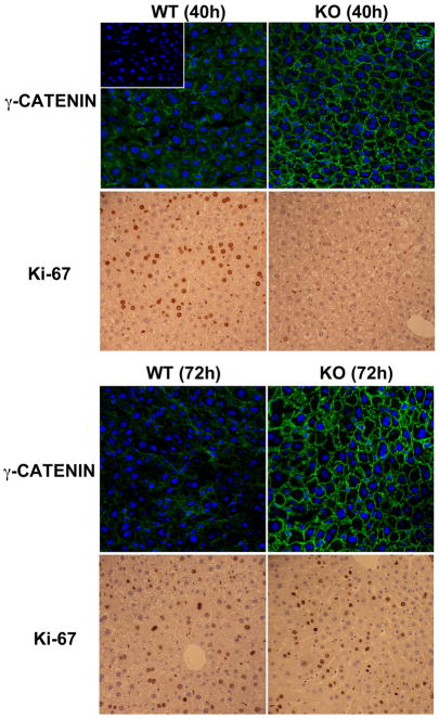
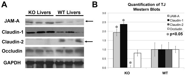
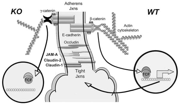
Results: We show association of E-cadherin with β-catenin in epithelial cells of WT livers, which is lost in the KOs. While total levels of α-catenin, E-cadherin, and F-actin were modestly decreased, KO livers show increased γ-catenin/plakoglobin. By co-immunoprecipitation, E-cadherin/β-catenin/F-actin association was observed in WT livers, while the association of E-cadherin/γ-catenin/F-actin was evident in KO livers. γ-Catenin was localized at the hepatocyte membrane at baseline in the KO liver. While γ-catenin gene expression remained unaltered, an increase in serine- and threonine-phosphorylated, but not tyrosine-phosphorylated γ-catenin was observed in KO livers. A continued presence of γ-catenin at the hepatocyte membrane, without any nuclear localization, was observed in liver regeneration after partial hepatectomy at 40 and 72 h, in both KO and WT. Analysis of TJ revealed lack of claudin-2 and increased levels of JAM-A and claudin-1 in KO livers.

Conclusions: β-Catenin adequately maintains AJ in the absence of β-catenin in hepatocytes; however, it lacks nuclear localization. Moreover, β-catenin/claudin-2 may be an important mechanism of crosstalk between the AJ and TJ.


Conflict of interest statement

Financial Conflict: None for any of the authors.

Figures

Figure 1
Figure 1. Assessment of β-catenin in KO and WT livers
A. β-Catenin (97kDa) and GAPDH (as loading control) protein levels in whole-cell lysates (WCL) and cytoskeleton-associated lysates (CAL) of three representative KO and age-and sex-matched WT livers by western blots (WB). B. Normalized (to GAPDH) densitometric analysis reveals significantly lower (p <0.05) average (+/−SD) β-catenin protein expression in KO than WT livers (n=3). C. Liver sections from KO and WT were analyzed for β-catenin expression by immunohistochemistry. D. WCLs were immunoprecipitated with anti-β-catenin antibody and probed for E-cadherin (120kDa), by WB.
Figure 2
Figure 2. Quantitative analysis of AJ proteins in KO and WT livers
A-E-cadherin (120kDa), γ-catenin (83kDa), α-catenin (100kDa) and F-actin (42kDa) protein levels were detected in whole cell lysates (WCL) and cytoskeleton-associated lysates (CAL) from two representative KO and age-matched WT livers by WB. WB for GAPDH verified comparable loading. B. Normalized (to GAPDH) densitometric analysis reveals significantly higher (P <0.05) average (+/−SD) protein expression of γ-catenin in KO than WT livers (n≥3), while other proteins showed insignificant differences between the two groups.
Figure 3
Figure 3. Mechanism of increased γ-catenin and its association with E-cadherin uniquely in KO livers
A. Whole-cell lysates were immunoprecipitated with anti-E-cadherin or anti-γ-catenin antibodies. WB performed for β-catenin (97kDa), γ-catenin (83kDa) and F-Actin (42kDa) (upper panel) show differential co-precipitation of β-catenin and F-actin with E-cadherin in WT or γ-catenin and F-actin and E-cadherin in WT livers. The conditions were reversed to show that γ-catenin also co-precipitates with E-cadherin (120kDa) in KO (lower panel). B. Increased membranous γ-catenin (green) associates with E-cadherin (red), prominently in the KO hepatocytes as shown by representative double immunofluorescence utilizing liver sections from KO and WT animals. C. Real-time PCR shown for γ-catenin gene (JUP) and cyclophilin-A reference gene shows insignificant difference in average mRNA expression (+/−SD) in KO versus WT livers (n=3). Similar results were obtained for all JUP and reference gene primer combinations. D. Whole-cell lysates immunoprecipitated with anti-γ-catenin antibody and western blotted for anti-phosphotyrosine, anti-phosphoserine, or anti-phosphothreonine show greater serine and threonine phosphorylation of γ-catenin in two representative KO livers as compared to WT, with no change in tyrosine phosphorylation.
Figure 4
Figure 4
Sustained membranous localization of γ-catenin in WT and KO livers during liver regeneration. While an increase in membrane signal of γ-catenin (green) is evident in KO as compared to WT, it was localized to hepatocyte membrane only at both 40 and 72 hours after partial hepatectomy, when several Ki-67+ve hepatocytes are observed in WT and KO livers, respectively. No signal is detected in negative control when primary antibody is omitted in the reaction as shown in the inset in upper left panel.
Figure 5
Figure 5. Changes in TJ protein expression in the KO livers
A. Examination of TJ protein expression by western blots using whole-cell lysates shows an increase in JAM-A (35kDa) and claudin-1 (22kDa), no change in occludin (60–82kDa) and absence of claudin-2 (22kDa) in KO livers. GAPDH served as the loading control. B. Normalized (to GAPDH) densitometric analysis reveals significantly (P <0.05) lower average (+/−SD) protein expression of claudin-2 and higher average (+/−SD) expression of JAM-A and claudin-1 in KO than WT livers (n≥3), while occludin remained unchanged.
Figure 6
Figure 6
A cartoon representing the configuration of AJ and TJ in β-catenin KO liver (left) and normal liver (right). While β-catenin bridges E-cadherin to F-actin in the WT livers, this function is compensated by γ-catenin in absence of β-catenin. β-Catenin and γ-catenin are both able to translocate to the nucleus, however β-catenin has robust nuclear interactions with TCF/LEF and hence transactivates expression of genes such as claudin-2, which is a means of crosstalk between AJ proteins and TJ components. However in KO, loss of claudin-2 impacts JAM-A and claudin-1 expression, which may affect TJ functions.

References

    1. Aberle H, Schwartz H, Kemler R. Cadherin-catenin complex: protein interactions and their implications for cadherin function. J Cell Biochem. 1996;61(4):514–523. - PubMed
    1. Behar J, Yeh TH, Krauland L, Otruba W, Cieply B, Hauth B, et al. Liver-specific beta-catenin knockout mice exhibit defective bile acid and cholesterol homeostasis and increased susceptibility to diet-induced steatohepatitis. Am J Pathol. 2010;176(2):744–753. - PMC - PubMed
    1. Benhamouche S, Decaens T, Godard C, Chambrey R, Rickman DS, Moinard C, et al. Apc tumor suppressor gene is the “zonation-keeper” of mouse liver. Dev Cell. 2006;10(6):759–770. - PubMed
    1. Bierkamp C, Schwarz H, Huber O, Kemler R. Desmosomal localization of beta-catenin in the skin of plakoglobin null-mutant mice. Development. 1999;126(2):371–381. - PubMed
    1. Birchmeier W, Weidner KM, Behrens J. Molecular mechanisms leading to loss of differentiation and gain of invasiveness in epithelial cells. J Cell Sci Suppl. 1993;17:159–164. - PubMed

Publication types

MeSH terms