. 2011 Dec;55(6):1263-71.
doi: 10.1016/j.jhep.2011.02.033. Epub 2011 Apr 13.

Treatment of newborn G6pc(-/-) mice with bone marrow-derived myelomonocytes induces liver repair

Affiliations

Treatment of newborn G6pc(-/-) mice with bone marrow-derived myelomonocytes induces liver repair

Roberta Resaz et al. J Hepatol. 2011 Dec.

Abstract

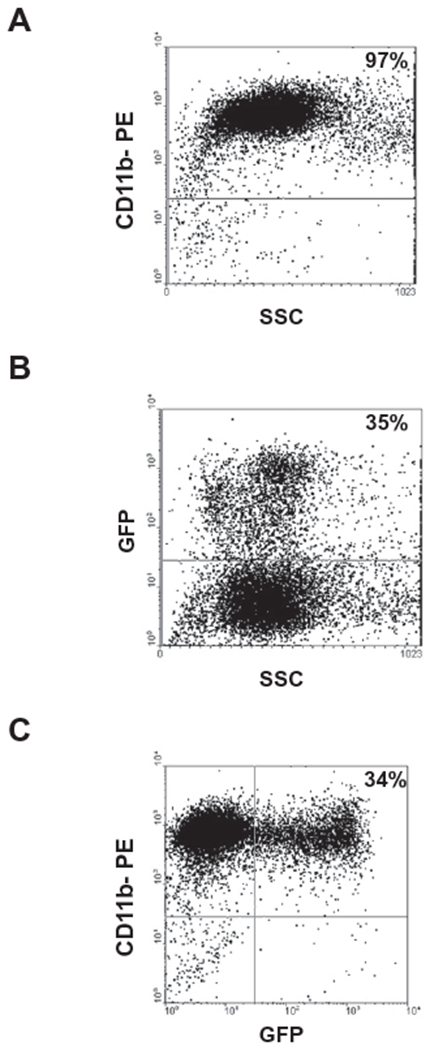
Background & aims: Several studies have shown that bone marrow-derived committed myelomonocytic cells can repopulate diseased livers by fusing with host hepatocytes and can restore normal liver function. These data suggest that myelomonocyte transplantation could be a promising approach for targeted and well-tolerated cell therapy aimed at liver regeneration. We sought to determine whether bone marrow-derived myelomonocytic cells could be effective for liver reconstitution in newborn mice knock-out for glucose-6-phosphatase-α.

Methods: Bone marrow-derived myelomonocytic cells obtained from adult wild type mice were transplanted in newborn knock-out mice. Tissues of control and treated mice were frozen for histochemical analysis, or paraffin-embedded and stained with hematoxylin and eosin for histological examination or analyzed by immunohistochemistry or fluorescent in situ hybridization.

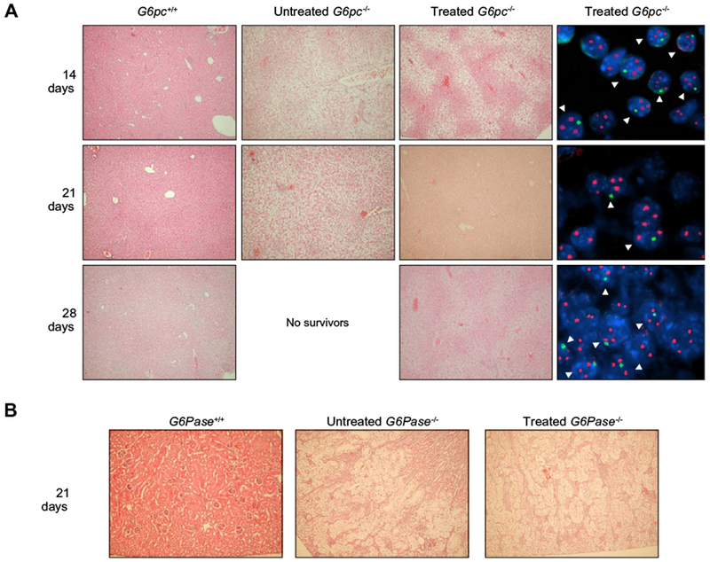
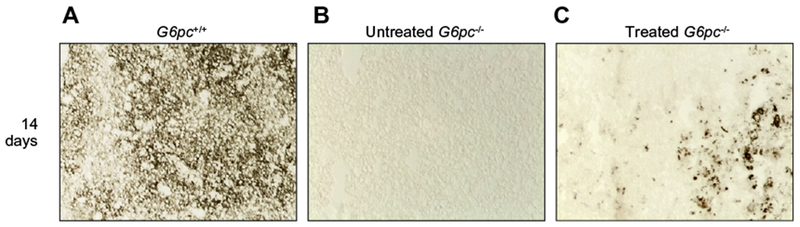
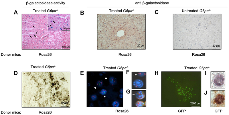
Results: Histological sections of livers of treated knock-out mice revealed areas of regenerating tissue consisting of hepatocytes of normal appearance and partial recovery of normal architecture as early as 1 week after myelomonocytic cells transplant. FISH analysis with X and Y chromosome paints indicated fusion between infused cells and host hepatocytes. Glucose-6-phosphatase activity was detected in treated mice with improved profiles of liver functional parameters.

Conclusions: Our data indicate that bone marrow-derived myelomonocytic cell transplant may represent an effective way to achieve liver reconstitution of highly degenerated livers in newborn animals.


Conflict of interest statement

Conflict of interest

The authors who have taken part in this study declared that they do not have anything to disclose regarding funding or conflict of interest with respect to this manuscript.

Figures

Fig. 1.
Fig. 1.. Isolation and CD11b expression of mouse bone marrow cells.
Myelomonocytic cells were purified from murine bone marrow cells by incubation with CD11 b (MAC-1) MicroBeads (as described in Materials and methods). (A) Purity of cell suspension was assessed by flow cytometric analysis after labeling cells for an individual staining with a PE-conjugated antibody against CD11b. (B) Myelomonocytic cells were purified from the bone marrow of GFP-Tg mice and purity of cell suspension was assessed by flow cytometric analysis for GFP+ expression. (C) Dot plot for two-color staining of CD11b+ and GFP+ is shown.
Fig. 2.
Fig. 2.. Histological analyses of tissues from G6pc−/− mice after BMM transplantation.
(A) G6pc−/− mice were transplanted with CD11b cells selected from the bone marrow of congenic G6pc+/+ mice. At 14 (n = 8), 21 (n = 10), and 28 (n = 7) days after treatment, mice were sacrificed and the livers processed. Control G6pc+/+ mice were sacrificed at 14 (n = 2), 21 (n = 2), and 28 (n = 2) days. Untreated G6pc−/− mice were sacrificed at 14 (n = 2) and 21 (n = 2). A very limited number of G6pc−/− could be analyzed since only 5% survived beyond 2 weeks of age and, due to their short lifespan, livers could not be collected beyond 3 weeks of age. Hematoxylin/eosin staining of livers of G6pc+/+ or untreated G6pc−/− mice is shown in the two left panels (20× magnification). Serial sections of livers from treated G6pc−/− mice were stained with hematoxylin/eosin (20× magnification) or analyzed with fluorescent in situ hybridization for detection of X and Y chromosome-positive nuclei (100× magnification). Representative results are shown for each time point in the two right panels. X chromosome signals are red and Y chromosome signals are green. (B) Hematoxylin- and eosin-stained kidneys from age-matched G6pc+/+ (n = 2), untreated G6pc−/− (n = 2), and BMM-treated G6pc−/− (n = 4) mice are shown. In the untreated G6pc−/− and BMM-treated G6pc−/− mice, the renal tubular epithelial cells are markedly enlarged by glycogen deposition, resulting in the compression of glomeruli. Glycogen deposits in renal epithelial cells are seen as clear areas. 40× magnification.
Fig. 3.
Fig. 3.. Qualitative histochemical analysis of G6Pase activity.
To evaluate the G6Pase activity, liver cryostat sections were treated as described in Material and methods. Colored lead sulfide was developed with ammonium sulfide. Representative results are shown. Liver sections were from (A) G6pc+/+, (B) untreated G6pc−/− and (C) treated G6pc−/− mice.
Fig. 4.
Fig. 4.. Injection of BMMs into livers of newborn and prenatal G6pc−/− mice.
Five-day-old G6pc−/− mice (n = 4) were treated by BMM injection directly into the liver median lobe (i.h.). Representative results obtained with livers of two treated mice are shown. Liver reconstitution can be appreciated in the treated liver median lobe (B and E) in comparison with an adjacent untreated lobe (A and D). In the treated areas, cell fusion can be detected (C and F), (arrowheads). Alternatively, BMMs were injected into the livers of 14/15-day-old fetuses (i.u.). Three pregnant females were utilized for intrauterine injections. Three G6pc−/− mice were born alive and analyzed at 3 weeks of age. (G and H) Hematoxylin/eosin staining of liver sections of two treated G6pc−/− mice are shown. Histologically normal liver areas are evident in animals injected at 15 days of gestation, where cell fusion can be visualized (I). (C, F, and I) 100× magnification.
Fig. 5.
Fig. 5.. Liver repopulation by Rosa 26- and GFP-Tg-derived BMMs.
G6pc−/− mice were injected into the temporal vein with BMMs derived from Rosa26 mice (n = 7) (A–G) or with BMMs derived from GFP-transgenic mice (n = 10) (H–J). A) Normal looking hepatocytes (black arrows) are visible after staining for β-galactosidase (visualized by the blue precipitate of X-gal) or (B) after immunostaining with anti-β-galactosidase antibody at 2 weeks after treatment. (C) A liver section from an untreated G6pc−/− mouse immunostained with anti-β-galactosidase antibody. (D) Histochemical analysis of G6Pase activity in a liver section from a mouse infused with BMMs derived from Rosa26 and (E) FISH analysis for X and Y chromosome detection in a liver section from the same animal. Presence of fused cells is indicated by arrowheads. (F) A liver section from a G6pc−/− mouse treated with BMMs derived from Rosa26 mice was analyzed by fluorescent in situ hybridization for detection of the X chromosome. A fused cell showing three X chromosomes is indicated by an arrow. (G) The same section was analyzed by immunofluorescence with an antibody anti-HNF1. A fused cell showing positivity for HNF1 is indicated by an arrow. (H) Whole mount of a liver from a 2-week-old G6pc−/− mouse with GFP-derived BMMs showing presence of fluorescent cells. (I) GFP-positive hepatocytes stained with anti-GFP and (J) double-stained with anti-albumin. (E, F, G, I, and J) 100× magnification.

References

    1. Almeida-Porada G, Zanjani ED, Porada CD. Bone marrow stem cells and liver regeneration. Exp Hematol 2010;38:574–580. - PMC - PubMed
    1. Gilchrist ES, Plevris JN. Bone marrow-derived stem cells in liver repair: 10 years down the line. Liver Transplant 2010;16:118–129. - PubMed
    1. Lagasse E, Connors H, Al Dhalimy M, Reitsma M, Dohse M, Osborne L, et al. Purified hematopoietic stem cells can differentiate into hepatocytes in vivo. Nat Med 2000;6:1229–1234. - PubMed
    1. Vassilopoulos G, Wang PR, Russell DW. Transplanted bone marrow regenerates liver by cell fusion. Nature 2003;422:901–904. - PubMed
    1. Wang X, Willenbring H, Akkari Y, Torimaru Y, Foster M, Al Dhalimy M, et al. Cell fusion is the principal source of bone-marrow-derived hepatocytes. Nature 2003;422:897–901. - PubMed

Publication types

MeSH terms

Substances