. 2011 Sep;231(1):171-80.
doi: 10.1016/j.expneurol.2011.06.005. Epub 2011 Jun 15.

Insulin resistance impairs nigrostriatal dopamine function

Affiliations

Insulin resistance impairs nigrostriatal dopamine function

J K Morris et al. Exp Neurol. 2011 Sep.

Abstract

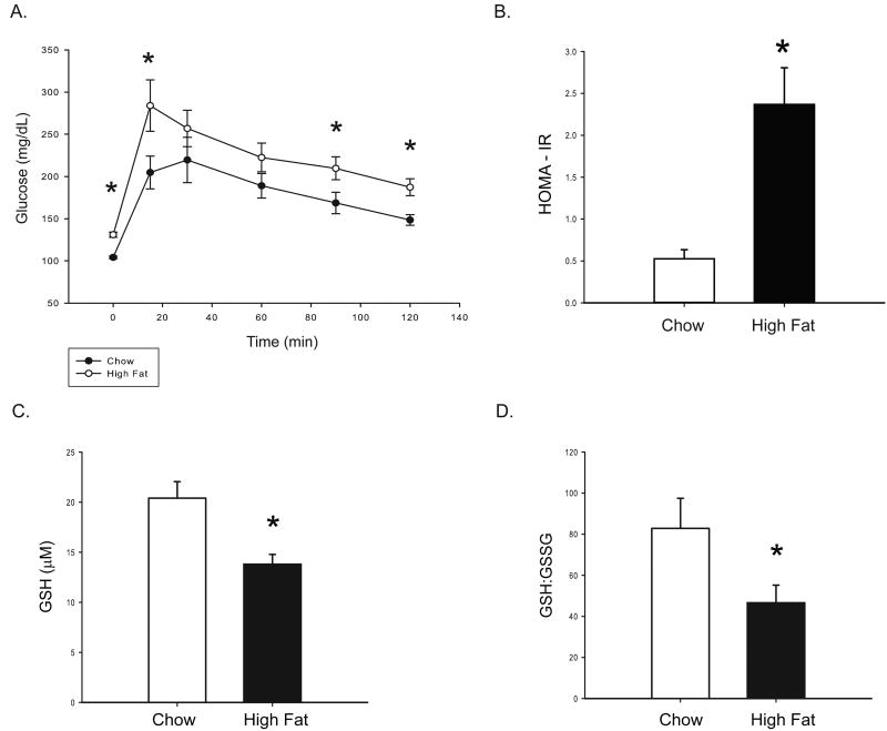
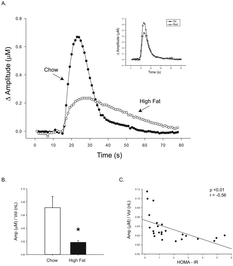
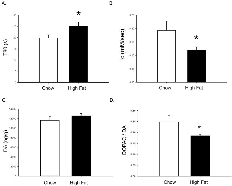
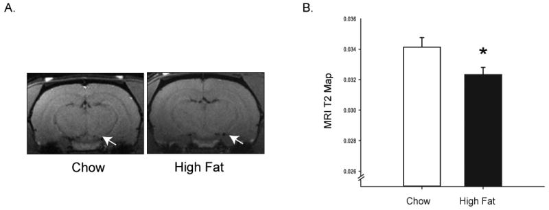
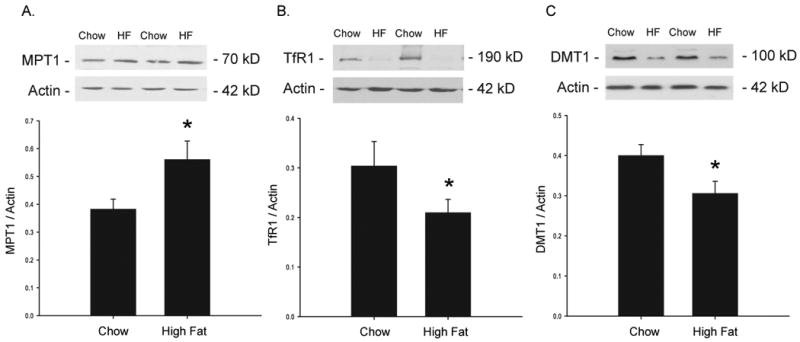
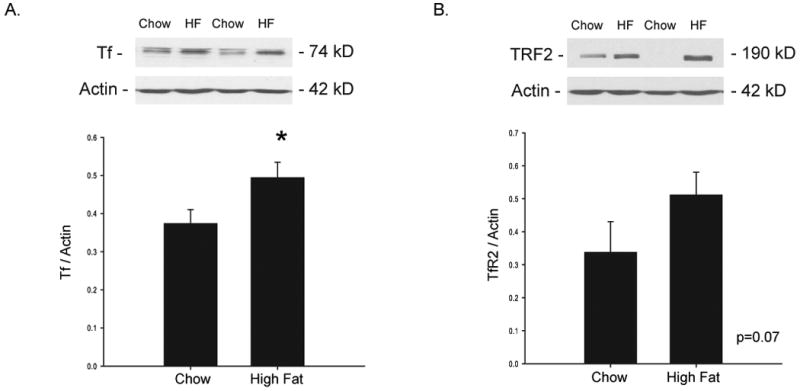
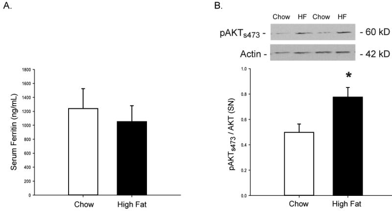
Clinical studies have indicated a link between Parkinson's disease (PD) and Type 2 Diabetes. Although preclinical studies have examined the effect of high-fat feeding on dopamine function in brain reward pathways, the effect of diet on neurotransmission in the nigrostriatal pathway, which is affected in PD and parkinsonism, is less clear. We hypothesized that a high-fat diet, which models early-stage Type 2 Diabetes, would disrupt nigrostriatal dopamine function in young adult Fischer 344 rats. Rats were fed a high fat diet (60% calories from fat) or a normal chow diet for 12 weeks. High fat-fed animals were insulin resistant compared to chow-fed controls. Potassium-evoked dopamine release and dopamine clearance were measured in the striatum using in vivo electrochemistry. Dopamine release was attenuated and dopamine clearance was diminished in the high-fat diet group compared to chow-fed rats. Magnetic resonance imaging indicated increased iron deposition in the substantia nigra of the high fat group. This finding was supported by alterations in the expression of several proteins involved in iron metabolism in the substantia nigra in this group compared to chow-fed animals. The diet-induced systemic and basal ganglia-specific changes may play a role in the observed impairment of nigrostriatal dopamine function.


Figures

Figure 1
Figure 1. Peripheral glucose tolerance and oxidative stress
After an overnight (12 hour) fast, an intraperitoneal glucose tolerance test was performed. A 60% glucose solution was administered intraperitoneally at 2g glucose/kg body weight. (A) Glucose was measured in tail blood at six time points: 0,15,45,60, 90, and 120 minutes after the glucose bolus (injection at t=0). Over the course of the test, glucose levels were higher in HF fed animals. HOMA-IR was also significantly increased (B), indicating insulin resistance. Serum levels of reduced glutathione (C) and the ratio of reduced to oxidized glutathione (D) were measured to gauge diet-induced oxidative stress. HF-feeding decreased reduced glutathione levels and decreased the ratio of reduced to oxidized glutathione. Values are means ± SE for 11-15 rats per group. *P<0.05 chow vs. HF.
Figure 2
Figure 2. Diet-induced insulin resistance attenuates striatal DA release
In vivo electrochemistry was used to measure potassium-evoked DA release in the striatum. A representative plot of evoked DA signals seen in response to a 50nL KCl stimulus in a HF vs. Chow animal is shown (A). A representative plot of redox ratios indicates specific detection of DA in our study (inset). HF-feeding significantly decreased the amount of DA released per volume of stimulus (B). The degree of insulin resistance (HOMA-IR) correlated negatively with the evoked DA concentration: more insulin resistant animals were less responsive to stimulus (C). Values are means ± SE for 11-15 rats per group. *p<0.05 chow vs. HF.
Figure 3
Figure 3. Uptake and turnover mechanics are affected by diet
The rate of DA uptake was also affected by diet. The amount of time required to clear DA (T80) was increased (A) and the rate of clearance (Tc) was decreased (B) with a HF diet. Although DA content was not affected (C), the DOPAC/DA ratio (D), which indicates decreased DA turnover, was significantly decreased in HF-fed animals. Values are means ± SE for 11-15 rats per group. *p<0.05 chow vs. HF.
Figure 4
Figure 4. Markers of iron deposition are increased with HF feeding
Magnetic resonance imaging (MRI) was performed on a subset of rats. MRI revealed hypointensity in the SN region, denoted by an arrow (A). T2 values were significantly decreased with a HF diet (B), indicating increased iron deposition. Values are means ± SE for a subset of 3 rats per group for MRI measures. *p<0.05 chow vs. HF.
Figure 5
Figure 5. Expression of IRE-regulated proteins in the SN
Expression of proteins involved in iron transport was also affected in the SN. Ferroportin was significantly increased (A), while TFR1 (B) and DMT1 (C) were significantly decreased, suggesting increased intracellular iron content. Values are means ± SE for 11-15 rats per group. *p<0.05 chow vs. HF.
Figure 6
Figure 6. Expression of non-IRE regulated proteins in the SN
The expression of other proteins involved in iron transport was affected as well. Tf was increased (A) and a trend for increased TfR2 was observed (B), indicating further effects of increased iron. Values are means ± SE for 11-15 rats per group. *p<0.05 chow vs. HF.
Figure 7
Figure 7. Mechanisms for increased iron deposition
(A) Ferritin levels were not different between groups, indicating that effects on iron transport may be tissue specific and not due to increased circulating iron. Activation of AKT via phosphorylation was analyzed using Western blot (B) and indicates upregulation of PI3-K pathway signaling, which has been implicated in iron transport. Serum ferritin levels were analyzed to determine if whole-body iron stores were affected by diet. Values are means ± SE for 11-15 rats per group. *p<0.05 chow vs. HF.

References

    1. Abbott RD, Ross GW, White LR, Nelson JS, Masaki KH, Tanner CM, Curb JD, Blanchette PL, Popper JS, Petrovitch H. Midlife adiposity and the future risk of Parkinson's disease. Neurology. 2002;59:1051–1057. - PubMed
    1. Alessi DR, Andjelkovic M, Caudwell B, Cron P, Morrice N, Cohen P, Hemmings BA. Mechanism of activation of protein kinase B by insulin and IGF-1. Embo J. 1996;15:6541–6551. - PMC - PubMed
    1. Anderson C, Checkoway H, Franklin GM, Beresford S, Smith-Weller T, Swanson PD. Dietary factors in Parkinson's disease: the role of food groups and specific foods. Mov Disord. 1999;14:21–27. - PubMed
    1. Anderson GJ, Vulpe CD. Mammalian iron transport. Cell Mol Life Sci. 2009;66:3241–3261. - PMC - PubMed
    1. Andrews ZB, Erion D, Beiler R, Liu ZW, Abizaid A, Zigman J, Elsworth JD, Savitt JM, DiMarchi R, Tschoep M, Roth RH, Gao XB, Horvath TL. Ghrelin promotes and protects nigrostriatal dopamine function via a UCP2-dependent mitochondrial mechanism. J Neurosci. 2009;29:14057–14065. - PMC - PubMed

Publication types

MeSH terms