. 2011 Sep;231(1):160-70.
doi: 10.1016/j.expneurol.2011.06.004. Epub 2011 Jun 15.

An angiogenic inhibitor, cyclic RGDfV, attenuates MPTP-induced dopamine neuron toxicity

Affiliations

An angiogenic inhibitor, cyclic RGDfV, attenuates MPTP-induced dopamine neuron toxicity

Aditiben Patel et al. Exp Neurol. 2011 Sep.

Abstract

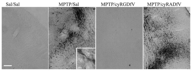
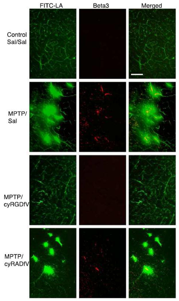
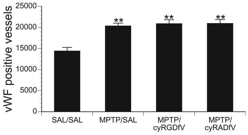
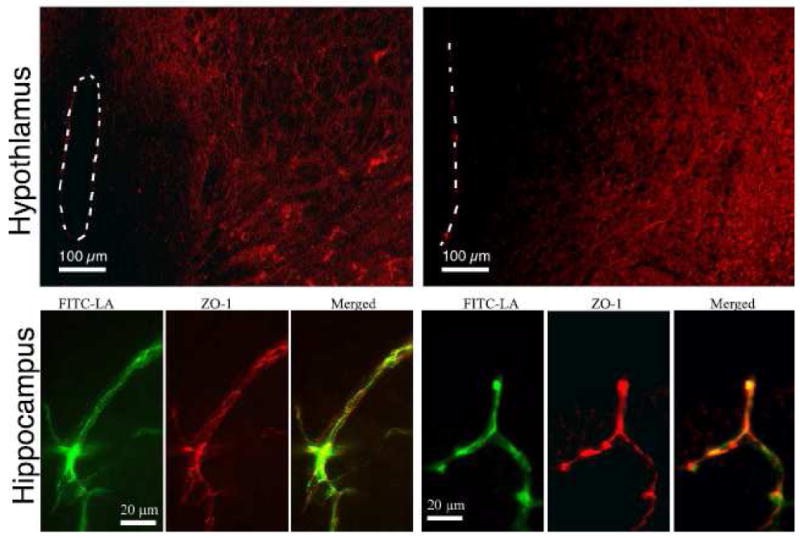
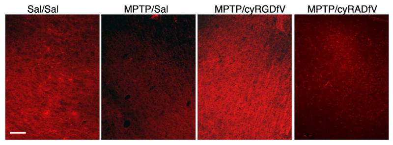
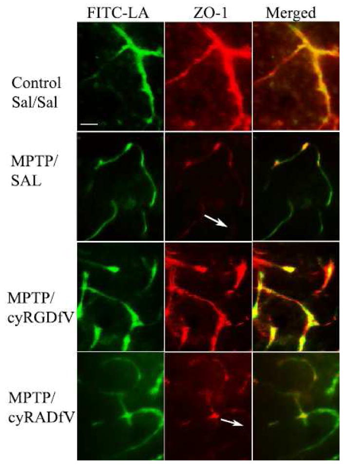
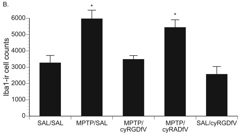
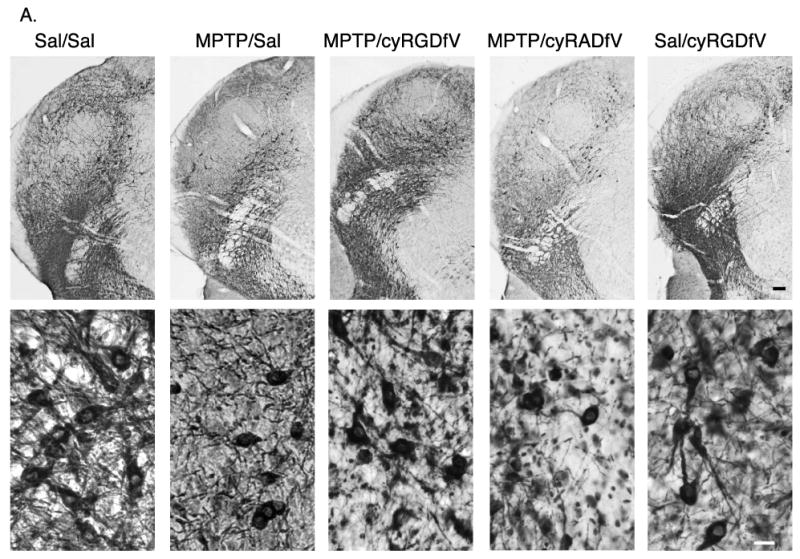
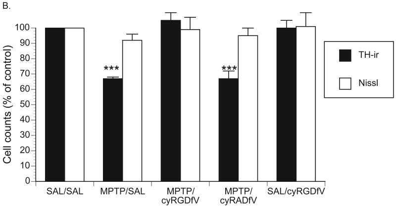
We previously demonstrated that several dopamine (DA) neurotoxins produced punctate areas of FITC-labeled albumin (FITC-LA) leakage in the substantia nigra and striatum suggesting blood brain barrier (BBB) dysfunction. Further, this leakage was co-localized with αvβ3 integrin up-regulation, a marker for angiogenesis. This suggested that the FITC-LA leakage might have been a result of angiogenesis. To assess the possible role of angiogenesis in DA neuron loss, we treated mice with 1-methyl-4-phenyl-1,2,3,6 tetrahydropyridine (MPTP) and on the following day treated with cyRGDfV, a cyclic peptide that binds to integrin αvβ3 and prevents angiogenesis. Post-treatment for 3 days (b.i.d.) with cyRGDfV blocked the MPTP-induced upregulation of integrin β3 immunoreactivity (a marker for angiogenesis), leakage of FITC-LA into brain parenchyma (a marker for BBB disruption) as well as the down regulation of Zona Occludin-1 (ZO-1; a marker for tight junction integrity). In addition, cyRGDfV also completely prevented tyrosine hydroxylase immunoreactive cell loss (a marker for DA neurons) and markedly attenuated the up-regulation of activated microglia (Iba1 cell counts and morphology). These data suggest that cyRGDfV, and perhaps other anti-angiogenic drugs, are neuroprotective following acute MPTP treatment and may suggest that compensatory angiogenesis and BBB dysfunction may contribute to inflammation and DA neuron loss.


Figures

Figure 1
Figure 1
Representative photomicrographs of β3 immunoreactivity in the substantia nigra of mice from the four treatment groups (scale bar = 100 μm). A representative higher magnification photomicrograph (scale bar = 20 μm) from an MPTP/Sal mouse reveals that the β3-ir was confined to vessels.
Figure 2
Figure 2
Representative photomicrographs of FITC-LA (green) and β3-ir (red) in the substantia nigra from the four treatment groups. Note in the merged figures that the β3 is generally co-localized in the center of areas of FITC-LA leakage suggesting a relationship between β3 up-regulation and leakage. (scale bar = 100 μm)
Figure 3
Figure 3
Stereological counts of von Willebrand factor (vWF) immunoreactive vessels in the substantia nigra from the four treatment groups (n=4/group). (Dunnett post hoc; ** = p<0.01)
Figure 4
Figure 4
Representative fluorescence photomicrographs of ZO-1 immunoreactivity in the hypothalamus and hippocampus of saline treated controls (left) and MPTP treated mice (right). The third ventricle is outlined. Note the characteristic halo effect around the ventricle where the BBB is absent (scale bar = 100 μm). In the hippocampus, note the extensive co-localization of ZO-1-ir on FITC-LA filled vessels (scale bar = 20 μm)
Figure 5
Figure 5
Representative fluorescence photomicrographs of ZO-1 immunoreactivity in the SN of the four treatment groups. (scale bar is 100 μm)
Figure 6
Figure 6
Representative fluorescence photomicrographs of FITC-LA vessels (green) and ZO-1 immunoreactivity (red) in the SNpc of the four treatment groups. Note the reduction in ZO-1-ir co-localization in the MPTP/Sal and MPTP/cyRADfV merged images and apparent areas where there is no ZO-1-ir on a FITC-LA filled vessel (arrows). (scale bar = 20 μm)
Figure 7
Figure 7
(A) Representative low-power (top row; scale bar = 100 μm) and higher power (bottom row; scale bar = 20 μm) photomicrographs of Iba1-ir cells in the SN of the five treatment groups. Note the larger cell bodies and thicker process indicative of microglial activation in the MPTP/Sal and MPTP/cyRADfV groups compared with controls. (B) Stereological Iba1-ir cell counts from the SN of the five treatment groups (n=5/group). (Tukey-Kramer post hoc * = p < 0.05)
Figure 7
Figure 7
(A) Representative low-power (top row; scale bar = 100 μm) and higher power (bottom row; scale bar = 20 μm) photomicrographs of Iba1-ir cells in the SN of the five treatment groups. Note the larger cell bodies and thicker process indicative of microglial activation in the MPTP/Sal and MPTP/cyRADfV groups compared with controls. (B) Stereological Iba1-ir cell counts from the SN of the five treatment groups (n=5/group). (Tukey-Kramer post hoc * = p < 0.05)
Figure 8
Figure 8
(A) Representative low-power (top row; scale bar = 100μm) and higher power (bottom row; scale bar = 20 μm) photomicrographs of TH-ir cells in the substantia nigra of the five treatment groups. (B) Stereological TH-ir and Nissl cell counts expressed as their respective % of Sal/Sal treated means (± SEM) in the five treatment groups (n=5/group). (Tukey-Kramer post hoc *** = p < 0.001)
Figure 8
Figure 8
(A) Representative low-power (top row; scale bar = 100μm) and higher power (bottom row; scale bar = 20 μm) photomicrographs of TH-ir cells in the substantia nigra of the five treatment groups. (B) Stereological TH-ir and Nissl cell counts expressed as their respective % of Sal/Sal treated means (± SEM) in the five treatment groups (n=5/group). (Tukey-Kramer post hoc *** = p < 0.001)

References

    1. Akiyama H, Kawamata T, Dedhar S, McGeer PL. Immunohistochemical localization of vitronectin, its receptor and beta-3 integrin in Alzheimer brain tissue. J Neuroimmunol. 1991;32:19–28. - PubMed
    1. Antonini A, Moresco RM, Gobbo C, De Notaris R, Panzacchi A, Barone P, Bonifati V, Pezzoli G, Fazio F. Striatal dopaminergic denervation in early and late onset Parkinson's disease assessed by PET and the tracer [11C]FECIT: preliminary findings in one patient with autosomal recessive parkinsonism (Park2) Neurol Sci. 2002;23 2:S51–52. - PubMed
    1. Astradsson A, Jenkins BG, Choi JK, Hallett PJ, Levesque MA, McDowell JS, Brownell AL, Spealman RD, Isacson O. The blood-brain barrier is intact after levodopa-induced dyskinesias in parkinsonian primates--evidence from in vivo neuroimaging studies. Neurobiol Dis. 2009;35:348–351. - PMC - PubMed
    1. Aumailley M, Gurrath M, Muller G, Calvete J, Timpl R, Kessler H. Arg-Gly-Asp constrained within cyclic pentapeptides. Strong and selective inhibitors of cell adhesion to vitronectin and laminin fragment P1. FEBS Lett. 1991;291:50–54. - PubMed
    1. Baluk P, Lee CG, Link H, Ator E, Haskell A, Elias JA, McDonald DM. Regulated angiogenesis and vascular regression in mice overexpressing vascular endothelial growth factor in airways. Am J Pathol. 2004;165:1071–1085. - PMC - PubMed

Publication types

MeSH terms

LinkOut - more resources