

Effects of transcranial direct current stimulation (tDCS) on human regional cerebral blood flow
- PMID: 21703350
- PMCID: PMC3155947
- DOI: 10.1016/j.neuroimage.2011.06.018
Effects of transcranial direct current stimulation (tDCS) on human regional cerebral blood flow
Abstract
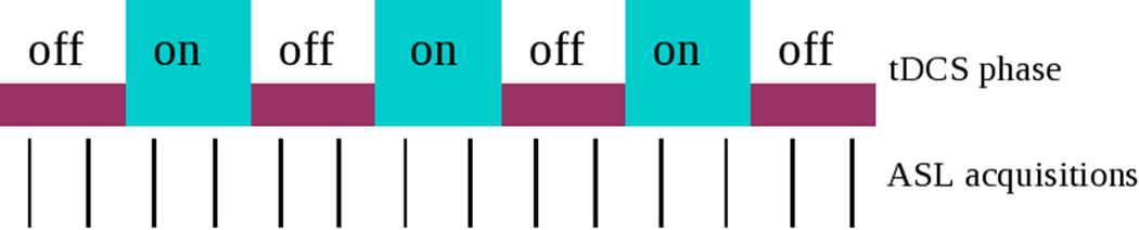
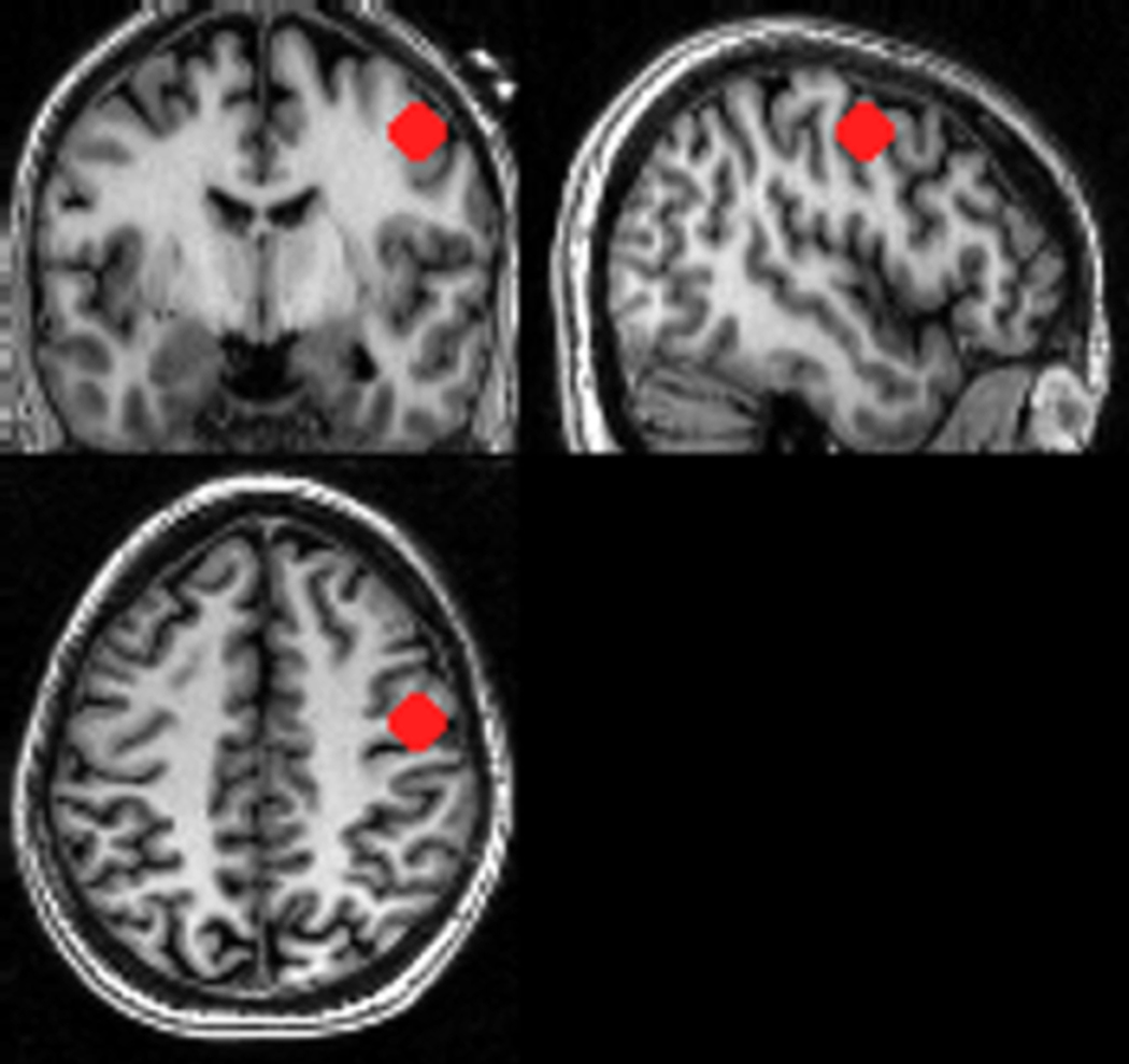
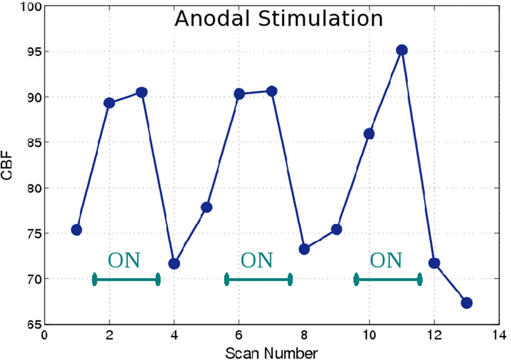
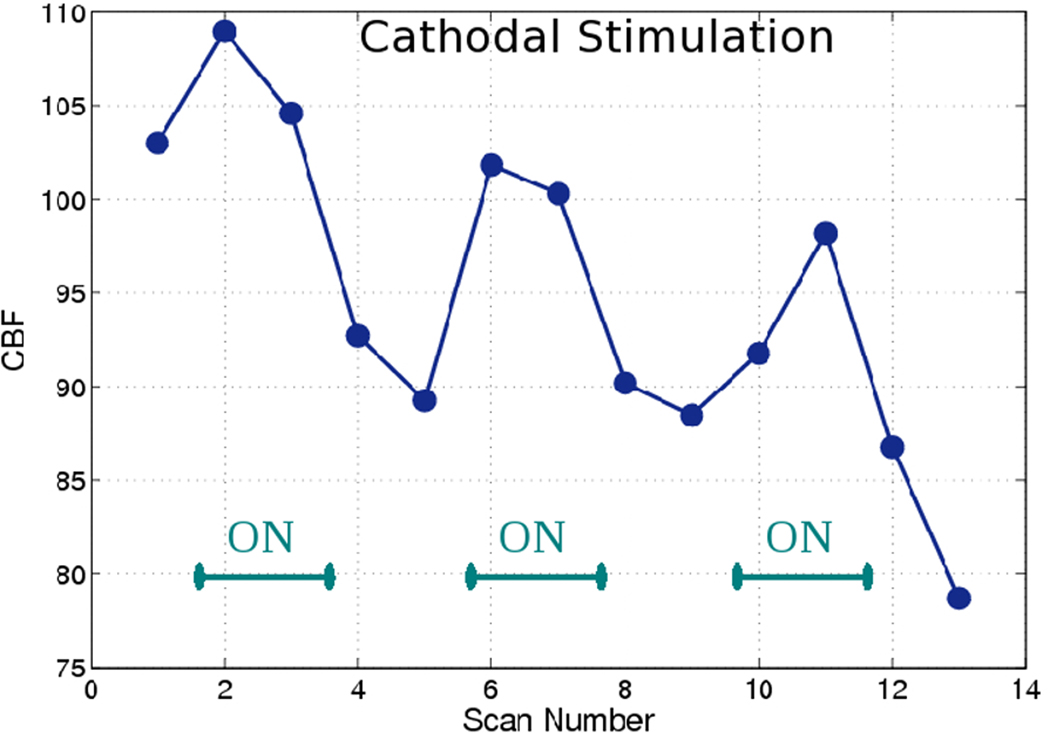
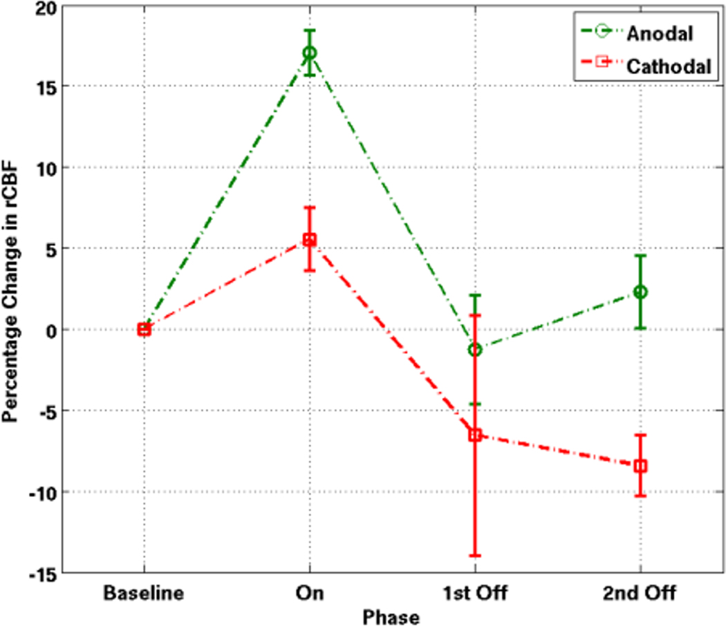
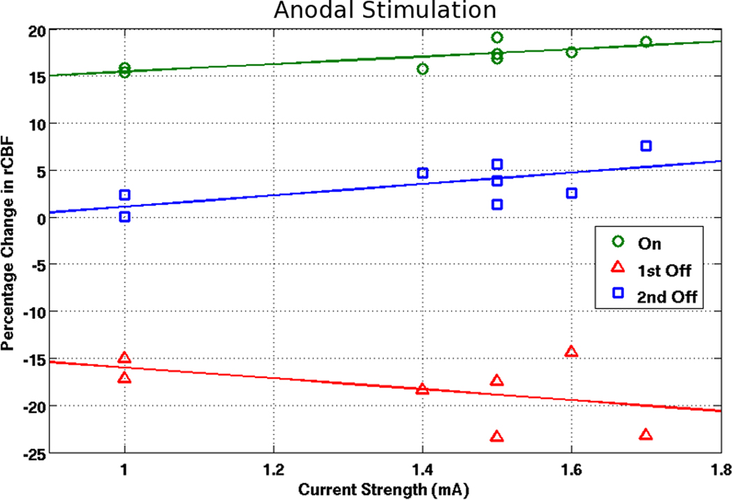
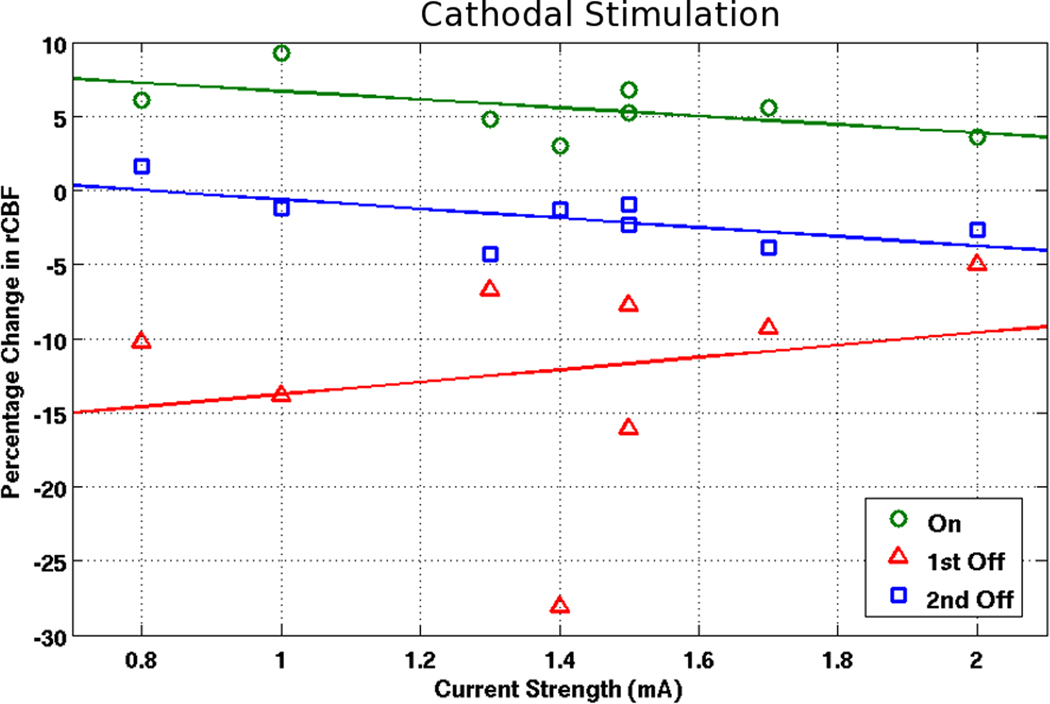
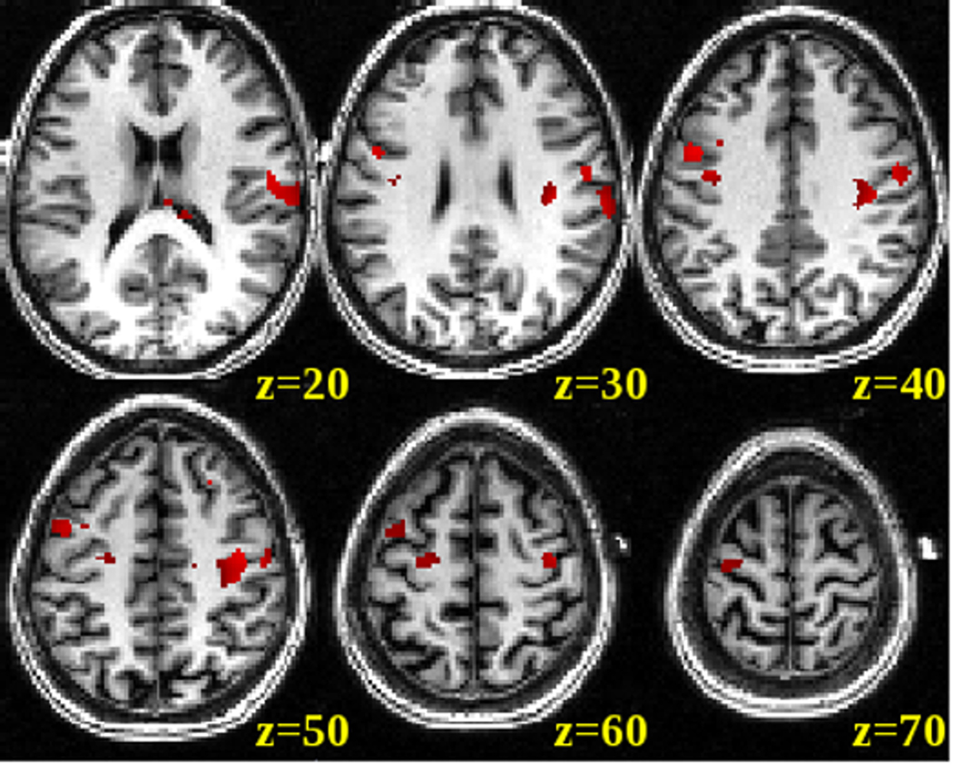
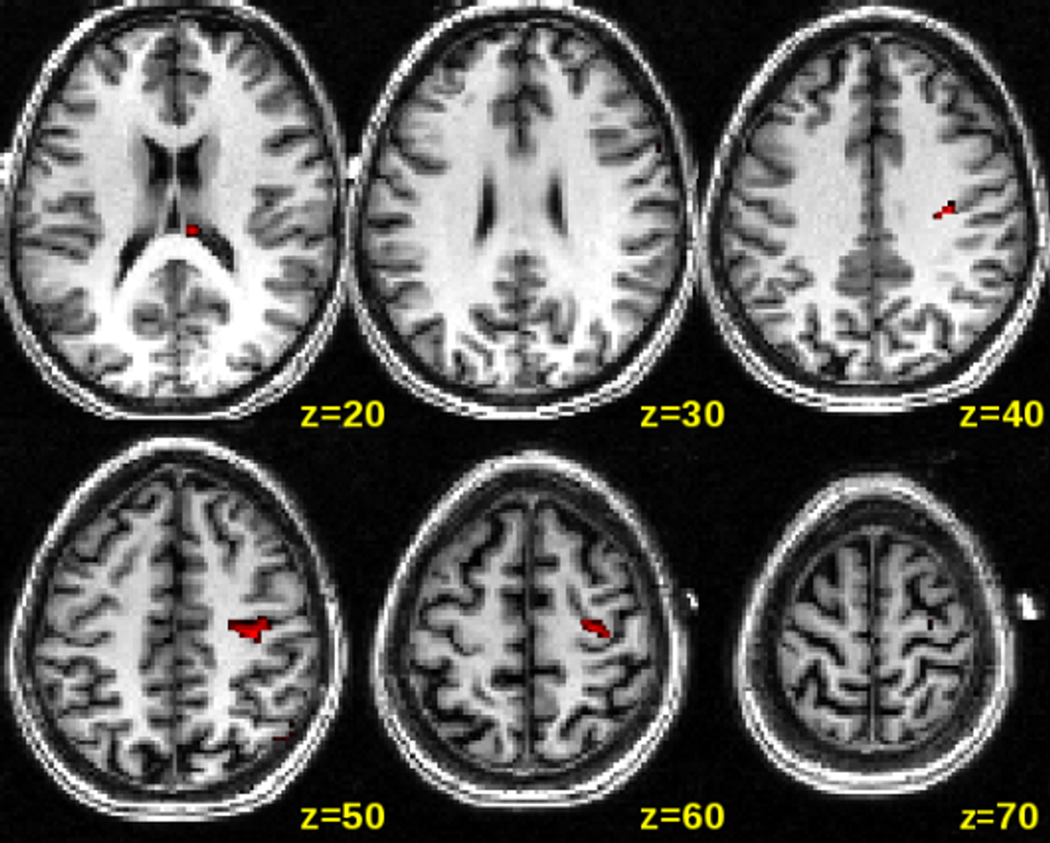
Transcranial direct current stimulation (tDCS) can up- and down-regulate cortical excitability depending on current direction, however our abilities to measure brain-tissue effects of the stimulation and its after-effects have been limited so far. We used regional cerebral blood flow (rCBF), a surrogate measure of brain activity, to examine regional brain-tissue and brain-network effects during and after tDCS. We varied the polarity (anodal and cathodal) as well as the current strength (0.8 to 2.0mA) of the stimulation. Fourteen healthy subjects were randomized into receiving either anodal or cathodal stimulation (two subjects received both, one week apart) while undergoing Arterial Spin Labeling (ASL) in the MRI scanner with an alternating off-on sampling paradigm. The stimulating, MRI-compatible electrode was placed over the right motor region and the reference electrode over the contralateral supra-orbital region. SPM5 was used to process and extract the rCBF data using a 10mm spherical volume of interest (VOI) placed in the motor cortex directly underneath the stimulating scalp electrode. Anodal stimulation induced a large increase (17.1%) in rCBF during stimulation, which returned to baseline after the current was turned off, but exhibited an increase in rCBF again in the post-stimulation period. Cathodal stimulation induced a smaller increase (5.6%) during stimulation, a significant decrease compared to baseline (-6.5%) after cessation, and a continued decrease in the post-stimulation period. These changes in rCBF were all significant when compared to the pre-stimulation baseline or to a control region. Furthermore, for anodal stimulation, there was a significant correlation between current strength and the increase in rCBF in the on-period relative to the pre-stimulation baseline. The differential rCBF after-effects of anodal (increase in resting state rCBF) and cathodal (decrease in resting state rCBF) tDCS support findings of behavioral and cognitive after-effects after cathodal and anodal tDCS. We also show that tDCS not only modulates activity in the brain region directly underlying the stimulating electrode but also in a network of brain regions that are functionally related to the stimulated area. Our results indicate that ASL may be an excellent tool to investigate the effects of tDCS and its stimulation parameters on brain activity.
Copyright © 2011 Elsevier Inc. All rights reserved.
Figures
Similar articles
-
Short periods of bipolar anodal TDCS induce no instantaneous dose-dependent increase in cerebral blood flow in the targeted human motor cortex.Sci Rep. 2022 Jun 10;12(1):9580. doi: 10.1038/s41598-022-13091-7. Sci Rep. 2022. PMID: 35688875 Free PMC article.
-
Effects of tDCS dose and electrode montage on regional cerebral blood flow and motor behavior.Neuroimage. 2021 Aug 15;237:118144. doi: 10.1016/j.neuroimage.2021.118144. Epub 2021 May 12. Neuroimage. 2021. PMID: 33991697 Free PMC article.
-
How does transcranial DC stimulation of the primary motor cortex alter regional neuronal activity in the human brain?Eur J Neurosci. 2005 Jul;22(2):495-504. doi: 10.1111/j.1460-9568.2005.04233.x. Eur J Neurosci. 2005. PMID: 16045502 Free PMC article. Clinical Trial.
-
Current intensity- and polarity-specific online and aftereffects of transcranial direct current stimulation: An fMRI study.Hum Brain Mapp. 2020 Apr 15;41(6):1644-1666. doi: 10.1002/hbm.24901. Epub 2019 Dec 20. Hum Brain Mapp. 2020. PMID: 31860160 Free PMC article.
-
tDCS polarity effects in motor and cognitive domains: a meta-analytical review.Exp Brain Res. 2012 Jan;216(1):1-10. doi: 10.1007/s00221-011-2891-9. Epub 2011 Oct 12. Exp Brain Res. 2012. PMID: 21989847 Review.
Cited by
-
Transcranial direct current stimulation in stroke rehabilitation: a review of recent advancements.Stroke Res Treat. 2013;2013:170256. doi: 10.1155/2013/170256. Epub 2013 Feb 27. Stroke Res Treat. 2013. PMID: 23533955 Free PMC article.
-
Development of Point of Care Testing Device for Neurovascular Coupling From Simultaneous Recording of EEG and NIRS During Anodal Transcranial Direct Current Stimulation.IEEE J Transl Eng Health Med. 2015 Jan 16;3:2000112. doi: 10.1109/JTEHM.2015.2389230. eCollection 2015. IEEE J Transl Eng Health Med. 2015. PMID: 27170897 Free PMC article.
-
Effects of Transcranial Direct Current Stimulation (tDCS) on Cognitive Performance and Cerebral Oxygen Hemodynamics: A Systematic Review.Front Hum Neurosci. 2021 Apr 7;15:623315. doi: 10.3389/fnhum.2021.623315. eCollection 2021. Front Hum Neurosci. 2021. PMID: 33897392 Free PMC article.
-
Short periods of bipolar anodal TDCS induce no instantaneous dose-dependent increase in cerebral blood flow in the targeted human motor cortex.Sci Rep. 2022 Jun 10;12(1):9580. doi: 10.1038/s41598-022-13091-7. Sci Rep. 2022. PMID: 35688875 Free PMC article.
-
Hemodynamic responses in rat brain during transcranial direct current stimulation: a functional near-infrared spectroscopy study.Biomed Opt Express. 2014 May 13;5(6):1812-21. doi: 10.1364/BOE.5.001812. eCollection 2014 Jun 1. Biomed Opt Express. 2014. PMID: 24940542 Free PMC article.
References
-
- Aguirre GK, Detre JA, et al. Experimental design and the relative sensitivity of BOLD and perfusion fMRI. Neuroimage. 2002;15(3):488–500. - PubMed
-
- Alsop DC, Detre JA. Multisection cerebral blood flow MR imaging with continuous arterial spin labeling. Radiology. 1998;208(2):410–416. - PubMed
-
- Angelone LM, Vasios CE, et al. On the effect of resistive EEG electrodes and leads during 7 T MRI: simulation and temperature measurement studies. Magn Reson Imaging. 2006;24(6):801–812. - PubMed
-
- Antal AM, Nitsche A, et al. Direct current stimulation over V5 enhances visuomotor coordination by improving motion perception in humans. J Cogn Neurosci. 2004;16(4):521–527. - PubMed
-
- Antal A, Polania R, et al. Transcranial direct current stimulation over the primary motor cortex during fMRI. Neuroimage. 2011 in press. - PubMed
Publication types
MeSH terms
Substances
Grants and funding
LinkOut - more resources
Full Text Sources
