Review
. 2011 Jul;179(1):12-22.
doi: 10.1016/j.ajpath.2011.03.050. Epub 2011 May 23.

Restoring dystrophin expression in duchenne muscular dystrophy muscle progress in exon skipping and stop codon read through

Affiliations
Review

Restoring dystrophin expression in duchenne muscular dystrophy muscle progress in exon skipping and stop codon read through

Eric P Hoffman et al. Am J Pathol. 2011 Jul.

Abstract

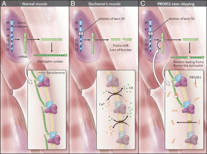
The identification of the Duchenne muscular dystrophy gene and protein in the late 1980s led to high hopes of rapid translation to molecular therapeutics. These hopes were fueled by early reports of delivering new functional genes to dystrophic muscle in mouse models using gene therapy and stem cell transplantation. However, significant barriers have thwarted translation of these approaches to true therapies, including insufficient therapeutic material (eg, cells and viral vectors), challenges in systemic delivery, and immunological hurdles. An alternative approach is to repair the patient's own gene. Two innovative small-molecule approaches have emerged as front-line molecular therapeutics: exon skipping and stop codon read through. Both approaches are in human clinical trials and aim to coax dystrophin protein production from otherwise inactive mutant genes. In the clinically severe dog model of Duchenne muscular dystrophy, the exon-skipping approach recently improved multiple functional outcomes. We discuss the status of these two methods aimed at inducing de novo dystrophin production from mutant genes and review implications for other disorders.


Figures

Figure 1
Figure 1
Mechanism of action of AO exon-skipping drugs. A: Dystrophin gene splicing in healthy muscle, in which all 79 exons are precisely spliced together to maintain the protein translational reading frame (only exons 48 to 53 are shown). B: A patient with DMD with a deletion of exon 50. The remaining exons are spliced together, but there is a disruption of the reading frame, disabling the ability of the mRNA to produce any dystrophin. Consequently, there is a dystrophin deficiency in muscle and unstable plasma membranes. CK indicates creatine kinase. C: The mechanism of action of PRO051, an AO drug targeting exon 51. The exon 51 sequence (adjacent to the missing exon 50 sequence) is skipped, so that the mRNA splices exon 49 to 52. The new deletion is able to be translated into semifunctional Becker-like dystrophin, resulting in partial repair of the myofiber plasma membrane.
Figure 2
Figure 2
Morpholino AOs achieve myofiber delivery through bulk flow across unstable plasma membranes. Many publications have shown that morpholinos delivered i.v. achieve unexpected efficacy for modulating splicing within dystrophic myofibers, presumably through bulk flow across unstable dystrophic plasma membranes. Herein, we test this model using i.v. versus i.m. delivery of a morpholino in healthy murine muscle. A: 0 mg (water) or 1 or 4 mg morpholino was given in an i.v. bolus in healthy mice, and drug delivery to myofibers was assayed by exon skipping in the Akt1 mRNA (skipped Akt1). No detectable exon skipping was observed in healthy skeletal muscle (0%). B: As a positive control, the same morpholino was delivered by i.m. injection in saline (0, 0.1, 1, 10, and 100 μg). The saline destabilizes the myofiber membranes, and efficient dose-related exon skipping is observed (skipped Akt1). GAPDH indicates glyceraldehyde-3-phosphate dehydrogenase.
Figure 3
Figure 3
Backbone chemistries of nucleic acids and antisense drugs. Normal DNA and RNA has ribose rings (sugar moieties) attached by phosphodiester linkages, and one of four bases (G, A, T, C for DNA and G, A, U, C for RNA) is attached to the ribose and participates in sequence-specific base pairing with other nucleic acid strands. The AO drug chemistries modify the backbone to make the drugs more stable and less toxic. The 2′OMe AO adds a methyl group to the ribose ring and a sulfur residue to the phosphodiester linkage. The morpholino (PMO) chemistry makes many more changes, replacing the ribose with a nitrogenous morpholine ring; amine groups replace the phosphodiester linkage. Despite the relatively dramatic chemical changes to the PMO backbone, the spacing between the bases is similar to DNA and RNA and does not disrupt base pairing with other nucleic acid strands.
Figure 4
Figure 4
Delivery of multiple PMO drugs to a dog model of DMD skips multiple exons and results in de novo dystrophin production. A: Schematic of dog gene structure. The sporadic golden retriever dystrophin gene mutation is a splice-site mutation of exon 7 (red symbol). This forces the exclusion of exon 7, whereby the dystrophic dog muscle splices exon 6 to 8, but these exons do not share the same reading frame (out of frame). AOs covering exons 6 and 8 were designed (AOs 1, 2, and 3) to block inclusion of exons 6 and 8, leading to in-frame rescued transcripts (exons 5 to 9 or 5 to 10). B: Histological features and matched dystrophin immunostaining of AO-treated dystrophic dogs (right) and controls [nontreated canine X linked muscular dystrophy (CXMD) muscle; left]. Nontreated muscle shows necrosis of myofibers and inflammatory cell infiltration, whereas AO-treated muscle shows no inflammation or necrosis. Dystrophin protein is absent in the nontreated muscle, whereas the AO-treated muscle shows high amounts of membrane dystrophin, comparable to healthy muscle.

Similar articles

Cited by

References

    1. Hoffman E.P., Brown R.H., Jr, Kunkel L.M. Dystrophin: the protein product of the Duchenne muscular dystrophy locus. Cell. 1987;51:919–928. - PubMed
    1. Hoffman E.P., Hudecki M.S., Rosenberg P.A., Pollina C.M., Kunkel L.M. Cell and fiber-type distribution of dystrophin. Neuron. 1988;1:411–420. - PubMed
    1. Koenig M., Hoffman E.P., Bertelson C.J., Monaco A.P., Feener C., Kunkel L.M. Complete cloning of the Duchenne muscular dystrophy (DMD) cDNA and preliminary genomic organization of the DMD gene in normal and affected individuals. Cell. 1987;50:509–517. - PubMed
    1. Hoffman E.P., Fischbeck K.H., Brown R.H., Johnson M., Medori R., Loike J.D., Harris J.B., Waterston R., Brooke M., Specht L., Kupsky W., Chamberlain J., Caskey C.T., Shapiro F., Kunkel L.M. Characterization of dystrophin in muscle-biopsy specimens from patients with Duchenne's or Becker's muscular dystrophy. N Engl J Med. 1988;318:1363–1368. - PubMed
    1. Koppanati B.M., Li J., Reay D.P., Wang B., Daood M., Zheng H., Xiao X., Watchko J.F., Clemens P.R. Improvement of the mdx mouse dystrophic phenotype by systemic in utero AAV8 delivery of a minidystrophin gene. Gene Ther. 2010;17:1355–1362. - PMC - PubMed

Publication types