. 2011 Jul;179(1):37-45.
doi: 10.1016/j.ajpath.2011.03.007. Epub 2011 May 3.

Unique ectopic lymph node-like structures present in human primary colorectal carcinoma are identified by immune gene array profiling

Affiliations

Unique ectopic lymph node-like structures present in human primary colorectal carcinoma are identified by immune gene array profiling

Domenico Coppola et al. Am J Pathol. 2011 Jul.

Abstract

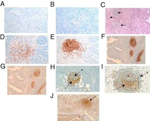
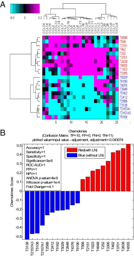
We hypothesized that immune gene-related signatures would predict the presence of unique histological features of lymphoid cell infiltrates in colorectal carcinoma (CRC) that correlate with clinical parameters. Metagene analysis with gene chip technology was performed on 326 CRCs, which were then sorted by low versus high gene scores. Microscopically, CRCs with a high gene score revealed a marked host immune response organized, remarkably, as lymphoid follicles. Proliferation involved both B and T cells. In every case, the presence of CD79a(+) B-cell precursors was identified, suggesting that the lymphoid follicles represent newly formed, ectopic lymph node-like structures. CD21(+) dendritic cells were present within the follicular germinal centers, and CD3(+) T cells were localized mainly in the parafollicular cortex zone surrounding the B-cell area of the follicles. A strong correlation between a 12-chemokine gene subset of the molecular profile and the presence of ectopic lymph node-like structures was associated with better patient survival independent of tumor staging, site location, microsatellite instability or stability, and patient treatment. These findings suggest beneficial, intratumoral immune cell priming and raise the possibility of immunotherapy intervention decisions based on molecular signatures that can identify the presence of tumor-localized, ectopic lymph node-like structures.


Figures

Figure 1
Figure 1
A: The observed range of the immune response as represented by the metagene 1 score. Shown are the CRCs that have the 10 highest and 11 lowest values of the mean score of selected immune metagenes, sorted by the mean metagene score. B: The relationship between patient overall survival (OS) and the immune response as quantified by the metagene 1 score on selected CRCs with known status of ectopic lymph node-like structures confirmed by IHC. CRCs with and without lymphoid structures (high and low metagene 1 scores, respectively) segregated into two groups: overall survival time of <2 years versus >2 years. Score for metagene 1 is plotted on the y axis.
Figure 2
Figure 2
Analysis of primary CRCs with H&E staining and IHC. All 11 of the lowest gene signature-scored CRCs revealed a lightly dispersed or absent lymphocytic peritumoral host response, and low to no appreciable expression of the B cell marker CD20 (A) and the T cell marker CD3 (B). All 10 of the highest gene signature-scored CRCs revealed a marked peritumoral lymphocytic host response, organized as ectopic lymph node-like structures (arrows), by H&E staining (C) and by IHC. (D–I). CD20+ B cells (E and F), CD79a+ B cells (G), and CD21+ follicular dendritic cells (J) are concentrated in the center of follicles, with CD3+ T cells (D, H, and I) appearing in the parafollicular cortex or marginal zones and with some dispersion into the follicles. In some cases, a fibrous stroma was observed to encapsulate a follicle (I). J: The arrow indicates a follicle at the front edge of the invading colonic adenocarcinoma. B, B cells; S, stroma; T, T cells (H and I) or tumor (J). Magnification: A, ×100; B, ×100; C, ×40; D, ×200; E, ×200; F, ×100; G, ×100; H, ×100; I, ×200; J, ×100.
Figure 3
Figure 3
Chemokines are up-regulated in tumors with ectopic lymph node structures. A: Heat map of mean-centered intensities, averaged within each probe set across all tumors shown. For each gene, a single representative probe set with the highest dynamic range across all profiled samples was picked up from all probe sets that mapped to a given gene symbol. Genes are clustered using Pearson's correlation distance metric; tumors are clustered using Euclidean distance metric. Ward's linkage criterion was applied for both tumors and genes. Along the y axis, red versus blue identifiers indicate CRCs with versus without lymphoid structures. B: Hierarchical clustering of tumors with and without lymphoid structures was done on a selected set of known chemokines. The chemokine score is the mean value of chemokines as averaged across all probe sets shown for the heat map.
Figure 4
Figure 4
The 12 selected chemokines that correlate with metagene 1. A: Heat map of 326 colorectal tumors and 12 genes comprising metagene 1 sorted by the metagene score (mean of probe sets that map to a given 12-gene set of genes). The profile below the heat map demarcates the 10 samples with the highest metagene score and the 11 samples with the lowest metagene score. B: The relationship between patient overall survival and the immune response as quantified by the score of chemokine genes on selected CRCs with known status of ectopic lymph node-like structures confirmed by IHC. CRCs with and without lymphoid structures (high and low metagene 1 scores, respectively) segregated into two groups: overall survival time of <2 years versus >2 years. Scores for the chemokine genes are plotted on the y axis.

Similar articles

Cited by

References

    1. Jemal A., Siegel R., Ward E., Hao Y., Xu J., Murray T., Thun M.J. Cancer Statistics. CA Cancer J Clin. 2008;58:71–96. - PubMed
    1. Coronella J.A., Spier C., Welch M., Trevor K.T., Stopeck A.T., Villar H., Hersh E.M. Antigen-driven oligoclonal expansion of tumor-infiltrating B cells in infiltrating ductal carcinoma of the breast. J Immunol. 2002;169:1829–1836. - PubMed
    1. Coronella-Wood J.A., Hersh E.M. Naturally occurring B-cell responses to breast cancer. Cancer Immunol Immunother. 2003;52:715–738. - PMC - PubMed
    1. Bell D., Chomarat P., Broyles D., Netto G., Harb G.M., Lebecque S., Valladeau J., Davoust J., Palucka K.A., Banchereau J. In breast carcinoma tissue, immature dendritic cells reside within the tumor, whereas mature dendritic cells are located in peritumoral areas. J Exp Med. 1999;190:1417–1426. - PMC - PubMed
    1. Aspord C., Pedroza-Gonzalez A., Gallegos M., Tindle S., Burton E.C., Su D., Marches F., Banchereau J., Palucka A.K. Breast cancer instructs dendritic cells to prime interleukin 13-secreting CD4+ T cells that facilitate tumor development. J Exp Med. 2007;204:1037–1047. - PMC - PubMed

Publication types

MeSH terms