. 2011 Jul;179(1):66-74.
doi: 10.1016/j.ajpath.2011.03.034. Epub 2011 May 7.

Abnormal cell properties and down-regulated FAK-Src complex signaling in B lymphoblasts of autistic subjects

Affiliations

Abnormal cell properties and down-regulated FAK-Src complex signaling in B lymphoblasts of autistic subjects

Hongen Wei et al. Am J Pathol. 2011 Jul.

Erratum in

  • Am J Pathol. 2011 Nov;179(5):2674

Abstract

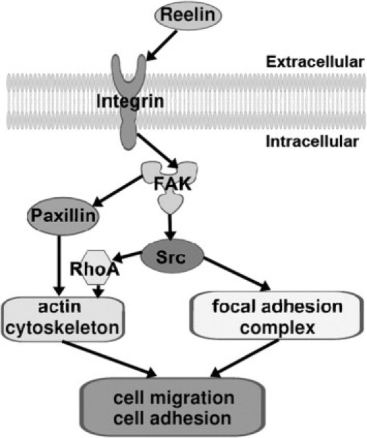
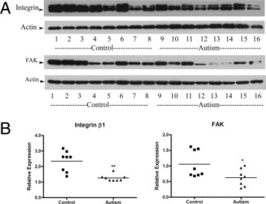
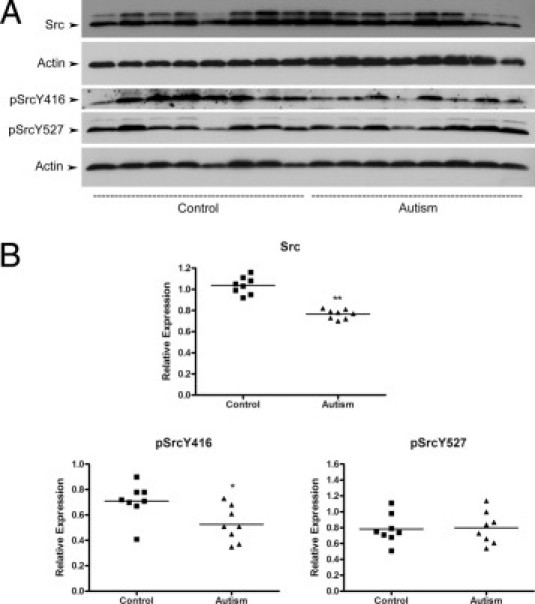
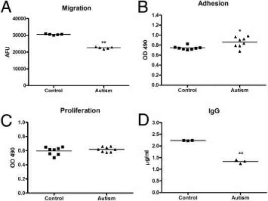
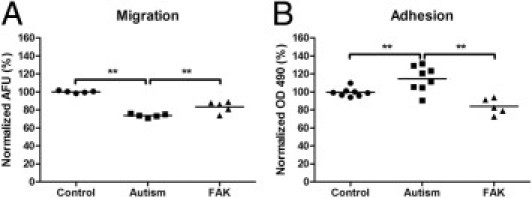
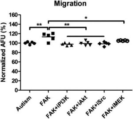
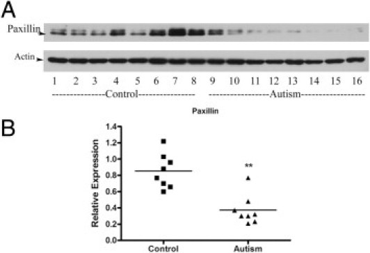
Recent studies suggest that one of the major pathways to the pathogenesis of autism is reduced cell migration. Focal adhesion kinase (FAK) has an important role in neural migration, dendritic morphological characteristics, axonal branching, and synapse formation. The FAK-Src complex, activated by upstream reelin and integrin β1, can initiate a cascade of phosphorylation events to trigger multiple intracellular pathways, including mitogen-activated protein kinase-extracellular signal-regulated kinase and phosphatidylinositol 3-kinase-Akt signaling. In this study, by using B lymphoblasts as a model, we tested whether integrin β1 and FAK-Src signaling are abnormally regulated in autism and whether abnormal FAK-Src signaling leads to defects in B-lymphoblast adhesion, migration, proliferation, and IgG production. To our knowledge, for the first time, we show that protein expression levels of both integrin β1 and FAK are significantly decreased in autistic lymphoblasts and that Src protein expression and the phosphorylation of an active site (Y416) are also significantly decreased. We also found that lymphoblasts from autistic subjects exhibit significantly decreased migration, increased adhesion properties, and an impaired capacity for IgG production. The overexpression of FAK in autistic lymphoblasts countered the adhesion and migration defects. In addition, we demonstrate that FAK mediates its effect through the activation of Src, phosphatidylinositol 3-kinase-Akt, and mitogen-activated protein kinase signaling cascades and that paxillin is also likely involved in the regulation of adhesion and migration in autistic lymphoblasts.


Figures

Figure 1
Figure 1
Signaling pathways of FAK and Src, controlling actin cytoskeletal rearrangement, and FAs. Integrin β1 can be activated by reelin and induces recruitment and stimulation of FAK and Src, which control cell migration and adhesion via regulation of the actin cytoskeleton and FA complex. Activated FAK can also recruit paxillin in the regulation of cell migration and adhesion. RhoA indicates Ras homolog gene family, member A.
Figure 2
Figure 2
Integrin β1 and FAK protein expression in autistic lymphoblasts. A: Western blot analyses of lymphoblast lysates using integrin β1 antibody and FAK antibody. B: The blots shown in A were quantitated after normalization by actin. Data are shown as mean ± SE (n = 8). *P < 0.05, **P < 0.01.
Figure 3
Figure 3
Src protein expression and phosphorylation in autistic lymphoblasts. A: Western blot analyses of lymphoblast lysates using Src antibody, phosphorylated Src (pSrc) (Y416) antibody, and pSrc (Y527) antibody. B: The blots shown in A were quantitated after normalization by actin. Data are shown as mean ± SE (n = 8). *P < 0.05, **P < 0.01.
Figure 4
Figure 4
The profile of lymphoblast migration, adhesion, proliferation, and IgG production in autistic subjects. A: Modified Boyden chamber migration assays on lymphoblasts from autistic and control subjects. The number of migrated cells was determined by fluorescence reading at 14 hours. The values are mean ± SE from five replicates. AFU indicates arbitrary fluorescence units. B: Comparison of 6-hour lymphoblast adhesion between the autistic and control groups. The values are mean ± SE from eight replicates. C: Comparison of 48-hour lymphoblast proliferation between the autistic and control groups. The values are mean ± SE from eight replicates. D: Comparison of IgG production between the autistic and control groups. The values are mean ± SE from three replicates. *P < 0.05, **P < 0.01.
Figure 5
Figure 5
Fluorescence staining of lymphoblasts: A representative image of lymphoblast cells from six autistic subjects and six controls after acridine orange (AO) plus propidium iodide (PI) double staining. Cells were examined at ×400 magnification with a fluorescence microscope (Nikon). AO stained live cells with intact cytoplasm (green) and nuclei (bright green), and PI stained dead/dying cells. The nuclear and cytoplasmic areas of viable lymphoblast cells were measured by Image-Pro Plus software.
Figure 6
Figure 6
The effect of FAK overexpression on autistic lymphoblast migration and adhesion. A: Migration assays on control lymphoblasts, autistic lymphoblasts, and autistic lymphoblasts treated with FAK. The number of migrated cells was determined by fluorescence reading at 14 hours. The values are mean ± SE from five replicates. B: Adhesion assay on control lymphoblasts, autistic lymphoblasts, and autistic lymphoblasts treated with FAK. Adherent cells were quantitated by the colorimetric aqueous MTS assay. The values are mean ± SE from five replicates. AFU indicates arbitrary fluorescence units. **P < 0.01.
Figure 7
Figure 7
Effect of selective inhibitors on FAK-mediated autistic lymphoblast migration. Migration assays on autistic lymphoblasts treated with FAK, FAK plus Src inhibitor (SU6656), FAK plus PI3K inhibitor (wortmannin), FAK plus Akt kinase inhibitor, or FAK plus MEK inhibitor (PD98059) (i indicates inhibitor). The number of migrated cells was determined by fluorescence reading at 14 hours. The values are mean ± SE from five replicates. AFU indicates arbitrary fluorescence units. *P < 0.05, **P < 0.01.
Figure 8
Figure 8
Paxillin protein expression in autistic lymphoblasts. A: Western blot analyses of lymphoblast lysates using paxillin antibody. B: The blot shown in A was quantitated after normalization by actin. Data are shown as mean ± SE (n = 8). **P < 0.01.

References

    1. Abrahams B.S., Geschwind D.H. Advances in autism genetics: on the threshold of a new neurobiology. Nat Rev Genet. 2008;9:341–355. - PMC - PubMed
    1. Li X., Chauhan A., Sheikh A.M., Patil S., Chauhan V., Li X.M., Ji L., Brown T., Malik M. Elevated immune response in the brain of autistic patients. J Neuroimmunol. 2009;207:111–116. - PMC - PubMed
    1. Molloy C.A., Morrow A.L., Meinzen-Derr J., Schleifer K., Dienger K., Manning-Courtney P., Altaye M., Wills-Karp M. Elevated cytokine levels in children with autism spectrum disorder. J Neuroimmunol. 2006;172:198–205. - PubMed
    1. Sheikh A.M., Li X., Wen G., Tauqeer Z., Brown W.T., Malik M. Cathepsin D and apoptosis related proteins are elevated in the brain of autistic subjects. Neuroscience. 2010;165:363–370. - PubMed
    1. Vargas D.L., Nascimbene C., Krishnan C., Zimmerman A.W., Pardo C.A. Neuroglial activation and neuroinflammation in the brain of patients with autism. Ann Neurol. 2005;57:67–81. - PubMed

Publication types

MeSH terms

LinkOut - more resources