Comparative Study
. 2011 Jul;179(1):167-79.
doi: 10.1016/j.ajpath.2011.03.040. Epub 2011 May 11.

Plexiform lesions in pulmonary arterial hypertension composition, architecture, and microenvironment

Affiliations
Comparative Study

Plexiform lesions in pulmonary arterial hypertension composition, architecture, and microenvironment

Danny Jonigk et al. Am J Pathol. 2011 Jul.

Abstract

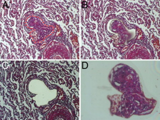
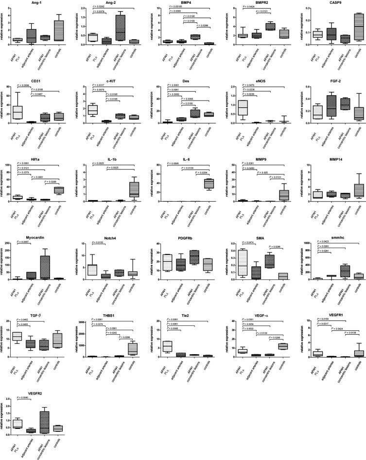
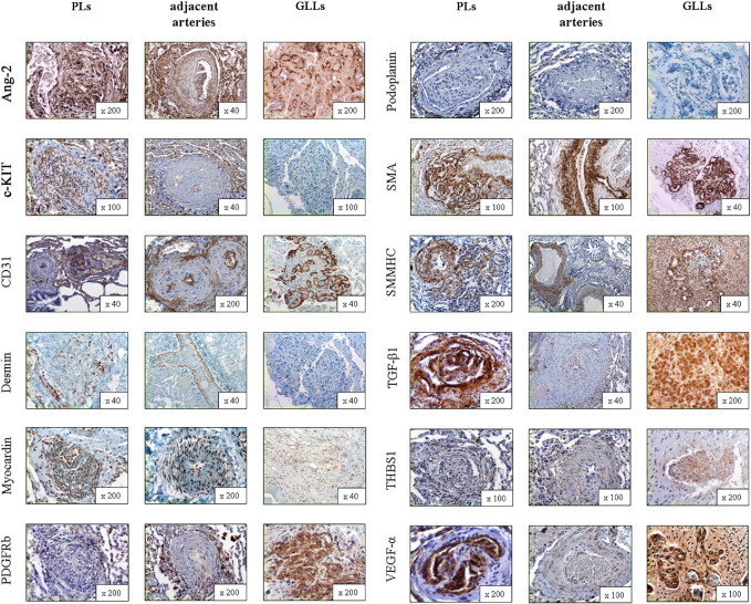
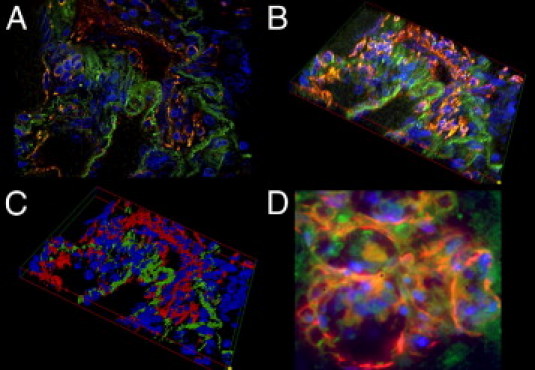
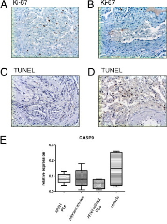
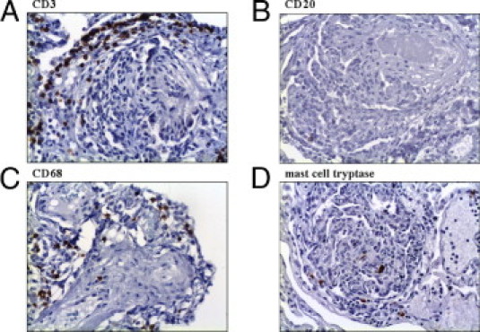
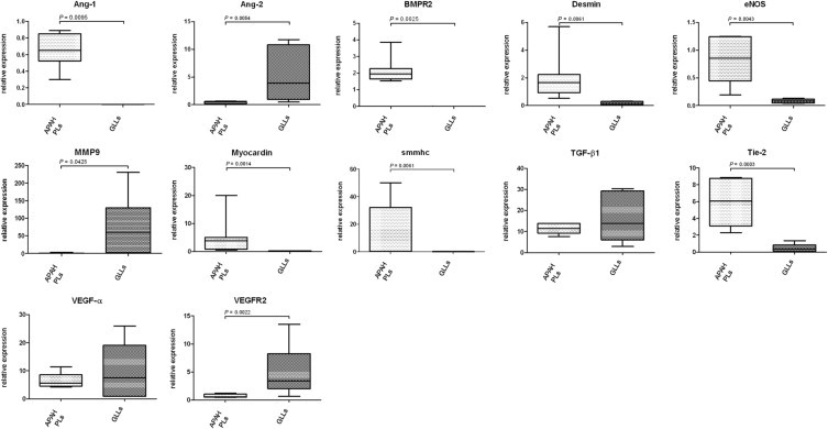
Pulmonary arterial hypertension (PAH) is a debilitating disease with a high mortality rate. A hallmark of PAH is plexiform lesions (PLs), complex vascular formations originating from remodeled pulmonary arteries. The development and significance of these lesions have been debated and are not yet fully understood. Some features of PLs resemble neoplastic disorders, and there is a striking resemblance to glomeruloid-like lesions (GLLs) in glioblastomas. To further elucidate PLs, we used in situ methods, such as (fluorescent) IHC staining, three-dimensional reconstruction, and laser microdissection, followed by mRNA expression analysis. We generated compartment-specific expression patterns in the lungs of 25 patients (11 with PAH associated with systemic shunts, 6 with idiopathic PAH, and 8 controls) and GLLs from 5 glioblastomas. PLs consisted of vascular channels lined by a continuously proliferating endothelium and backed by a uniform myogenic interstitium. They also showed up-regulation of remodeling-associated genes, such as HIF1a, TGF-β1, VEGF-α, VEGFR-1/-2, Ang-1, Tie-2, and THBS1, but also of cKIT and sprouting-associated markers, such as NOTCH and matrix metalloproteinases. The cellular composition and signaling seen in GLLs in neural neoplasms differed significantly from those in PLs. In conclusion, PLs show a distinct cellular composition and microenvironment, which contribute to the plexiform phenotype and set them apart from other processes of vascular remodeling in patients with PAH. Neoplastic models of angiogenesis seem to be of limited use in further study of plexiform vasculopathy.


Figures

Figure 1
Figure 1
Laser-assisted microdissection. A–D: Isolation of a PL from the surrounding unagitated lung tissue by laser-assisted microdissection (the laser cut is visible in B; D shows the isolated PL in the cap). Note the adjacent artery from which the PL originates in the lower right corner in A–C. Original magnification: ×100 (A–C); ×200 (D). The IX71 microscope (Olympus Europa GmbH) with the CellCut Plus system was used for laser-assisted microdissection.
Figure 2
Figure 2
mRNA expression of target genes in PLs in APAH in alphabetical order. Comparison of mRNA expression characteristics of PLs in patients with congenital shunts between the systemic and pulmonary circulation (APAH), the adjacent arteries from which they sprout, concentric lesions in patients with APAH without PLs, and controls. Expression of target genes was calculated using the ΔCT method (see Materials and Methods for details). Middle horizontal lines indicate medians; error bars, percentiles. PDGFRb, β-type platelet-derived growth factor receptor.
Figure 3
Figure 3
Protein expression of structural composition and tissue remodeling–associated markers in PLs, the adjacent arteries from which PLs originate, and GLLs in high-grade primary neural malignomas (in alphabetical order). Ang-2 showed strong cytoplasmic positivity in the endothelial/luminal compartment of PLs and also in the endothelial layer of the adjacent arteries and GLLs. c-KIT (CD117) stained positive in the endothelial/luminal compartment of PLs and in the endothelium of the adjacent arteries, whereas there was no delimitable staining in GLLs. CD31 staining was pronounced in the endothelial/luminal layers in PLs, adjacent arteries, and GLLs alike. Desmin showed only speckled cytoplasmic positivity in the interstitial/myogenic layer of PLs, whereas the media of the adjacent arteries was continuously positive and GLLs showed no staining reaction. Myocardin showed moderate cytoplasmic positivity in vascular structures in PLs and adjacent arteries, whereas GLLs stained only faintly. β-Type platelet-derived growth factor receptor (PDGFRb) stained only faintly in PLs, whereas the directly adjacent arteries showed moderate positivity, mainly at the border between the actual vascular wall and the neighboring connective tissue. GLLs showed only faint perivascular staining. Podoplanin showed no delimitable signal in PLs but a slender network of lymphatic vessels around them. There was also no positivity in the adjacent arteries or in GLLs. Staining for SMA showed slim but continuous positivity between the luminal/endothelial layers in PLs, whereas there was a prominent and homogenous staining pattern in the media of the adjacent arteries. In GLLs, the walls of the neoplastic blood vessels also showed strong cytoplasmic positivity, although it was less well organized compared with the nonneoplastic specimens/compartments. Smmhc stained weakly in the interstitium of PLs, whereas there was strong staining in the media of the adjacent arteries. GLLs displayed inhomogenous positivity of the neoplastic blood vessels. TGF-β1 stained strongly in the luminal and the interstitial compartment of PLs, whereas the adjacent arteries showed only faint positivity of the endothelium. Staining in GLLs was strong in the vascular structures. THBS1 showed inhomogenous cytoplasmic positivity in PLs, whereas the adjacent arteries showed only a faint reaction. GLLs showed inhomogenous moderate positivity. VEGF-α showed marked luminal/endothelial positivity, with only a faint endothelial signal in the adjacent arteries. The vessels in GLLs showed strong positivity. Original magnification is included in the lower right corner of each histologic image.
Figure 4
Figure 4
The-3-D reconstruction of a PL. A: A fluorescent double-stained PL: the luminal vascular channels stain positive for CD31 (red) and the adjacent interstitium shows homogenous positivity for SMA (green). B: Systematically scanned images of the whole width of the PL were merged into 3-D shapes. C: Images were skeletonized into a voxel model for increased clarity. Note the distinct separation of the endothelial layer and the interstitium in the PL. D: GLLs in high-grade neural tumors show a rather disorganized composition without an equally clear separation of endothelial layer and interstitium. Original magnification: ×630 (A–C); ×1000 (D).
Figure 5
Figure 5
Proliferation and apoptosis in PLs in APAH. IHC staining for the proliferation marker Ki-67 in PLs (A) and the adjacent arteries (B). Whereas there is marked proliferation in PLs, only single cells of the endothelium stain positive in the adjacent arteries. Terminal deoxynucleotidyl transferase–mediated dUTP nick-end labeling (TUNEL) assays do not show significant degrees of apoptosis in PLs (C) or the adjacent arteries (D), and neither is there significant mRNA up-regulation of apoptosis-associated caspase 9 (CASP9) (E). Middle horizontal lines indicate medians; error bars, percentiles. Original magnification, ×100 (A–D).
Figure 6
Figure 6
Inflammatory cells in PLs. Most inflammatory cells in PLs are CD3-positive T lymphocytes, which also accumulate around PLs (A). CD20-positive B lymphocytes account for only a few leukocytes in PLs (B). CD68-positive macrophages (C) and mast cells (D) show an almost equal distribution in PLs. Original magnification, ×100 (A–D).
Figure 7
Figure 7
mRNA expression of target genes in PLs in APAH and GLLs in alphabetical order. Comparison of selected mRNA expression characteristics of PLs in patients with congenital shunts between the systemic and pulmonary circulation (APAH) and GLLs in high-grade primary neural tumors. Expression of target genes was calculated using the ΔCT method (see Materials and Methods for details). Lines indicate medians; error bars, percentiles.

References

    1. Simonneau G., Galie N., Rubin L.J., Langleben D., Seeger W., Domenighetti G., Gibbs S., Lebrec D., Speich R., Beghetti M., Rich S., Fishman A. Clinical classification of pulmonary hypertension. J Am Coll Cardiol. 2004;43:5S–12S. - PubMed
    1. Firth A.L., Mandel J., Yuan J.X. Idiopathic pulmonary arterial hypertension. Dis Model Mech. 2010;3:268–273. - PMC - PubMed
    1. Sakao S., Tatsumi K., Voelkel N.F. Endothelial cells and pulmonary arterial hypertension: apoptosis, proliferation, interaction and transdifferentiation. Respir Res. 2009;10:95. - PMC - PubMed
    1. Pietra G.G., Edwards W.D., Kay J.M., Rich S., Kernis J., Schloo B., Ayres S.M., Bergofsky E.H., Brundage B.H., Detre K.M. Histopathology of primary pulmonary hypertension: a qualitative and quantitative study of pulmonary blood vessels from 58 patients in the National Heart, Lung, and Blood Institute, Primary Pulmonary Hypertension Registry. Circulation. 1989;80:1198–1206. - PubMed
    1. Stevens T. Molecular and cellular determinants of lung endothelial cell heterogeneity. Chest. 2005;128:558S–564S. - PubMed

Publication types

MeSH terms