. 2011 Jul;179(1):189-98.
doi: 10.1016/j.ajpath.2011.03.049. Epub 2011 May 19.

Migration of fibrocytes in fibrogenic liver injury

Affiliations

Migration of fibrocytes in fibrogenic liver injury

David Scholten et al. Am J Pathol. 2011 Jul.

Abstract

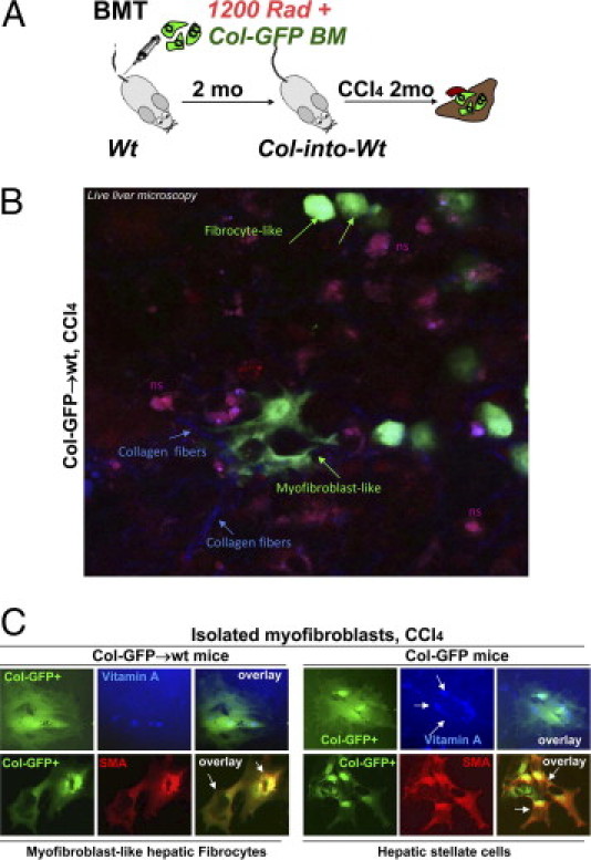
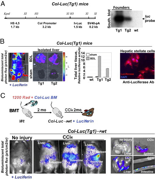
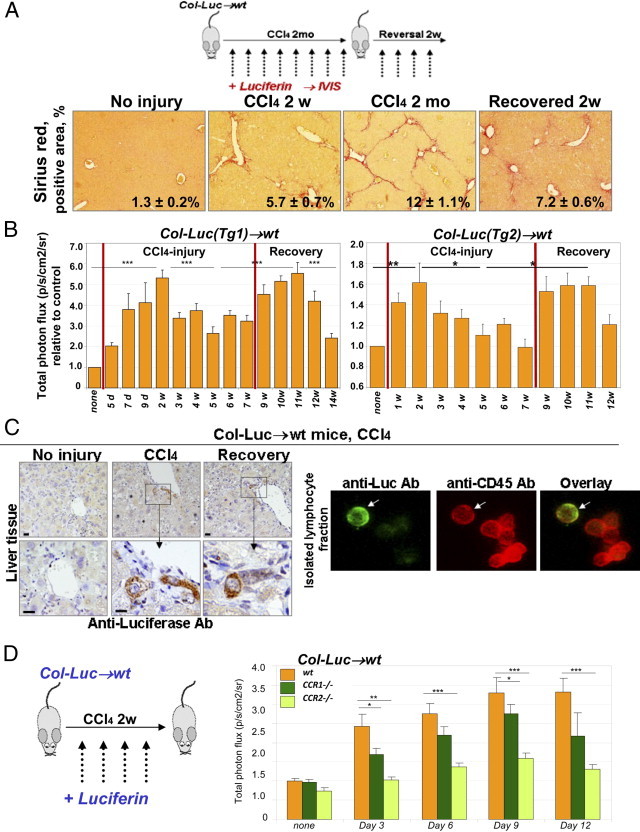
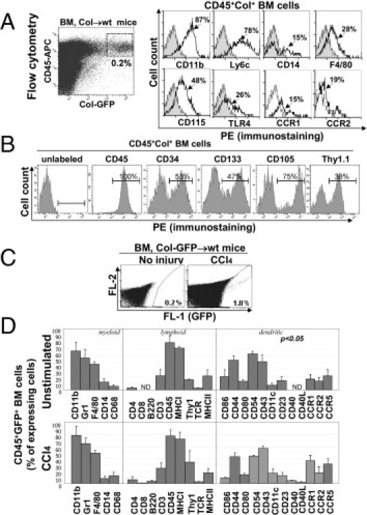
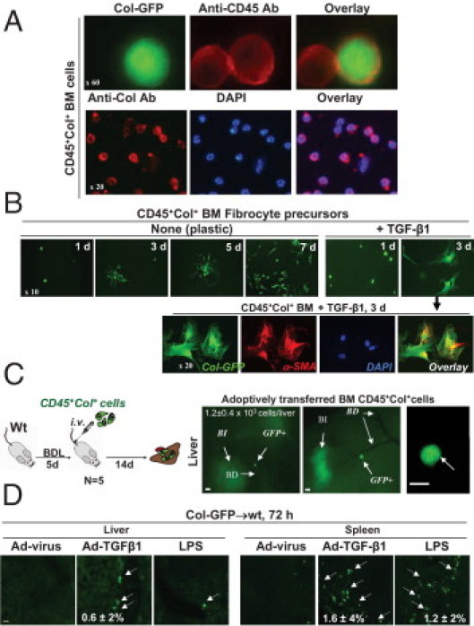
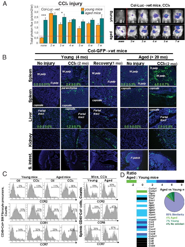
CD45(+) and collagen I-positive (Col(+)) fibrocytes are implicated in fibrogenesis in skin, lungs, and kidneys. Fibrocyte migration in response to liver injury was investigated using bone marrow (BM) from chimeric mice expressing luciferase (Col-Luc→wt) or green fluorescent protein (Col-GFP→wt) under control of the α1(I) collagen promoter and enhancer, respectively. Monitored by luciferase expression, recruitment of fibrocytes was detected in CCl(4)-damaged liver and in spleen. Migration of CD45(+)Col(+) fibrocytes was regulated by chemokine receptors CCR2 and CCR1, as demonstrated, respectively, by 50% and 25% inhibition of fibrocyte migration in Col-Luc(CCR2-/-)→wt and Col-Luc(CCR1-/-)→wt mice. In addition to CCR2 and CCR1, egress of BM CD45(+)Col(+) cells was regulated by transforming growth factor-β1 (TGF-β1) and liposaccharide in vitro and in vivo, which suggests that release of TGF-β1 and increased intestinal permeability have important roles in fibrocyte trafficking. In the injured liver, fibrocytes gave rise to (myo)fibroblasts. In addition, a BM population of CD45(+)Col(+) cells capable of differentiation into fibrocytes in culture was identified. Egress of CD45(+)Col(+) cells from BM was detected in the absence of injury or stress in aged mice but not in young mice. Development of liver fibrosis was also increased in aged mice and correlated with high numbers of liver fibrocytes. In conclusion, in response to liver injury, fibrocytes migrate from BM to the liver. Their migration is regulated by CCR2 and CCR1 but is compromised with age.


Figures

Figure 1
Figure 1
Liver fibrocytes have potential to differentiate into (myo)fibroblasts. A: Study design. BMT chimeric Col-GFP→wt mice were subjected to CCl4 for 2 months. B: Round and (myo)fibroblast-like collagen α1(I)–GFP+ cells (green arrows) were detected using fluorescent microscopy in fibrotic livers of CCl4-treated Col-GFP→wt mice (n = 4). Collagen fibers (blue arrows) are shown. ns, Nonspecific autofluorescence of fibrotic livers. C: GFP+ (myo)fibroblasts from CCl4-treated Col-into-wt mice were compared with GFP+ HSCs from CCl4-treated Col-GFP mice using fluorescent microscopy for expression of vitamin A (blue) and α-SMA (red).
Figure 2
Figure 2
Generation of BM chimeric Col-Luc→wt mice. A: Transgenic construct consists of collagen α1(I) enhancer (1.7 kb), promoter (3.2 kb), and hlucP+ (1.5 kb) (left panel). Southern blot analysis with Luc-specific probe (luc, 1 kb) identified Tg1 and Tg2 transgenic founders (right panel). B: Luc expression is detected in Col-Luc mice at i.v. administration of D-Luc using Ivis-200 by up-regulation of bioluminescent signal (p/s/cm2/sr) and conformed by luciferase assay and immunostaining of activated HSCs with anti-Luc antibody. C: Generation of chimeric Col-Luc→wt mice. Migration of Luc+ cells in abdominal area is detected in liver, spleen, and intestine of CCl4-treated Col-Luc→wt mice. Data are given as p/s/cm2/sr. Representative images are shown.
Figure 3
Figure 3
CCR2 and CCR1 facilitate fibrocyte recruitment in response to CCl4. A: Study design (top). Col-Luc→wt mice were treated with CCl4 and recovered from fibrosis. Sirius red staining (bottom) determined the total collagen deposition at chosen times (mean ± SEM; P < 0.05). B: CCl4-treated Col-Luc→wt mice were monitored using bioluminescence. Luc+ cells egress BM in two phases corresponding to acute injury and recovery (n = 10). *P < 0.05, **P < 0.01, and ***P < 0.001. Groups compared are none and 2w, 2w and 5w, 5w and 11w, and 11w and 14w for Col-Luc(Tg1)-Luc mice. Graphic representation of the biphastic egress of fibrocytes and precursors in response to CCl4. C: Livers and fibrocyte-containing fraction from Col-Luc→wt mice are stained with anti-Luc and anti-CD45 antibodies. Scale bars = 50 μm. D: Recruitment of Luc+ cells was studied in LucCCR2−/−→wt mice (n = 10) and LucCCR1−/−→wt mice (n = 10) mice and compared with Lucwt-into-wt mice in response to CCl4 (2 weeks). Bars represent mean ± SEM. *P < 0.05, **P < 0.01, and ***P < 0.001. Groups compared are wt and LucCCR2−/−→wt mice, and wt and LucCCR1−/−→wt mice.
Figure 4
Figure 4
BM CD45+Col+ cells represent precursors of liver fibrocytes. A: Flow cytometry. BM CD45+Col+ cells constitute 0.2% of total BM cells in untreated Col-GFP→wt mice (dot plot) and express monocyte CD11b+Ly6c+CCR2+CD115+ markers (histograms). B: BM CD45+Col+ cells from untreated Col-GFP→wt mice express markers of hematopoietic progenitors. C: The number of CD45+Col+ cells is increased in Col-GFP→wt mice in response to CCl4. D: Comparative analysis of antigen profile of BM CD45+Col+ cells with and without CCl4 liver injury. Bars represent mean ± SEM fluorescent intensity.
Figure 5
Figure 5
BM CD45+Col+ FPs is regulated by TGF-β1 and LPS. A: Isolated BM GFP+ cells expressed CD45 and collagen type I, detected at IHC. B: BM CD45+Col+ cells, cultured on plastic in RPMI plus 10% fetal calf serum with or without 5 ng/mL TGF-β1 were stained using anti–α-SMA antibody. C: Adoptively transferred BM CD45+Col+ cells migrated specifically to fibrotic livers (n = 6) in BDL mice. Whole liver sections, bile ducts (BD), bile infarcts (BI), and adoptively transferred GFP+ are shown (arrows). Scale bars = 50 μm. D: Col-GFP→wt mice (n = 5 mice per group) are infected i.v. with TGF-β1–expressing adenovirus (Ad–TGF-β1, 1 × 10⁸ PFU) or control adenovirus (Ad-virus), or injected with 6 μg/g LPS. In vivo migration of fibrocytes (arrows) is examined at 72 hours after injury.
Figure 6
Figure 6
Egress of fibrocyte-like cells to spleen occurs without injury in aged mice. A: Migration of Luc+ cells was compared in young (6 to 8 weeks, n = 10) and aged (older than 20 months, n = 10) Col-Luc→wt mice before and during CCl4-induced liver injury. Data are given as mean ± SEM p/s/cm2/sr. *P < 0.05, ***P < 0.01. Young and aged mice were compared at each time point. B: Migration of fibrocytes into peripheral organs in Col-GFP→wt mice was demonstrated using fluorescent microscopy for GFP+ and CD45+ cells. The mean ± SEM percentage of GFP+ cells was calculated for total splenocytes (n = 4 per group). P < 0.05. C: Flow cytometry analysis of BM and splenic CD45+Col+ cells from young and aged Col-GFP mice revealed increased expression of CCR2 and CCR7. D: CD45+Col+ cells isolated from spleens of LPS-treated young and aged Col-GFP mice were analyzed using the mRNA microarray.

References

    1. Kisseleva T., Brenner D.A. Fibrogenesis of parenchymal organs. Proc Am Thorac Soc. 2008;5:338–342. - PMC - PubMed
    1. Seki E., De Minicis S., Osterreicher C.H., Kluwe J., Osawa Y., Brenner D.A., Schwabe R.F. TLR4 enhances TGF-beta signaling and hepatic fibrosis. Nat Med. 2007;13:1324–1332. - PubMed
    1. Bucala R., Spiegel L.A., Chesney J., Hogan M., Cerami A. Circulating fibrocytes define a new leukocyte subpopulation that mediates tissue repair. Mol Med. 1994;1:71–81. - PMC - PubMed
    1. Kisseleva T., Uchinami H., Feirt N., Quintana-Bustamante O., Segovia J.C., Schwabe R.F., Brenner D.A. Bone marrow-derived fibrocytes participate in pathogenesis of liver fibrosis. J Hepatol. 2006;45:429–438. - PubMed
    1. Strieter R.M., Gomperts B.N., Keane M.P. The role of CXC chemokines in pulmonary fibrosis. J Clin Invest. 2007;117:549–556. - PMC - PubMed

Publication types

MeSH terms