. 2011 Jul;179(1):248-58.
doi: 10.1016/j.ajpath.2011.03.003. Epub 2011 May 3.

IL-17-induced pulmonary pathogenesis during respiratory viral infection and exacerbation of allergic disease

Affiliations

IL-17-induced pulmonary pathogenesis during respiratory viral infection and exacerbation of allergic disease

Sumanta Mukherjee et al. Am J Pathol. 2011 Jul.

Abstract

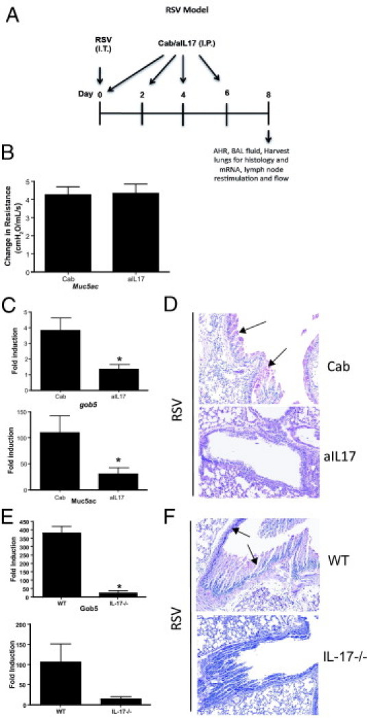
Severe respiratory syncytial virus (RSV) infections are characterized by airway epithelial cell damage, mucus hypersecretion, and Th2 cytokine production. Less is known about the role of IL-17. We observed increased IL-6 and IL-17 levels in tracheal aspirate samples from severely ill infants with RSV infection. In a mouse model of RSV infection, time-dependent increases in pulmonary IL-6, IL-23, and IL-17 expression were observed. Neutralization of IL-17 during infection and observations from IL-17(-/-) knockout mice resulted in significant inhibition of mucus production during RSV infection. RSV-infected animals treated with anti-IL-17 had reduced inflammation and decreased viral load, compared with control antibody-treated mice. Blocking IL-17 during infection resulted in significantly increased RSV-specific CD8 T cells. Factors associated with CD8 cytotoxic T lymphocytes, T-bet, IFN-γ, eomesodermin, and granzyme B were significantly up-regulated after IL-17 blockade. Additionally, in vitro analyses suggest that IL-17 directly inhibits T-bet, eomesodermin, and IFN-γ in CD8 T cells. The role of IL-17 was also investigated in RSV-induced exacerbation of allergic airway responses, in which neutralization of IL-17 led to a significant decrease in the exacerbated disease, including reduced mucus production and Th2 cytokines, with decreased viral proteins. Taken together, our data demonstrate that IL-17 plays a pathogenic role during RSV infections.


Figures

Figure 1
Figure 1
RSV infection induces IL-17 production in mice and humans, and CD4 T cells contribute significantly to peak IL-17 production. A: Total RNA was extracted from the lungs of RSV-infected BALB/c mice on days 1, 2, 4, 6, 8, and 12 after infection, and the relative expression of IL-17, IL-17F, IL-6, and IL-23p19 was analyzed by real-time PCR. The expression levels were normalized to the housekeeping gene GAPDH and fold inductions were compared with naïve mice. B: IL-17 production in the lungs on days 2, 4, 6, 8, and 12 after RSV infection. The data were analyzed by a Bio-Plex system. C: Draining lymph nodes from naïve and RSV-infected mice were harvested on day 8 after infection and were rechallenged with RSV. Supernatants were collected 48 hours later, and IL-17 production was analyzed by a Bio-Plex system. D: Intracellular IL-17 levels were measured by flow cytometry in different IL-17 producing cells in the lungs of mice at day 8 after RSV infection and were compared with naïve lungs. Each experiment was repeated at least twice, with four to five mice per group. Human IL-6 (E) and IL-17 (F) were measured in tracheal aspirate samples from infants with or without RSV infection. Data are reported as means ± SE. *P < 0.04, **P = 0.036.
Figure 2
Figure 2
Neutralization of IL-17 inhibits mucus-associated genes and mucus accumulation in the lungs after RSV infection. A: Schematic representation of the primary RSV infection model. B: Airway resistance was measured at 8 days after RSV infection in mice treated with control or anti-IL-17 antibody (aIL17) after a single dose of methacholine. Data are represented as mean AHR in cmH2O/mL per second ± SE. Relative expression of mucus-associated genes in the lungs of mice treated with Cab versus anti-IL-17 antibody (C) or of WT versus IL-17−/− mice (E) was analyzed by real-time PCR. Data are reported as means ± SE. *P < 0.01. Lungs from Cab versus aIL17-treated mice (D) or WT versus IL-17−/− mice (F) were harvested and stained with PAS. Arrows indicate PAS positive staining. Original magnification, ×200. Each experiment was repeated three times, with five mice in each group.
Figure 3
Figure 3
Blockade of IL-17 inhibits neutrophilic infiltration in bronchoalveolar lavage fluid (BALF). A: Differential count of BALF cellularity for RSV-infected mice treated with anti-IL-17 antibody (aIL17) or Cab. B: Relative expression of chemokines in the lungs analyzed by real-time PCR from RSV-infected mice treated with anti-IL-17 antibody (aIL-17) or Cab. The experiment was repeated twice with five mice in each group. Data are reported as means ± SE. *P < 0.05.
Figure 4
Figure 4
RSV-induced IL-17 reduces viral clearance but does not alter Th2 cytokines. A: RSV nuclear protein N expression in the lungs was measured by real-time PCR on days 2, 4, 8, 10, and 12 after RSV infection and treatment with Cab or anti-IL-17 antibody (aIL-17). The relative expression of RSV-N in Cab and aIL-17 treated groups was normalized to RSV-infected mice at day 1. B: Viral plaque-forming units (PFU) per wet weight of lung were measured 4 days after RSV infection and after treatment with anti-IL-17 antibody or Cab. Data are reported as means ± SE. *P < 0.05. The experiment was repeated twice with similar results. C: RSV antigens were measured by ELISA. O.D., optical density. D: Relative expression of cytokines IL-4, IL-13, IFN-γ, and IL-17A was analyzed by real-time PCR in the lungs of RSV-infected mice treated with Cab or aIL-17 on day 8 after infection. Cytokine mRNA expression levels were normalized to those of naïve mice. E: Lung DLNs from the same mice were restimulated with RSV for 48 hours and the supernatants were analyzed for cytokine production by a Bio-Plex system. Cytokine responses to RSV in WT and IL-17−/− mice were compared in the lungs (F) and lymph nodes (G). The experiment was repeated twice, with four to five mice per group. Data are reported as means ± SE. *P < 0.05.
Figure 5
Figure 5
IL-17 inhibits CD8 T cell effector responses during RSV infection. Flow cytometry in RSV-infected mice treated with Cab or with anti-IL-17 antibody was performed at 8 days after infection using five dispersed lung DLNs (A) or whole-lung digests (B) The results from one experiment of three repeats are shown with total number of CD4 (*P < 0.04), CD4CD69 (*P < 0.02), CD8 (*P < 0.05), and CD8CD69 T cells (*P < 0.05). C: The number of CD8 T cells positive for IL-17 receptor A chain (IL-17RA) in naïve or RSV-infected mice. D: RSV-specific immunodominant T-cell receptor tetramer surface staining (left), expressed as a percentage of CD3+ cells in the lungs (right). E–I: Total tetramer-positive cells (E) IFN-γ (F), Eomes (G), T-bet (H), and granzyme B (I) CD8 T cells in the lungs of RSV-infected mice (*P < 0.05). J: Relative expression of T-bet, Eomes, and IFN-γ was analyzed by real-time PCR in magnetic-cell-sorting–purified CD8 T cells from the spleen of naïve BALB/c mice that were stimulated with anti-CD3 (2 μg/mL) and anti-CD28 (2 μg/mL) in the presence (+rIL-17) or absence (−rIL-17) of recombinant IL-17 for 6 hours in vitro. The data were normalized to GAPDH expression levels and the fold induction was compared with unstimulated CD8 T cell values. N.D. indicates not detected.
Figure 6
Figure 6
RSV exacerbation of allergic airway disease is mediated by IL-17 by altering mucus production, activating CD8 T cell effector functions, and decreasing viral clearance. A: Schematic representation of RSV exacerbation of existing CRA-induced allergic airway disease. B: Lung DLN cells were isolated from animals sensitized to CRA, RSV, or both CRA and RSV and then were restimulated with either CRA or RSV for 48 hours. The supernatants were analyzed for IL-17 by a Bio-Plex system. C: Lungs from CRA sensitized, RSV-infected mice treated with Cab or anti-IL-17 antibody with H&E or PAS staining. Original magnification, ×400. Insets: Higher-magnification view of boxed area. Original magnification, ×1000. D: Relative expression of mucus-associated genes (Muc5ac and Gob5) in the lungs was analyzed by real-time PCR. *P < 0.05, **P < 0.01. E: IFN-γ, IL-4, IL-5, and IL-13 production was analyzed by a Bio-Plex system. F: Flow cytometry was performed using whole-lung digests. Data are reported as total number of CD4, CD4CD69, CD8, and CD8CD69 T cells per lobe of the lung. *P < 0.05. G: RSV-specific immunodominant T-cell receptor tetramer surface staining and analysis by flow cytometry. H: Relative expression of RSV proteins (G, F, and N) in the lungs was analyzed by real-time PCR. Data are reported as means ± SE of five mice per group, representative of one of the two independent experiments. N.D. indicates not detected.

References

    1. Openshaw P.J., Dean G.S., Culley F.J. Links between respiratory syncytial virus bronchiolitis and childhood asthma: clinical and research approaches. Pediatr Infect Dis J. 2003;22(2 Suppl):S58–S64. discussion S64–S65. - PubMed
    1. Black C.P. Systematic review of the biology and medical management of respiratory syncytial virus infection. Respir Care. 2003;48:209–231. discussion 231–233. - PubMed
    1. Stensballe L.G., Devasundaram J.K., Simoes E.A. Respiratory syncytial virus epidemics: the ups and downs of a seasonal virus. Pediatr Infect Dis J. 2003;22(2 Suppl):S21–S32. - PubMed
    1. Jafri H.S., Chavez-Bueno S., Mejias A., Gomez A.M., Rios A.M., Nassi S.S., Yusuf M., Kapur P., Hardy R.D., Hatfield J., Rogers B.B., Krisher K., Ramilo O. Respiratory syncytial virus induces pneumonia, cytokine response, airway obstruction, and chronic inflammatory infiltrates associated with long-term airway hyperresponsiveness in mice. J Infect Dis. 2004;189:1856–1865. - PubMed
    1. Schwarze J., Cieslewicz G., Hamelmann E., Joetham A., Shultz L.D., Lamers M.C., Gelfand E.W. IL-5 and eosinophils are essential for the development of airway hyperresponsiveness following acute respiratory syncytial virus infection. J Immunol. 1999;162:2997–3004. - PubMed

Publication types

MeSH terms