. 2011 Jul;179(1):281-8.
doi: 10.1016/j.ajpath.2011.03.043. Epub 2011 May 14.

Cavia porcellus as a model for experimental infection by Trypanosoma cruzi

Affiliations

Cavia porcellus as a model for experimental infection by Trypanosoma cruzi

Yagahira E Castro-Sesquen et al. Am J Pathol. 2011 Jul.

Abstract

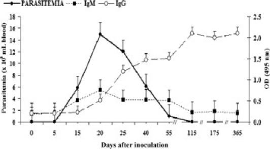
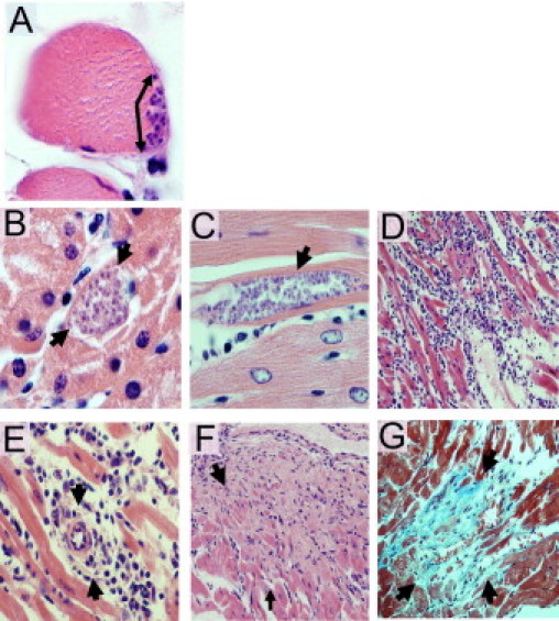
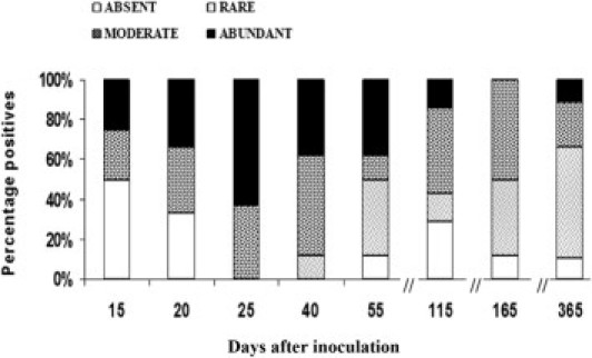
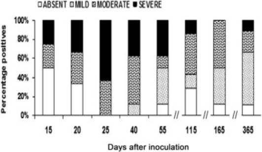
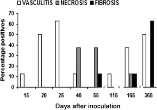
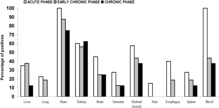
The guinea pig (Cavia porcellus) is a natural reservoir for Trypanosoma cruzi but has seldom been used as an experimental infection model. We developed a guinea pig infection model for acute and chronic Chagas disease. Seventy-two guinea pigs were inoculated intradermally with 10(4) trypomastigotes of T. cruzi strain Y (experimental group); 18 guinea pigs were used as control group. Eight animals from the experimental group and two from the control group were sacrificed 5, 15, 20, 25, 40, 55, 115, 165, and 365 days after inoculation. During the acute phase (15 to 55 days), we observed parasitemia (with a peak on day 20) and positive IgM and IgG Western blots with anti-shed acute-phase antigen bands. The cardiac tissue showed vasculitis, necrosis (on days 40 to 55), moderate to severe inflammation, and abundant amastigote nests. Smaller numbers of amastigote nests were also present in kidney, brain, and other organs. In the early chronic phase (115 to 165 days), parasitemia disappeared and anti-T. cruzi IgG antibodies were still detectable. In cardiac tissue, the number of amastigote nests and the grade of inflammation decreased. In the chronic phase (365 days), the cardiac tissue showed vasculitis and fibrosis; detectable parasite DNA was associated with higher grades of inflammation. The experimental T. cruzi infection model in guinea pigs shows kinetics and pathologic changes similar to those of the human disease.


Figures

Figure 1
Figure 1
Detection of parasitemia using the microhematocrit technique and IgM and IgG to T. cruzi by TESA-ELISA during experimental T. cruzi infection of C. porcellus. Eight animals were used per group. The points and error bars represent median ± SE.
Figure 2
Figure 2
Detection of IgM and IgG by TESA blot analysis during experimental T. cruzi infection of C. porcellus. A: IgM detection. The classic six SAPA bands (130 to 200 kDa) were seen during the acute phase (until 55 days after inoculation). The bottom band is slightly smeared due to prolonged run time. On day 55 after inoculation, a single 150- to 160-kDa band was also detected. From 115 days after inoculation until the end of the experiment, IgM was undetectable. B: IgG detection. SAPA bands (130 to 200 kDa) were observed from 20 until 25 days after inoculation. Subsequently, the 150- to 160-kDa band was also detected until the end of the experiment. Lane 1: day 0 (before experimental inoculation). Serum samples of guinea pig after inoculation: lane 2, day 5; lane 3, day 15; lane 4, day 20; lane 5, day 25; lane 6, day 40; lane 7, day 55; lane 8, day 115; lane 9, day 165; and lane 10, day 365.
Figure 3
Figure 3
Histopathologic analysis in C. porcellus after inoculation of T. cruzi strain Y. Amastigote nests (arrows) were seen 25 days after inoculation in skeletal muscle (A) and kidney (B). Original magnification: ×1000. Histopathologic analysis in cardiac tissue demonstrates amastigote nests (arrow) between myocardial fibers 25 days after inoculation (C); severe and diffuse inflammatory infiltrate 25 days after inoculation (D); perivascular inflammation (arrows) 25 days after inoculation (E); areas of necrosis 56 days after inoculation (F); and foci of interstitial fibrosis (arrows) and lymphocytic infiltrate (arrows) 365 days after inoculation (G). Original magnification: ×1000 (C); ×200 (D, F, and G); ×500 (E). Staining: H&E (A–F); Masson's trichrome (G).
Figure 4
Figure 4
Percentage of animals that presented rare, moderate, or abundant amastigote nests in cardiac tissue of C. porcellus during experimental infection by T. cruzi. Quantification of parasite number was the mean number of parasites seen in two entire sections: rare, 1 nest; moderate, 2 to 10 nests; abundant, >10 nests. H&E stain. Eight animals per group.
Figure 5
Figure 5
Percentage of animals that presented mild, moderate, or severe inflammation in cardiac tissue of C. porcellus during experimental T. cruzi infection. Semiquantitative analysis of the degree of inflammation was evaluated in two entire sections: mild, lymphocytes in 2% to 15% of the entire section with focal distribution; moderate, lymphocytes in 20% to 60% of the entire section; severe, lymphocytes in >70% of the entire section. For moderate and severe inflammation, the distribution was either multifocal or diffuse. H&E stain. Eight animals per group.
Figure 6
Figure 6
Percentage of animals that presented vasculitis, necrosis, and fibrosis in cardiac tissue of C. porcellus during experimental infection by T. cruzi. Qualitative analysis was evaluated in two total sections. H&E or Masson's trichrome stain. Eight animals per group.
Figure 7
Figure 7
Detection of kinetoplast DNA by PCR in blood and tissue samples during experimental infection of C. porcellus by T. cruzi. Acute phase: 5 to 55 days after inoculation. n = 40 animals. Early chronic phase: 115 to 165 days after inoculation. n = 16 animals. Chronic phase: 365 days after inoculation. n = 8 animals.

Similar articles

Cited by

References

    1. Organización Panamericana de la Salud . Organización Panamericana de la Salud; Montevideo, Uruguay: 2006. Estimación cuantitativa de la enfermedad de Chagas en las Américas.
    1. Prata A. Clinical and epidemiological aspects of Chagas disease. Lancet Infect Dis. 2001;1:92–100. - PubMed
    1. Rassi A., Jr, Rassi A., Little W.C. Chagas' heart disease. Clin Cardiol. 2000;23:883–889. - PMC - PubMed
    1. Rassi A., Jr, Rassi S.G., Rassi A. Sudden death in Chagas' disease. Arq Bras Cardiol. 2001;76:75–96. - PubMed
    1. de Oliveira R.B., Troncon L.E., Dantas R.O., Menghelli U.G. Gastrointestinal manifestations of Chagas' disease. Am J Gastroenterol. 1998;93:884–889. - PubMed

Publication types

MeSH terms