. 2011 Jul;179(1):302-14.
doi: 10.1016/j.ajpath.2011.03.047. Epub 2011 May 10.

Dual functions of prostaglandin D2 in murine contact hypersensitivity via DP and CRTH2

Affiliations

Dual functions of prostaglandin D2 in murine contact hypersensitivity via DP and CRTH2

Yoshihiro Yamamoto et al. Am J Pathol. 2011 Jul.

Erratum in

  • Am J Pathol. 2012 Jan;180(1):429-30

Abstract

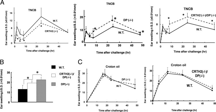
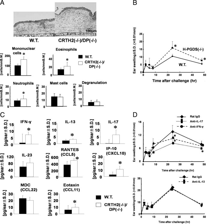
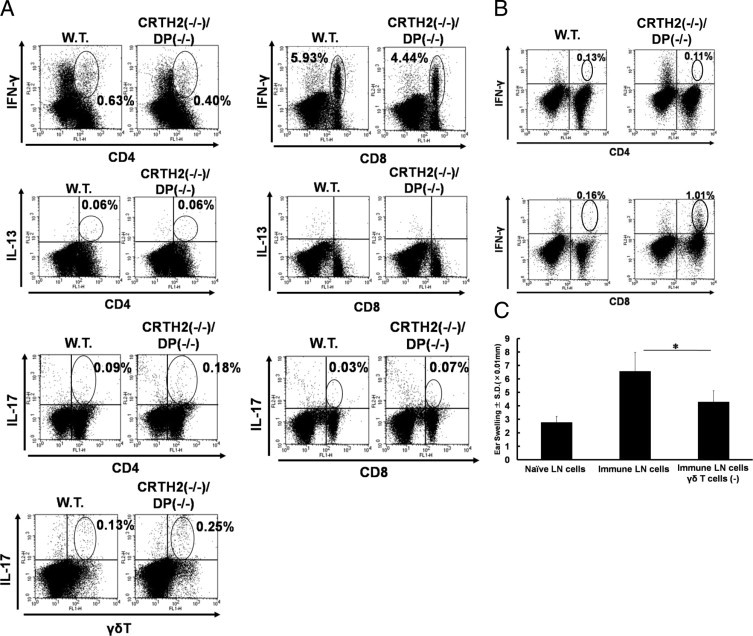
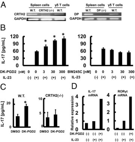
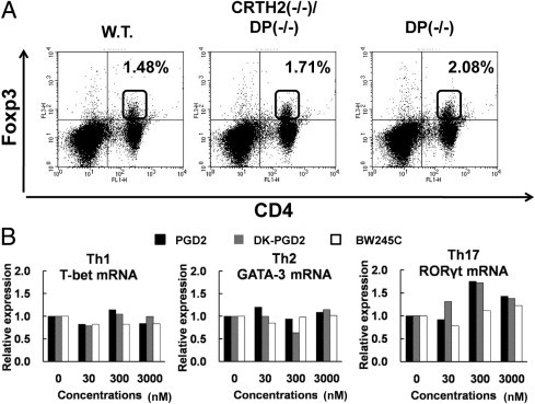
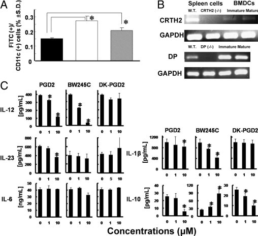
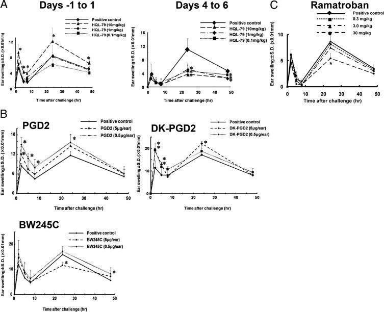
Prostaglandin D2 (PGD2) exerts its effects through two distinct receptors: the chemoattractant receptor-homologous molecule expressed on Th2 cells (CRTH2) and the D prostanoid (DP) receptor. Our previous study demonstrated that CRTH2 mediates contact hypersensitivity (CHS) in mice. However, the function of DP receptor remains to be fully established. In this study, we examine the pathophysiological roles of PGD2 using DP-deficient (DP(-/-)) and CRTH2/DP-deficient (CRTH2(-/-)/DP(-/-)) mice to elucidate receptor-mediated PGD2 action in CHS. We observed profound exacerbation of CHS in DP(-/-) mice. CRTH2(-/-)/DP(-/-) mice showed similar exacerbation, but to a lesser extent. These symptoms were accompanied by increased production of interferon-γ and IL-17. The increase in IL-17 producing γδ T cells was marked and presumably contributed to the enhanced CHS. DP deficiency promoted the in vivo migration of dendritic cells to regional lymph nodes. A DP agonist added to DCs in vitro was able to inhibit production of IL-12 and IL-1β. Interestingly, production of IL-10 in dendritic cells was elevated via the DP pathway, but it was lowered by the CRTH2 pathway. Collectively, PGD2 signals through CRTH2 to mediate CHS inflammation, and conversely, DP signals to exert inhibitory effects on CHS. Thus, we report opposing functions for PGD2 that depend on receptor usage in allergic reactions.


Figures

Figure 1
Figure 1
Contact hypersensitivity (CHS) responses in prostaglandin D2 (PGD2) receptor-deficient mice. A: Ear thickness in CHS responses to TNCB. WT, CRTH2−/−, D prostanoid (DP)−/− and CRTH2−/−/DP−/− mice were challenged with 2,4,6-trinitrochlorobenzene (TNCB). Ear thickness was measured at the times indicated. Numbers of mice in each group were four to six. Experiments were independently repeated at least three times. Means ± SD. B: Simultaneous comparison of ear thickness in CHS responses. Wild-type (W.T.), DP−/− receptor and chemoattractant receptor-homologous molecule expressed on Th2 cells CRTH2−/−/DP−/− mice were challenged with TNCB. Ear thickness was measured 24 hours after challenge. C: Ear thickness in irritation dermatitis to croton oil. DP−/− mice and CRTH2−/−/DP−/− mice were painted with croton oil and ear thickness was measured at the times indicated. *P < 0.05.
Figure 2
Figure 2
Modulation of parameters in contact hypersensitivity (CHS) in mutant mice. A: Histological features and cell populations in inflammatory skin. Wild-type (W.T.) and chemoattractant receptor-homologous molecule expressed on Th2 cells (CRTH2−/−)/D prostanoid (DP−/−) mice were challenged with 2,4,6-trinitrochlorobenzene (TNCB) and inflammatory ears at 24 hours after challenge were subjected to histological observation and cell counting. Cells were counted in five distinct areas and determined as numbers per 1 mm of basement membrane (B.M.). Means ± SD. B: CHS responses in mice lacking the H-PGDS gene. W.T. and hematopoietic-type PGD synthase (H-PGDS−/−) mice were challenged with TNCB. Ear thickness was measured at the times indicated. C: Cytokine and chemokine production in CHS. W.T. and CRTH2−/−/DP−/− mice were challenged with TNCB. Ear lobes were punched 24 hours after challenge and levels of cytokines and chemokines were determined by enzyme-linked immunosorbent assay. Means ± SD. D: Ear thickness in CHS treated with anti-cytokine antibodies. CRTH2−/−/DP−/− mice were sensitized with TNCB. Blocking Abs against cytokines interferon [(IFN)-γ, IL-13, and IL-17 at doses of 200 μg/mouse] were administered i.p. 1 day before challenge. Ear thickness was measured at the times indicated. *P < 0.05. Numbers of mice in each group were four to six. Representative results from at least two independent experiments are shown.
Figure 3
Figure 3
Profiles of cytokine production in T-cell subpopulations. A, B: Cytokine production by T cells. Wild-type (W.T.) and chemoattractant receptor-homologous molecule expressed on CRTH2−/−/DP−/− mice were sensitized with 2,4,6-trinitrochlorobenzene (TNCB) on day 0. Abdominal (A) and cervical (B) lymph node (LN) cells were collected on day 5 (A) and 24 hours after challenge (B), respectively. They were incubated with trinitrophenyl-spleen cells for 48 hours followed by stimulation with phorbol 12-myristate 13-acetate and ionomycin in the presence of brefeldin A for a further 12 hours. Intracellular cytokines in CD4, CD8, and γδT cells of LNs were separately analyzed by flow cytometry. C: LN cells from CRTH2−/−/DP−/− mice sensitized with TNCB were transferred into ear lobes of W.T. mice. Transferred mice were challenged with TNCB and examined for ear thickness at 24 hours after challenge. Depletion of γδ T cells was performed with the MACS system using a biotin-conjugated γδ TCR (T cell receptor) Ab (GL-3). *P < 0.05. Numbers of mice in each group were 4. Representative results from at least two independent experiments are shown. IFN, interferon.
Figure 4
Figure 4
IL-17 production by γδ T cells. A: Expression of chemoattractant receptor-homologous molecule expressed on Th2 cells (CRTH2) and D prostanoid (DP) in γδ T cells. γδ T cells were isolated from wild-type (W.T.) mice and examined for their expression of both CRTH2 and DP mRNA by RT-PCR. The purity of γδ T cells was always >95% as assessed by γδ TCR-FITC antibody staining (UC7-13D5). Spleen cells from W.T. mice DP−/− and CRTH2−/− mice were used as positive and negative controls, respectively. B and C: γδ T cells were isolated from W.T. (B, C) and CRTH2−/− mice (C), and stimulated with DK-PGD2 (B, C) or BW245C (B) in the presence of IL-23 (50 ng/mL). IL-17 levels were determined by enzyme-linked immunosorbent assay. Error bars indicate SD. D: Expression of IL-17 and RORγt mRNA in γδ T cells. γδ T cells were isolated from W.T. mice and stimulated with DK-PGD2 and IL-23. Levels of mRNA for IL-17 and RORγt were determined by real-time RT-PCR. *P < 0.05. Representative results from at least three independent experiments are shown.
Figure 5
Figure 5
Effects of prostaglandin D2 (PGD2) signals on T-cell development. A: Foxp3 expression in CD4(+) T cells of PGD2 receptor-deficient mice. Immune lymph node (LN) cells were prepared from wild-type (W.T.), D prostanoid (DP)−/− and chemoattractant receptor-homologous molecule expressed on Th2 cells (CRTH2)−/−/DP−/− mice sensitized with 2,4,6-trinitrochlorobenzene (TNCB) and then analyzed by flow cytometry after staining with anti-Foxp3 and CD4 antibodies. B: Effects of PGD2 on the expression of T-cell transcription factors. Th1, Th2, or Th17 cells were induced as described in Materials and Methods and incubated for 4 hours with the respective PGD2 agonists. mRNA was extracted and examined for T-bet, GATA-3, and RORγt by real-time RT-PCR. Representative results from at least three independent experiments are shown.
Figure 6
Figure 6
Modulation of dendritic cell (DC) function by prostaglandin D2 (PGD2) receptor signals. A: Migration of DC in mutant mice. Wild-type (WT; black bar), DP−/− (grey bar), and chemoattractant receptor-homologous molecule expressed on CRTH2−/−/DP−/− (white bar) mice were sensitized with fluorescein isothiocyanate (FITC), and 24 hours later, draining lymph node (LN) cells were isolated. FITC(+)/CD11c(+) DCs in LN cells were assessed by flow cytometry. B: Expression in CRTH2 and DP in bone marrow-derived dendritic cells (BMDC). The BMDC were subjected to mRNA extraction, followed by real-time PCR with primers specific to CRTH2, DP, and GADPH. Spleen cells from W.T. and CRTH2−/−/DP−/− mice were used as positive and negative controls, respectively. Maturation of DC induced by lipopolysaccharide (LPS) (1 μg/mL, 20 hours) was assessed by the increased expression of major histocompatibility complex class II and CD86 (data not shown). C: Effects of PGD2 on cytokine production. BMDC (5 × 10⁵ cells/mL) were incubated with PGD2, BW245C, or DK-PGD2 for 4 hours, followed by stimulation with LPS (1 μg/mL) for an additional 16 hours. Enzyme-linked immunosorbent assay was used to measure cytokines in the supernatants. Error bars indicate standard deviation. *P < 0.05. Representative results from at least three independent experiments are shown.
Figure 7
Figure 7
Effects of agonists and inhibitors on contact hypersensitivity (CHS) in wild-type (W.T.) mice. A: Effects of the hematopoietic-type PGD synthase (H-PGDS) inhibitor on CHS. W.T. mice were sensitized and challenged with 2,4,6-trinitrochlorobenzene (TNCB) along with p.o. administration of HQL-79 dissolved in 0.5% methylcellulose (an H-PGDS inhibitor) during either the sensitization phase (days −1, 0, and 1) or the elicitation phase (days 4, 5, and 6). Ear thickness was measured at the times indicated. B: Effects of prostaglandin D2 (PGD2) receptor agonists on CHS. PGD2 agonists were directly administered s.c. into challenged ear lobes (20 μL/ear) immediately before challenge. Ear thickness in response to TNCB was measured. C: Effects of ramatroban [a chemoattractant receptor-homologous molecule expressed on Th2 cells (CRTH2) antagonist] on CHS. Ramatroban dissolved in 0.5% methylcellulose was administered p.o. on days 4, 5, and 6. Ear thickness in response to TNCB was measured. *P < 0.05. Numbers of mice in each group were four to six. Representative results from at least two independent experiments are shown.

Similar articles

Cited by

References

    1. Beasley C.R., Robinson C., Featherstone R.L., Varley J.G., Hardy C.C., Church M.K., Holgate S.T. 9 alpha,11 beta-prostaglandin F2, a novel metabolite of prostaglandin D2 is a potent contractile agonist of human and guinea pig airways. J Clin Invest. 1987;79:978–983. - PMC - PubMed
    1. Nagoshi H., Uehara Y., Kanai F., Maeda S., Ogura T., Goto A., Toyo-oka T., Esumi H., Shimizu T., Omata M. Prostaglandin D2 inhibits inducible nitric oxide synthase expression in rat vascular smooth muscle cells. Circ Res. 1998;82:204–209. - PubMed
    1. Narumiya S., Toda N. Different responsiveness of prostaglandin D2-sensitive systems to prostaglandin D2 and its analogues. Br J Pharmacol. 1985;85:367–375. - PMC - PubMed
    1. Whittle B.J., Moncada S., Vane J.R. Comparison of the effects of prostacyclin (PGI2), prostaglandin E1 and D2 on platelet aggregation in different species. Prostaglandins. 1978;16:373–388. - PubMed
    1. Kanaoka Y., Urade Y. Hematopoietic prostaglandin D synthase. Prostaglandins Leukot Essent Fatty Acids. 2003;69:163–167. - PubMed

Publication types

MeSH terms