

P-cadherin promotes liver metastasis and is associated with poor prognosis in colon cancer
- PMID: 21703417
- PMCID: PMC3123784
- DOI: 10.1016/j.ajpath.2011.03.046
P-cadherin promotes liver metastasis and is associated with poor prognosis in colon cancer
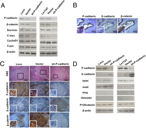
Abstract
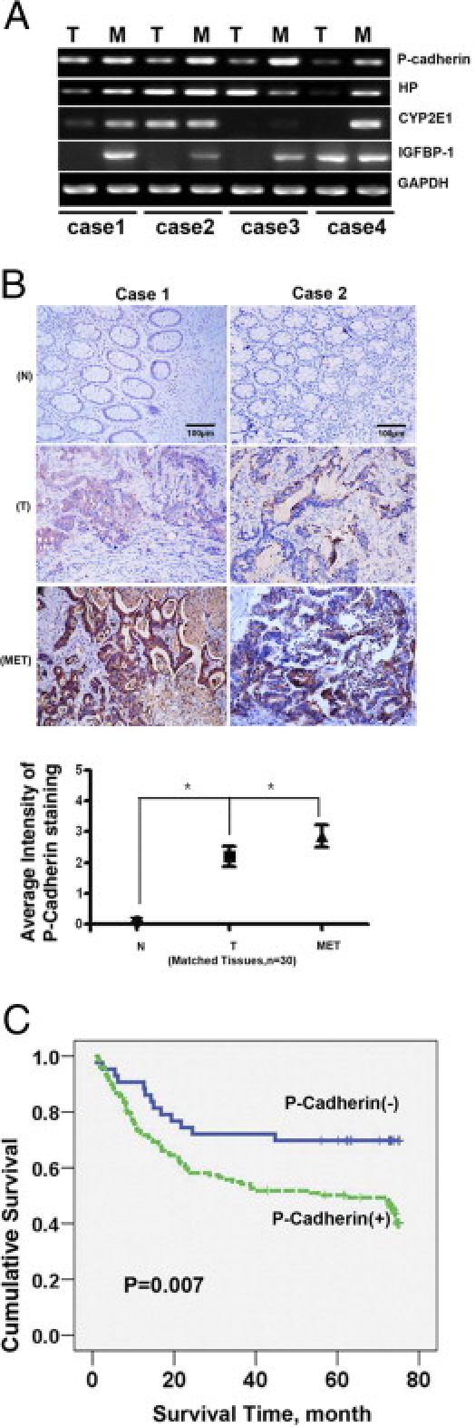
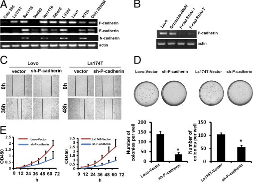
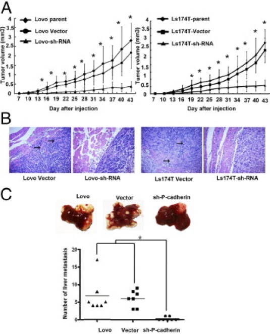
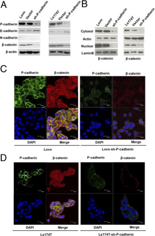
P-cadherin belongs to the family of classic cadherins, which is important for maintaining cellular localization and tissue integrity. Recently, it has become evident that P-cadherin contributes to the oncogenesis of many tumor types, including melanoma, prostate, breast, and colon carcinomas. Although cadherin switching is a crucial step in metastasis, the role of P-cadherin in colon cancer metastasis to the liver is unknown. In this study, we performed gene expression analysis and found that the level of P-cadherin was higher in tissue from liver metastases of colon cancer than in the corresponding primary colon cancer tissues. IHC analysis also showed that P-cadherin expression was significantly higher in liver metastases than in paired primary colorectal cancer tumors. Knockdown of P-cadherin in colon cancer cells inhibited wound healing, proliferation, and colony formation and resulted in developing fewer liver metastatic foci and reducing the tumor burden in vivo. Inhibition of P-cadherin expression also induced the up-regulation of E-cadherin and the down-regulation of β-catenin and its downstream target molecules, including survivin and c-Myc. In summary, these results uncover a novel function of P-cadherin in the regulation of colon cancer metastasis to the liver, suggesting that blocking the activity of P-cadherin or its associated signaling may be a valuable target for the treatment of hepatic metastases of colon carcinomas.
Copyright © 2011 American Society for Investigative Pathology. Published by Elsevier Inc. All rights reserved.
Figures
References
-
- Wicherts D.A., de Haas R.J., Borel Rinkes I.H., Voest E.E., van Hillegersberg R. Better treatment for patients with colorectal liver metastases. Ned Tijdschr Geneeskd. 2006;150:345–351. - PubMed
-
- Ochiai H., Nakanishi Y., Fukasawa Y., Sato Y., Yoshimura K., Moriya Y., Kanai Y., Watanabe M., Hasegawa H., Kitagawa Y., Kitajima M., Hirohashi S. A new formula for predicting liver metastasis in patients with colorectal cancer: immunohistochemical analysis of a large series of 439 surgically resected cases. Oncology. 2008;75:32–41. - PubMed
-
- Bakalakos E.A., Kim J.A., Young D.C., Martin E.W., Jr Determinants of survival following hepatic resection for metastatic colorectal cancer. World J Surg. 1998;22:399–404. - PubMed
Publication types
MeSH terms
Substances
LinkOut - more resources
Full Text Sources
Other Literature Sources
Medical
