. 2011 Jul;179(1):477-86.
doi: 10.1016/j.ajpath.2011.03.038.

RREB1 transcription factor splice variants in urologic cancer

Affiliations

RREB1 transcription factor splice variants in urologic cancer

Matthew D Nitz et al. Am J Pathol. 2011 Jul.

Abstract

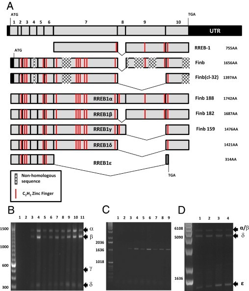
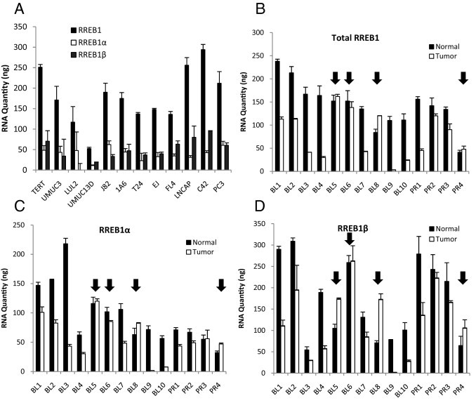
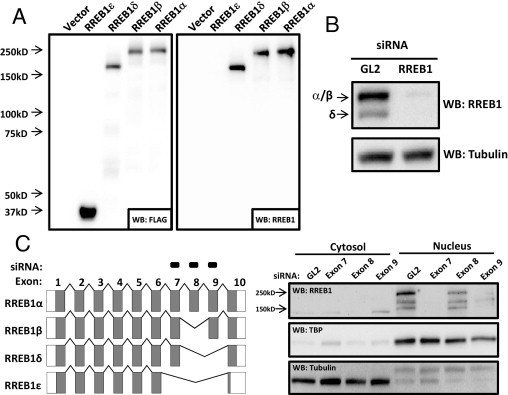
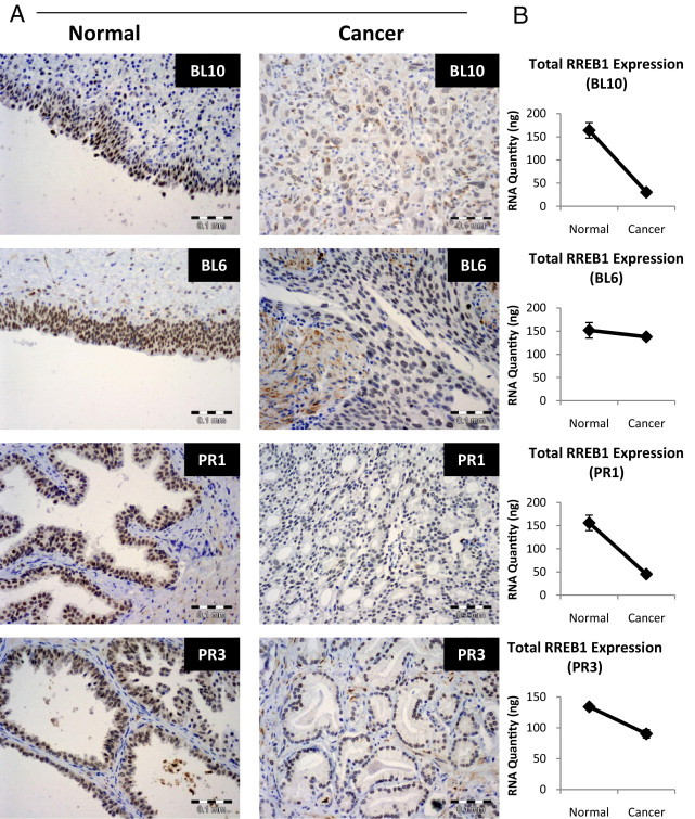
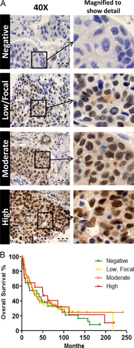
RREB1 is an alternatively spliced transcription factor implicated in Ras signaling and cancer. Little is known about the expression of RREB1 isoforms in cell lines or human tumors, or about the clinical relevance of the latter. We have developed tools for IHC of RREB1 protein isoform-specific amplification of RREB1 mRNA and selective knockdown of RREB1 isoforms and use these to provide new information by characterizing RREB1 expression in bladder and prostate cancer cell lines and human tissue samples. Previously described splice variants RREB1α, RREB1β, RREB1γ, and RREB1δ were identified, as well as the novel variant RREB1ε. Total and isoform-specific mRNA expression was lower in most but not all tumors, compared with normal tissues. RREB1 IHC performed on a bladder cancer TMA did not indicate a relationship between total RREB1 expression and overall survival after radical cystectomy for invasive bladder cancer. In contrast, in vitro proliferation studies using the UMUC-3 bladder cancer cell line after selective isoform-specific knockdown of expression indicate that RREB1α is not necessary for proliferation, but that RREB1β may be required. These contributions should accelerate progress in the nascent RREB1 field by providing new reagents while also providing clues to the role of RREB1 isoforms in human cancer and raising the possibility of isoform-specific roles in human carcinogenesis and progression.


Figures

Figure 1
Figure 1
RREB1 alternative splicing. A: 10 coding exons of RREB and previously described RREB1 sequences were aligned. Finb and Finb (cl-32) contained several regions of frame shifts in the cDNA that resulted in nonhomologous protein sequence. These sequences also described an alternative translation start site that results in a protein without the N-terminal 19 amino acids. We propose using Greek lettering to describe RREB1 alternative splicing: RREB1α, RREB1β, RREB1γ, RREB1δ, and RREB1ε. B: Primers designed to span the region of known RREB1 alternative splicing (exon 7 to exon 10) were interrogated on the following bladder and prostate cancer cell lines: 1, negative PCR control; 2, negative RT control; 3, TERT; 4, 293T; 5, UMUC-3; 6, LUL2; 7, UMUC13D; 8, J82; 9, 1A6; 10, PC3; and 11, LNCAP. C: Primers designed to span from exon 1 to exon 7 of RREB1 were used to amplify cDNA from the following samples: 1, negative PCR control; 2, negative RT control; 3, TERT; 4, 293T; 5, UMUC-3; 6, LUL2; 7, UMUC13D; 8, J82; and 9, 1A6. D: Primers designed to span exon 1 to exon 10 of RREB1 were used to amplify cDNA from the following samples: 1, UMUC-3; 2, LUL2; 3, LNCaP; and 4, J82. Arrows and Greek letters indicate the RREB1 splice variants.
Figure 2
Figure 2
RREB1 transcripts in bladder tumors and cancer cell lines. A: mRNA expression of total RREB1, RREB1α, and RREB1β was measured in bladder and prostate cancer cell lines. RNA quantities cannot be compared among total RREB1, RREB1α, and RREB1β, because the levels were calculated on unique standard curves for each primer pair. B: Total RREB1 expression was measured in paired normal and cancer (tumor) urothelial bladder (BL) and prostate (PR) tissues, using primers that span exons 2 and 3 of RREB1, an area lacking alternative splicing. Arrows indicate samples in which RREB1 expression is greater in the tumor than in the paired normal tissue. C and D: RNA expression of the RREB1α and RREB1β splice variants was measured using primers targeting base pairs at unique splice junctions.
Figure 3
Figure 3
Detection of RREB1 protein isoforms in human bladder cancer cells. A: RREB1 isoforms were either isolated from UMUC-3 cells (Isoforms δ and ε) or received as a gift from Dr. Akiyoshi Fukamizu (University of Tsukuba, Tsukuba, Japan) (isoforms α and β). The splice variants were cloned into a C-terminal 3XFLAG-tagged expression construct and expressed in 293T cells. Detection with anti-FLAG (1:1000) or anti-RREB1 (1:1000) (Sigma-Aldrich) antibody is shown. B: RREB1 (total) siRNA, designed to knock down all splice variants, was transfected into UMUC-3 cells. RREB1 antibody (Sigma-Aldrich) detection (1:1000) on Western blot is shown. C: siRNAs targeting exons 7, 8, or 9 were transfected into UMUC-3 cells. Cytoplasmic and nuclear fractions were isolated after 96 hours and RREB1 protein splice variants were detected on Western blot (1:1000) (Sigma-Aldrich).
Figure 4
Figure 4
IHC of RREB1 in human carcinoma. A: RREB1 IHC (1:100) (Sigma-Aldrich) of paired normal and cancer tissues for urothelial bladder (BL10 and BL6) and prostate (PR1 and PR3). B: Total RREB1 RNA levels by qRT-PCR from the same tissue as in A. Scale bars: 0.1 mm.
Figure 5
Figure 5
Depletion of RREB1β decreases UMUC-3 growth in vitro. A: UMUC-3 cells were treated with 25 nmol/L siRNA of control (GL2), RREB1 (all isoforms), and three unique siRNAs targeting exon 8 (8-1, 8-2, and 8-3). Growth was measured 96 hours after siRNA depletion using Alamar Blue and normalized to the GL2 control. B: Real-time PCR for the RREB1α splice variant was performed for the experiment described in panel A to confirm knockdown of RREB1α. RNA levels were normalized to the GL2 control siRNA. C: UMUC-3 cells were treated with 25 nmol/L siRNA of control (GL2), RREB1 (all isoforms), and three unique siRNAs targeting exon 9 (9-1, 9-2, and 9-3). D: RREB1α and RREB1β-specific real-time PCR was used to confirm RREB1α and RREB1β knockdown.
Figure 6
Figure 6
Total RREB1 expression in bladder cancer does not predict overall survival. A: A TMA of archived bladder cancers (n = 142) was used to assay for RREB1 expression. Representative examples of RREB1 expression grading are shown, as described under Materials and Methods (negative, low/focal, moderate, and high). B: Survival data of patients whose tumors comprised the bladder cancer TMA was used to generate a Kaplan-Meier analysis comparing RREB1 staining versus overall survival. RREB1 staining did not significantly correlate with this clinical parameter (P = 0.86, log rank).

References

    1. Traynor P., McGlynn L.M., Mukhergee R., Grimsley S.J., Bartlett J.M., Edwards J. An increase in N-Ras expression is associated with development of hormone refractory prostate cancer in a subset of patients. Dis Markers. 2008;24:157–165. - PMC - PubMed
    1. Erlich S., Tal-Or P., Liebling R., Blum R., Karunagaran D., Kloog Y., Pinkas-Kramarski R. Ras inhibition results in growth arrest and death of androgen-dependent and androgen-independent prostate cancer cells. Biochem Pharmacol. 2006;72:427–436. - PubMed
    1. Weber M.J., Gioeli D. Ras signaling in prostate cancer progression. J Cell Biochem. 2004;91:13–25. - PubMed
    1. Min J., Zaslavsky A., Fedele G., McLaughlin S.K., Reczek E.E., DE Raedt T., Guney I., Strochlic D.E., Macconaill L.E., Beroukhim R., Bronson R.T., Ryeom S., Hahn W.C., Loda M., Cichowski K. An oncogene-tumor suppressor cascade drives metastatic prostate cancer by coordinately activating Ras and nuclear factor-kappaB. Nat Med. 2010;16:286–294. - PMC - PubMed
    1. Oxford G., Theodorescu D. The role of Ras superfamily proteins in bladder cancer progression. J Urol. 2003;170:1987–1993. - PubMed

Publication types

MeSH terms