

Normalization of cortical gray matter deficits in nonpsychotic siblings of patients with childhood-onset schizophrenia
- PMID: 21703497
- PMCID: PMC3289252
- DOI: 10.1016/j.jaac.2011.03.016
Normalization of cortical gray matter deficits in nonpsychotic siblings of patients with childhood-onset schizophrenia
Abstract
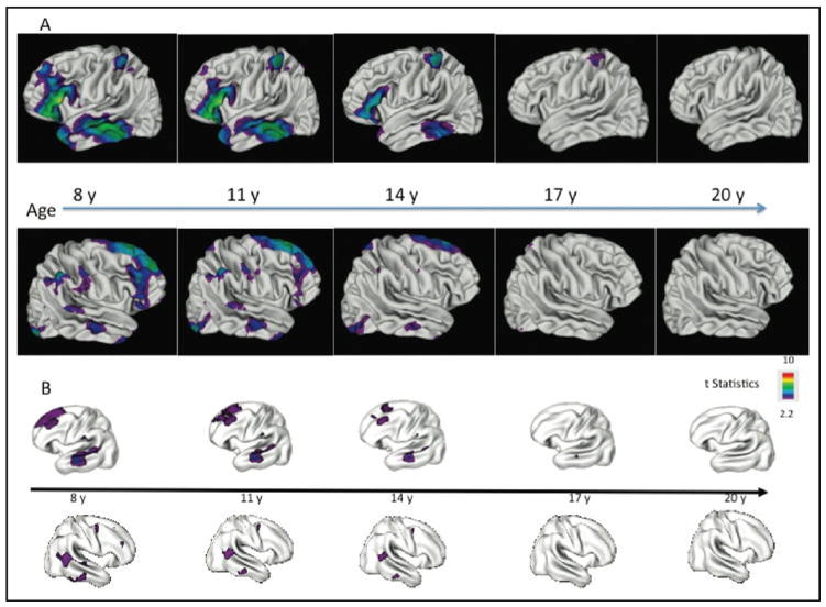
Objective: Cortical gray matter (GM) abnormalities in patients with childhood-onset schizophrenia (COS) progress during adolescence ultimately localizing to prefrontal and temporal cortices by early adult age. A previous study of 52 nonpsychotic siblings of COS probands had significant prefrontal and temporal GM deficits that appeared to "normalize" by age 17 years. Here we present a replication with nonoverlapping groups of healthy full siblings and healthy controls.
Method: Using an automated measure and prospectively acquired anatomical brain magnetic resonance images, we mapped cortical GM thickness in nonpsychotic full siblings (n = 43, 68 scans; ages 5 through 26 years) of patients with COS, contrasting them with age-, gender-, and scan interval-matched healthy controls (n = 86, 136 scans). The false-discovery rate procedure was used to control for type I errors due to multiple comparisons.
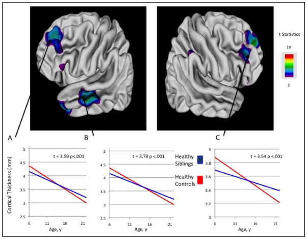
Results: As in our previous study, young nonpsychotic siblings (<17 years) showed significant GM deficits in bilateral prefrontal and left temporal cortices and, in addition, smaller deficits in the parietal and right inferior temporal cortices. These deficits in nonpsychotic siblings normalized with age with minimal abnormalities remaining by age 17.
Conclusions: Our results support previous findings showing nonpsychotic siblings of COS probands to have early GM deficits that ameliorate with time. At early ages, prefrontal and/or temporal loss may serve as a familial/trait marker for COS. Late adolescence appears to be a critical period for greatest localization of deficits in probands or normalization in nonpsychotic siblings.
Copyright © 2011 American Academy of Child and Adolescent Psychiatry. Published by Elsevier Inc. All rights reserved.
Conflict of interest statement
Disclosure: Drs. Mattai, Greenstein, Clasen, Miller, Tossell, Rapoport, and Gogtay, and Mr. Weisinger and Ms Stidd report no biomedical financial interests or potential conflicts of interest.
Figures
Similar articles
-
Delayed white matter growth trajectory in young nonpsychotic siblings of patients with childhood-onset schizophrenia.Arch Gen Psychiatry. 2012 Sep;69(9):875-84. doi: 10.1001/archgenpsychiatry.2011.2084. Arch Gen Psychiatry. 2012. PMID: 22945617 Free PMC article.
-
Cortical brain development in nonpsychotic siblings of patients with childhood-onset schizophrenia.Arch Gen Psychiatry. 2007 Jul;64(7):772-80. doi: 10.1001/archpsyc.64.7.772. Arch Gen Psychiatry. 2007. PMID: 17606811
-
Delayed Development of Brain Connectivity in Adolescents With Schizophrenia and Their Unaffected Siblings.JAMA Psychiatry. 2015 Sep;72(9):900-8. doi: 10.1001/jamapsychiatry.2015.0226. JAMA Psychiatry. 2015. PMID: 26176706
-
Cortical brain development in schizophrenia: insights from neuroimaging studies in childhood-onset schizophrenia.Schizophr Bull. 2008 Jan;34(1):30-6. doi: 10.1093/schbul/sbm103. Epub 2007 Sep 29. Schizophr Bull. 2008. PMID: 17906336 Free PMC article. Review.
-
Neuroimaging findings from childhood onset schizophrenia patients and their non-psychotic siblings.Schizophr Res. 2016 Jun;173(3):124-131. doi: 10.1016/j.schres.2015.03.003. Epub 2015 Mar 26. Schizophr Res. 2016. PMID: 25819937 Free PMC article. Review.
Cited by
-
Age-associated network controllability changes in first episode drug-naïve schizophrenia.BMC Psychiatry. 2022 Jan 10;22(1):26. doi: 10.1186/s12888-021-03674-5. BMC Psychiatry. 2022. PMID: 35012507 Free PMC article.
-
Dysplasticity, metaplasticity, and schizophrenia: Implications for risk, illness, and novel interventions.Dev Psychopathol. 2015 May;27(2):615-35. doi: 10.1017/S095457941500019X. Dev Psychopathol. 2015. PMID: 25997775 Free PMC article. Review.
-
Delayed white matter growth trajectory in young nonpsychotic siblings of patients with childhood-onset schizophrenia.Arch Gen Psychiatry. 2012 Sep;69(9):875-84. doi: 10.1001/archgenpsychiatry.2011.2084. Arch Gen Psychiatry. 2012. PMID: 22945617 Free PMC article.
-
Neurodevelopmental trajectories, polygenic risk, and lipometabolism in vulnerability and resilience to schizophrenia.BMC Psychiatry. 2023 Mar 9;23(1):153. doi: 10.1186/s12888-023-04597-z. BMC Psychiatry. 2023. PMID: 36894907 Free PMC article.
-
Differential neurodevelopmental trajectories in patients with early-onset bipolar and schizophrenia disorders.Schizophr Bull. 2014 Mar;40 Suppl 2(Suppl 2):S138-46. doi: 10.1093/schbul/sbt198. Epub 2013 Dec 26. Schizophr Bull. 2014. PMID: 24371326 Free PMC article.
References
-
- Wright IC, Rabe-Hesketh S, Woodruff PW, David AS, Murray RM, Bullmore ET. Meta-analysis of regional brain volumes in schizophrenia. Am J Psychiatry. 2000;157:16–25. - PubMed
-
- Lawrie SM, Abukmeil SS. Brain abnormality in schizophrenia. A systematic and quantitative review of volumetric magnetic resonance imaging studies. Br J Psychiatry. 1998;172:110–120. - PubMed
-
- Honea R, Crow TJ, Passingham D, Mackay CE. Regional deficits in brain volume in schizophrenia: a meta-analysis of voxel-based morphometry studies. Am J Psychiatry. 2005;162:2233–2245. - PubMed
-
- Kuperberg GR, Broome MR, McGuire PK, et al. Regionally localized thinning of the cerebral cortex in schizophrenia. Arch Gen Psychiatry. 2003;60:878–888. - PubMed
-
- Narr KL, Bilder RM, Toga AW, et al. Mapping cortical thickness and gray matter concentration in first episode schizophrenia. Cereb Cortex. 2005;15:708–719. - PubMed
MeSH terms
Grants and funding
LinkOut - more resources
Full Text Sources
Research Materials
