. 2011 Jun 24;34(6):843-53.
doi: 10.1016/j.immuni.2011.02.022.

NLRX1 negatively regulates TLR-induced NF-κB signaling by targeting TRAF6 and IKK

Affiliations

NLRX1 negatively regulates TLR-induced NF-κB signaling by targeting TRAF6 and IKK

Xiaojun Xia et al. Immunity. .

Abstract

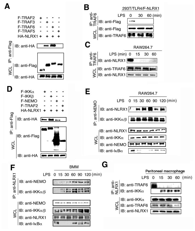
Tight regulation of NF-κB signaling is essential for innate and adaptive immune responses, yet the molecular mechanisms responsible for its negative regulation are not completely understood. Here, we report that NLRX1, a NOD-like receptor family member, negatively regulates Toll-like receptor-mediated NF-κB activation. NLRX1 interacts with TRAF6 or IκB kinase (IKK) in an activation signal-dependent fashion. Upon LPS stimulation, NLRX1 is rapidly ubiquitinated, disassociates from TRAF6, and then binds to the IKK complex, resulting in inhibition of IKKα and IKKβ phosphorylation and NF-κB activation. Knockdown of NLRX1 in various cell types markedly enhances IKK phosphorylation and the production of NF-κB-responsive cytokines after LPS stimulation. We further provide in vivo evidence that NLRX1 knockdown in mice markedly enhances susceptibility to LPS-induced septic shock and plasma IL-6 level. Our study identifies a previously unrecognized role for NLRX1 in the negative regulation of TLR-induced NF-κB activation by dynamically interacting with TRAF6 and the IKK complex.


Figures

Figure 1
Figure 1. NLRX1 inhibits TLR-induced NF-κB activation
(A) 293T cells were transfected with NF-κB-luciferase (luc) reporter plasmid, TLR4 or TLR7 plasmids, together with an pcDNA3.1 empty vector or NLRX1 construct, and analyzed for NF-κB-dependent luciferase activity (fold induction) after treatment with LPS (TLR4 ligand) or CL-097 (TLR7 ligand) for 18 h. (B) 293T cells were transfected with NF-κB-luc, MyD88, TRAF6, TAK1+TAB1, IKKα, IKKβ, or p65, along with NLRX1, NOD1 or NOD2 and analyzed for NF-κB-dependent luciferase activity at 36 h post-transfection. (C) WT and MAVS−/− MEFs were transfected with an empty vector or murine NLRX1 expression vector, followed by LPS or poly (I:C) treatment. Cell supernatants were used to measure IL-6 release by ELISA. (D) MEFs and human THP-1 cells were tranfected with the NF-κB-luc reporter plasmid, along with a empty vector, murine NLRX1 or human NLRX1, and then analyzed for NF-κB-dependent luciferase activity after LPS treatment. (E) 293T cells were tranfected with IKKβ or p65 plasmids, together with an empty vector or NLRX1 plasmid. Nuclear extracts were used to detect endogenous NF-κB DNA binding activity by a gel-mobility shift assay. OCT-1/DNA complexes served as a loading control. (F) RAW264.7 cells were transfected with an empty vector or murine NLRX1 plasmid. The localization of p65 was determined by immunostaining after 15 min of LPS treatment. Data from (A–D) are plotted as means ± s.d. * P < 0.05, ** P<0.01, *** P<0.001, versus controls. All experiments were performed at least three times.
Figure 2
Figure 2. NLRX1 dynamically interacts with TRAF6 and IKK complex upon LPS treatment
(A) 293T cells were transfected with F-TRAF2, F-TRAF3, F-TRAF5, F-TRAF6 and HA-NLRX1. Flag-tagged proteins was immunoprecipitated with anti-Flag followed by anti-HA immunoblotting. IP, IB and WCL denote immunoprecipitation, immunoblotting and whole cell lysate, respectively. (B–C), Cell extracts of 293T/TLR4 cells tranfected with Flag-NLRX1 (B) and RAW264.7 cells (C) were immunoprecipitated with anti-TRAF6 respectively, and then analyzed together with WCL by IB with indicated antibodies. (D) 293T cells were transfected with F-IKKα, F-IKKβ, F-NEMO, F-TRAF2 and HA-NLRX1. Flag-tagged proteins was immunoprecipitated with anti-Flag and blotted with anti-HA. (E–G) RAW264.7 cells (E), Bone marrow-derived macrophages (BMMs) (F) or peritoneal macrophages (G) were treated with LPS and the cell lysates were collected at the indicated time points and used for immunoprecipitation with anti-NEMO or anti-NLRX1, followed by immunoblot with the indicated antibodies. Results are representative of three independent experiments.
Figure 3
Figure 3. NLRX1 ubiquitination and its involvement in the interaction between NEMO and NLRX1
(A) 293T cells were transfected with HA- ubiquitin, HA-ubiquitin (K48 only), HA-ubiquitin (K63 only) and F-NLRX1. F-tagged NLRX1 was immunoprecipitated with anti-Flag beads, and blotted with anti-HA. (B) 293T/TLR4 cells transfected with Flag-NLRX1 were collected at the indicated time points after LPS treatment. Flag- NLRX1 was immunoprecipitated with anti-Flag beads, and then analyzed by immunoblot with indicated antibodies. (C) MEF lysates were collected at the indicated time points after LPS treatment. Endogenous NLRX1 was immunoprecipitated with anti-NLRX1, and then analyzed by immunoblot with anti-K63-linked polyubiquitin antibody. (D) Schematic diagram showing deletion constructs of NLRX1 containing different domains. (E) 293T cells were co-tranfected with Flag-NEMO and the indicated Myc-NRLX1 deletion constructs. Flag-tagged NEMO was immunoprecipitated with anti-Flag beads, and blotted with anti-Myc. (F) 293T cells were co-transfected with HA-NLRX1 and the indicated Flag-NEMO deletion constructs. Flag-tagged proteins were immunoprecipitated with anti-Flag beads, and blotted with anti-HA. Results are representative of three independent experiments.
Figure 4
Figure 4. Mapping the interaction domains of NLRX1 and IKK subunits
(A–B) 293T cells were tranfected with HA-IKKα (A) or HA-IKKβ (B) and the indicated F-NLRX1 deletion constructs. HA-tagged IKKα or HA-IKKβ was immunoprecipitated with anti-HA beads, and blotted with anti-Flag. (C) 293T cells were transfected with NF-κB-luc, IKKβ, along with F-NLRX1 and its deletion constructs and analyzed for NF-κB-dependent luciferase activity. Data are plotted as means ± s.d. (D) Experiment performed as in (B) except IKKβ WT was replaced by IKKβ EE mutant form. (E) Identification of the kinase domain of IKKβ interacting with NLRX1. Upper panel, schematic diagram showing protein domain structures of the IKKβ deletions. KD, kinase domain; LZ, leucine zipper; HLH, helix-loop-helix. Lower panel, 293T cells were co-tranfected with HA-NLRX1 with Flag-IKKβ and its deletion constructs indicated. Flag-tagged proteins were immunoprecipitated with anti-Flag beads, and then blotted with anti-HA. (F) Experiment performed as in (B) except IKKβ WT form was replaced by IKKβ KD mutant. Results are representative of three independent experiments.
Figure 5
Figure 5. NLRX1 inhibits IKK phosphorylation
(A) 293T cells transfected with HA-IKKα, HA-IKKβ and HA-p38 with or without F-NLRX1 were used to analyze the phosphorylation of IKKα/β and p38. (B) 293T/TLR4 cells transfected with increasing amount of F-NLRX1 plasmid were used to analyze the phosphorylation of IKKα/β after LPS treatment. (C) RAW264.7 cells were treated with LPS, and cell lystaes were collected at the indicated time points for immunoprecipitation with anti-NLRX1 or anti-NEMO, followed by immunoblot with indicated antibodies or kinase assay (KA). (D) 293T cells were transfected with HA-IKKβ, together with an empty vector, F-NLRX1 or its deletion constructs and the phosphorylation of IKK was determined. Results are representative of three independent experiments.
Figure 6
Figure 6. Knockdown of NLRX1 enhances IKK phosphorylation and NF-κB- responsive cytokine gene expression
(A) RAW264.7 cells were transfected with control siRNA or NLRX1-specific siRNA, and then treated with LPS for the indicated time points. LPS-induced IKK, IRF3 and MAPK (p38, JNK and ERK) activation were measured by IB with phospho-specific antibodies and IKK activity was measured by KA. (B) Quantitative comparison of signaling activation between NLRX1 knockdown and control cells by density scanning of the blots in (A). (C) RAW264.7 cells were transfected with control siRNA or NLRX1-specific siRNA, and then treated with LPS. The cytokine Tnf-α and Il-6 gene expression in RAW264.7 cells induced by LPS at different time points were determined by real-time PCR. (D) Production of cytokine TNF-α and IL-6 in culture medium of RAW264.7 cells transfected NLRX1-specific and control siRNAs after LPS treatment. Data in (C–D) are plotted as means ± s.d. * P < 0.05, ** P<0.01, versus controls. All experiments were performed at least three times with similar results.
Figure 7
Figure 7. NLRX1 negatively regulates NF-κB signaling and cytokine responsein vivo
(A) Knockdown of murine NLRX1 expression in different tissues of NLRX1-KD mice as measured by quantitative real-time PCR. (B) NLRX1 protein expression was determined by immunoblot in WT and NLRX1-KD MEFs. WT and NLRX1-KD MEFs were infected by VSV-eGFP, and was visualized by fluorescent microscopy. (C) WT and NLRX1-KD mice (n=10 per group) were injected intravenously with poly(I:C) (200 μg per mouse) and then sera were collected at indicated times for IFN-β. measurement. (D) LPS-induced IKK and MAPK activation in peritoneal macrophages from WT and NLRX1-KD mice. The quantified results are shown after band density scanning. (E) IL-6 production in WT and NLRX1-KD MEFs after treatment with LPS, Pam3CSK4 or infected with VSV-eGFP. (F) IL-6 and TNF-α production in peritoneal macrophages from NLRX1-KD or WT mice after treatment with LPS. (G) Survival of NLRX1-KD and WT mice (n=9 per group) after peritoneal injection with LPS (30 mg/kg). (H) Plasma IL-6 levels from WT and NLRX1-KD mice (n=9 per group) 3 h after peritoneal injection with LPS (30 mg/kg). Data in (A, C, E and F) are plotted as means ± s.d. * P<0.05, ** P<0.01, versus controls. All experiments were performed at least three times with similar results,

Comment in

References

    1. Akira S, Uematsu S, Takeuchi O. Pathogen recognition and innate immunity. Cell. 2006;124:783–801. - PubMed
    1. Chen ZJ. Ubiquitin signalling in the NF-kappaB pathway. Nat Cell Biol. 2005;7:758–765. - PMC - PubMed
    1. Cui J, Zhu L, Xia X, Wang HY, Legras X, Hong J, Ji J, Shen P, Zheng S, Chen ZJ, et al. NLRC5 negatively regulates the NF-kappaB and type I interferon signaling pathways. Cell. 2010;141:483–496. - PMC - PubMed
    1. Ea CK, Deng L, Xia ZP, Pineda G, Chen ZJ. Activation of IKK by TNFalpha requires site-specific ubiquitination of RIP1 and polyubiquitin binding by NEMO. Mol Cell. 2006;22:245–257. - PubMed
    1. Gough DJ, Corlett A, Schlessinger K, Wegrzyn J, Larner AC, Levy DE. Mitochondrial STAT3 supports Ras-dependent oncogenic transformation. Science. 2009;324:1713–1716. - PMC - PubMed

Publication types

MeSH terms