. 2011 Jun 24;34(6):854-65.
doi: 10.1016/j.immuni.2011.03.026.

NLRX1 protein attenuates inflammatory responses to infection by interfering with the RIG-I-MAVS and TRAF6-NF-κB signaling pathways

Affiliations

NLRX1 protein attenuates inflammatory responses to infection by interfering with the RIG-I-MAVS and TRAF6-NF-κB signaling pathways

Irving C Allen et al. Immunity. .

Abstract

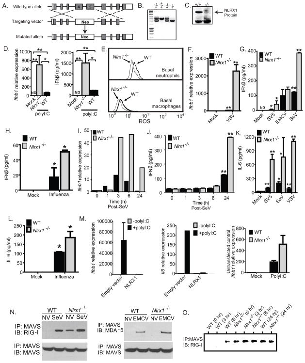
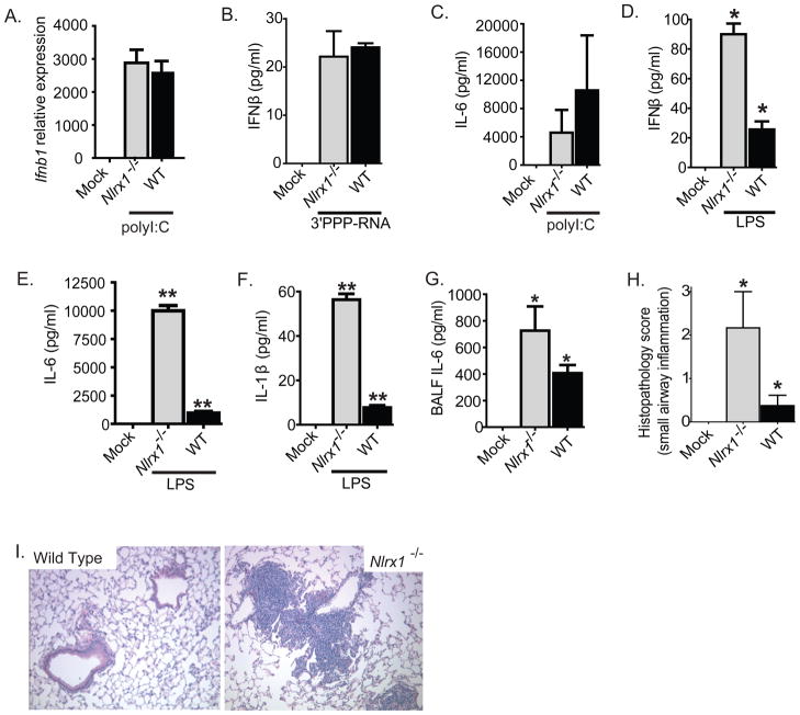
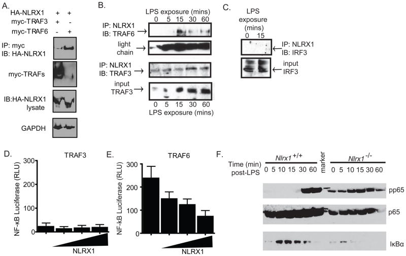
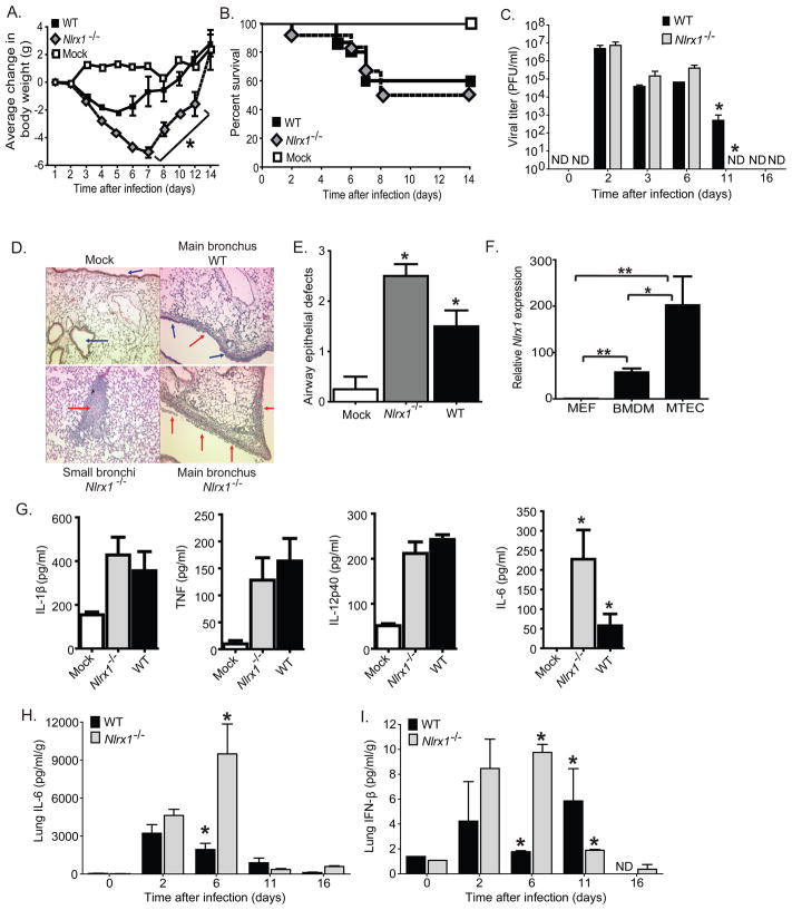
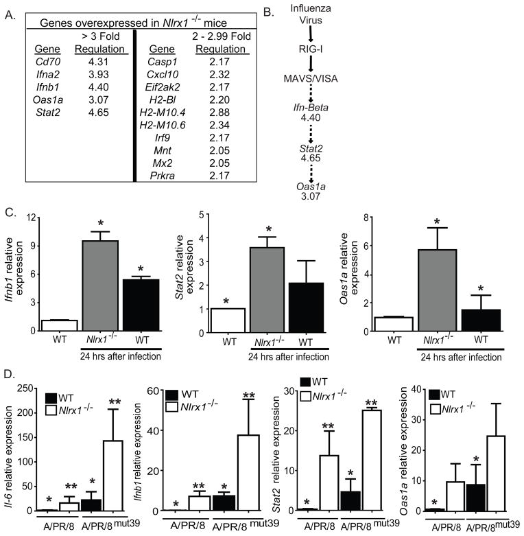
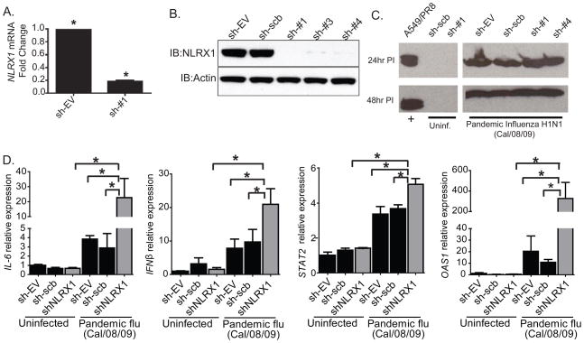
The nucleotide-binding domain and leucine-rich-repeat-containing (NLR) proteins regulate innate immunity. Although the positive regulatory impact of NLRs is clear, their inhibitory roles are not well defined. We showed that Nlrx1(-/-) mice exhibited increased expression of antiviral signaling molecules IFN-β, STAT2, OAS1, and IL-6 after influenza virus infection. Consistent with increased inflammation, Nlrx1(-/-) mice exhibited marked morbidity and histopathology. Infection of these mice with an influenza strain that carries a mutated NS-1 protein, which normally prevents IFN induction by interaction with RNA and the intracellular RNA sensor RIG-I, further exacerbated IL-6 and type I IFN signaling. NLRX1 also weakened cytokine responses to the 2009 H1N1 pandemic influenza virus in human cells. Mechanistically, Nlrx1 deletion led to constitutive interaction of MAVS and RIG-I. Additionally, an inhibitory function is identified for NLRX1 during LPS activation of macrophages where the MAVS-RIG-I pathway was not involved. NLRX1 interacts with TRAF6 and inhibits NF-κB activation. Thus, NLRX1 functions as a checkpoint of overzealous inflammation.


Figures

Figure 1
Figure 1. NLRX1 functions as a negative regulator of IFN-I production
A. Schematic of the generation of Nlrx1−/− mice through homologous recombination. B. PCR genotyping of offspring from heterozygous matings. C. Confirmation of the loss of NLRX1 protein in Nlrx1−/− offspring by immunoblot. D. Ifnb1 transcript and IFN-β protein were assessed in MEFs that were transfected with polyI:C. E. Neutrophils and macrophages were purified from the bone marrow of WT and Nlrx1−/− mice and were activated for 1 hour in the presence of DHR. Fluorescence intensity was used to measure ROS production. F. Ifnb1 transcription was measured in primary fibroblasts isolated from Nlrx1−/− mice following challenges with Vesicular Stomatitis Virus (VSV). G–H. IFNβ protein levels were determined by ELISA in the supernatants from Nlrx1−/− fibroblasts following stimulation with Simian Virus 5 (SV5), Encephalomyocarditis Virus (EMCV), Sendai Virus (SeV) or influenza A (PR/8/34) virus. I–J. Ifnβ gene transcription and IFNβ protein levels were measured over a timecourse SeV infection in primary ear fibroblasts isolated from Nlrx1−/− mice. K–L. IL-6 concentrations were assessed by ELISA in the supernatants from primary Nlrx1−/− MEFS following SV5, SeV, VSV and influenza virus infection. M. Nlrx1−/− MEFS were reconstituted with constructs containing full length human NLRX1 and assessed for Ifnb1 and Il6 gene transcription after polyI:C stimulation. N. The association between MAVS, RIG-I and MDA-5 were assessed by co-immunoprecipitation in primary ear fibroblasts isolated from WT and Nlrx1−/− mice. Data shown are at time 0 and 24 hours post-infection with either SeV (RIG-I) or EMCV (MDA-5). O. Primary ear fibroblasts were challenged with SeV and samples were harvested over a time course. The RIG-I-MAVS association was detected 6 hours after SeV challenge in WT fibroblasts and constitutive interaction was detected in Nlrx1−/− fibroblasts. *p < 0.05;**p < 0.005. See also Supplemental Figure S1.
Figure 2
Figure 2. NLRX1 attenuates LPS mediated IFNβ and cytokine production in mouse macrophages
A–B. Ifnβ transcript levels and IFNβ protein levels were measured in bone marrow macrophages that were isolated from WT and Nlrx1−/− mice following transfection with either polyI:C or 3′PPP-RNA. C. IL-6 production was measured in bone marrow macrophages isolated from WT and Nlrx1−/− mice following transfection with polyI:C. D–F. IFN-β, IL-6 and IL-1β protein levels were assessed in bone marrow derived macrophages that were challenged with LPS. G–I. Mice were challenged with LPS via i.t. instillation and monitored for changes in morbidity and innate immune responses. G. IL-6 levels were assessed by ELISA in BALF from WT and Nlrx1−/− mice, 24 hours following LPS challenge. H. Semiquantitative histopathological scoring was utilized to quantify lung histopathology 24 hours following LPS challenge. I. Peribroncholar and perivascular cuffing localized around the small/medium sized airways was a characteristic feature of the Nlrx1−/− histopathology.
Figure 3
Figure 3. NLRX1 functions as a novel inhibitor of TRAF6 and NF-κB
A. Overexpression systems were utilized to assess NLRX1 interactions with TRAF6. HEK293T cells were transfected with HA-NLRX1 and either myc-TRAF-3 or myc-TRAF-6. Myc-TRAFs were immunoprecipitated and the blot was probed with HA-HRP antibody, myc-HRP and GAPDH-HRP. B–C. NLRX1 associations with TRAF6, TRAF3 and IRF3 were determined following LPS stimulation in primary bone marrow derived macrophages. D–E. NF-kB luciferase activity was determined in transfected 293T cells in the presence of increasing concentrations of input NLRX1 following stimulation with TRAF3 and TRAF6. F. Phosphorylation of p65 and levels of the inhibitor IκBα were assessed throughout a timecourse of LPS stimulation in MEFS. All studies were repeated a minimum of 3 times.*p < 0.05;**p < 0.005. See also Supplemental Figure S1.
Figure 4
Figure 4. Characterization of the in vivo host immune response in Nlrx1−/− mice following influenza virus A/PR/8/34 infection
A–B. Morbidity (weight loss) and mortality were measured for 14 days in Nlrx1−/− and WT mice following in vivo intranasal administration of influenza virus A/PR/8/34. C. Lungs were harvested at specific time points and homogenized to assess influenza viral titers, which were determined by standard plaque assay. D. Lungs were harvested 3 dpi and fixed by inflation and immersion in buffered PFA. Sections through the main bronchiole of the left lobe revealed increased airway epithelial cell denuding (red arrows) and small airway obstruction throughout the lungs of Nlrx1−/− mice. E. Histological scoring of H&E stained lung sections revealed a significant increase in airway epithelial defects. F. High basel levels of Nlrx1 transcription was observed in primary bone marrow derived macrophages (BMDM) and mouse tracheal epithelial cells (MTECs) from naïve WT mice. G. Serum was harvested from Nlrx1−/− and WT mice 3 dpi with influenza A/PR/8/34 virus. Influenza virus infection induced increased serum levels of IL-1β, TNFα, IL-12p40, and IL-6. H–I. Whole lungs were harvested from WT and Nlrx1−/− mice at specific time points after i.n. infection with influenza A/PR/8/34 virus. IL-6 and IFN-β levels were determined in cell free supernatants by ELISA. All studies were repeated a minimum of 3 times with 5–7 mice per group for each individual study.*p < 0.05;**p < 0.005. See also Supplemental Figures S2 and S3.
Figure 5
Figure 5. NLRX1 functions in vivo as a negative regulator of the IFN-I response to influenza virus challenge
A. Gene expression was profiled for genes associated with IFN-I and viral innate immunity. Whole lungs were harvested from WT and Nlrx1−/− mice, 20 hours post-intratracheal (i.t.) inoculation with the influenza A/PR/8/34 virus. RNA was extracted and pooled in equal quantities for similarly treated mice from each genotype. Genes that demonstrated at least 2-fold changes in transcription between WT and Nlrx1−/− mice were considered significant. B. The expression profile data was analyzed using Ingenuity Pathways Analysis software, which revealed several genes in the RIG-I/MAVS pathway that were significantly up-regulated in the lungs of Nlrx1−/− mice. C. Realtime PCR was used to confirm increased Ifnβ, Stat2, and Oas1a gene expression in individual samples (un-pooled and distinct from the samples used in the array). D. NLRX1 and the viral NS1 protein have non-overlapping targets. WT and Nlrx1−/− mice were challenged with the influenza A/PR/8mut39 virus, which carries a mutant NS1 protein that attenuates the virus’s ability to inhibit the IFN-I response. IL-6, Ifnβ, Stat2, and Oas1a gene expression was assessed by realtime PCR. Expression profiling was repeated 2 times with samples pooled from 3 individual mice. All other studies were repeated 3 times with 3–7 mice per group for each individual study.*p < 0.05;**p < 0.005. See also Supplemental Figures S4 and S5.
Figure 6
Figure 6. NLRX1 attenuates IL-6 and the IFNβ-STAT2-OAS1 axis in human cells in response to the 2009 pandemic influenza A/California/08/09 virus
A–B. NLRX1 was efficiently knocked down by shRNA in 293T cells as shown by realtime PCR and western blot of three different shRNAs targeting NLRX1 (sh#1 to sh#3). C. Western blot for the influenza virus NS1 protein was used to confirm that cells were infected with the 2009 pandemic influenza A/California/08/09 virus at MOI=0.1. D. IL-6, IFNβ, STAT2 and OAS1A gene expression were assessed in 293T cells following the targeted knockdown of NLRX1 after infection with the 2009 pandemic influenza A/California/08/09 virus (MOI=0.1). Experiments were repeated 2 times in all three NLRX1 knockdown cell lines and scrambled control cell lines. *p < 0.05.

Comment in

References

    1. Abdul-Sater AA, Said-Sadier N, Lam VM, Singh B, Pettengill MA, Soares F, Tattoli I, Lipinski S, Girardin SE, Rosenstiel P, et al. Enhancement of reactive oxygenspecies production and chlamydial infection by the mitochondrial Nod like family member NLRX1. J Biol Chem. 2010;285:41637–41645. - PMC - PubMed
    1. Allen IC, Scull MA, Moore CB, Holl EK, McElvania-TeKippe E, Taxman DJ, Guthrie EH, Pickles RJ, Ting JP. The NLRP3 inflammasome mediates in vivo innate immunity to influenza A virus through recognition of viral RNA. Immunity. 2009;30:556–565. - PMC - PubMed
    1. Balachandran S, Roberts PC, Kipperman T, Bhalla KN, Compans RW, Archer DR, Barber GN. Alpha/beta interferons potentiate virus-induced apoptosis through activation of the FADD/Caspase-8 death signaling pathway. J Virol. 2000;74:1513–1523. - PMC - PubMed
    1. Benko S, Magalhaes JG, Philpott DJ, Girardin SE. NLRC5 limits the activation of inflammatory pathways. J Immunol. 2010;185:1681–1691. - PubMed
    1. Buchweitz JP, Harkema JR, Kaminski NE. Time-dependent airway epithelial and inflammatory cell responses induced by influenza virus A/PR/8/34 in C57BL/6 mice. Toxicol Pathol. 2007;35:424–435. - PubMed

Publication types

MeSH terms