. 2011 Jul 15;89(1):44-55.
doi: 10.1016/j.ajhg.2011.05.023. Epub 2011 Jun 23.

Deficiency of the cytoskeletal protein SPECC1L leads to oblique facial clefting

Affiliations

Deficiency of the cytoskeletal protein SPECC1L leads to oblique facial clefting

Irfan Saadi et al. Am J Hum Genet. .

Abstract

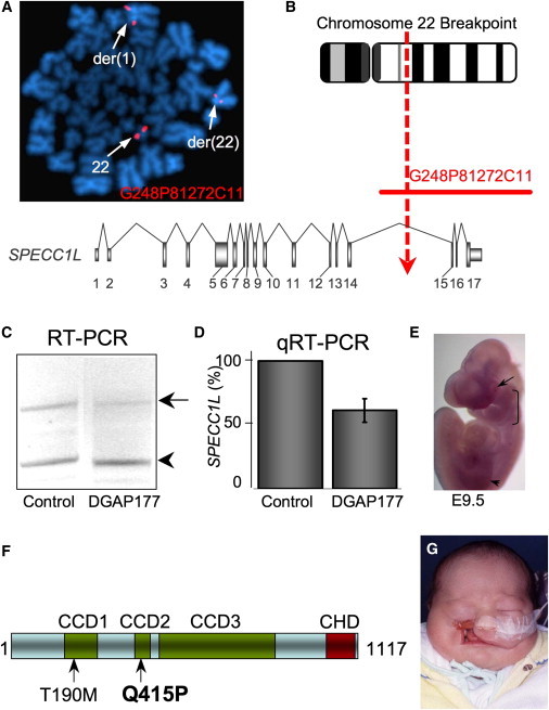
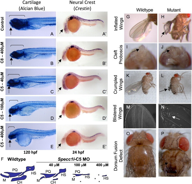
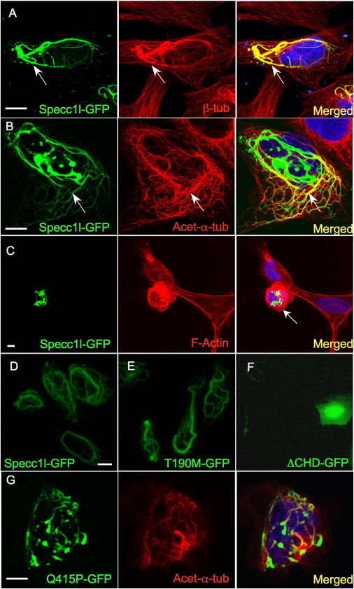
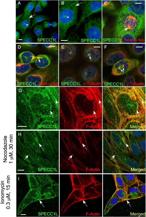
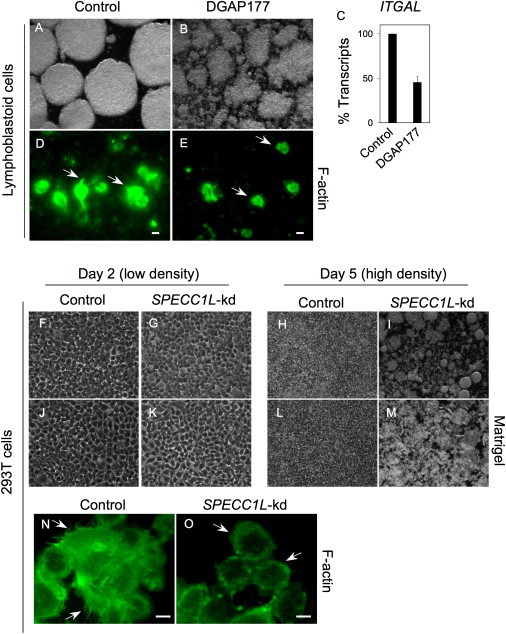
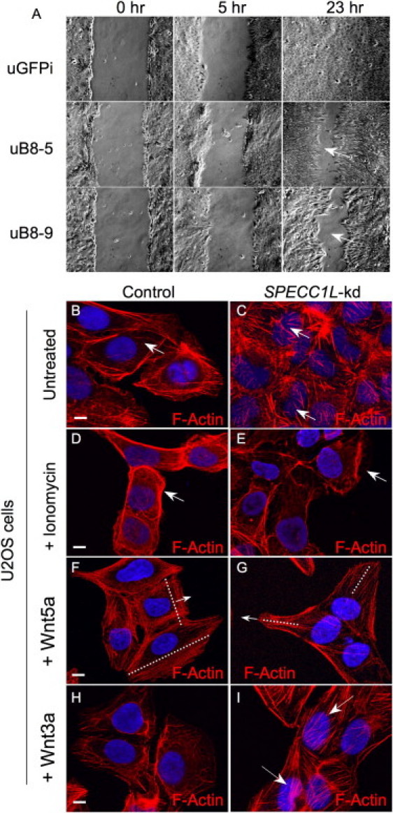
Genetic mutations responsible for oblique facial clefts (ObFC), a unique class of facial malformations, are largely unknown. We show that loss-of-function mutations in SPECC1L are pathogenic for this human developmental disorder and that SPECC1L is a critical organizer of vertebrate facial morphogenesis. During murine embryogenesis, Specc1l is expressed in cell populations of the developing facial primordial, which proliferate and fuse to form the face. In zebrafish, knockdown of a SPECC1L homolog produces a faceless phenotype with loss of jaw and facial structures, and knockdown in Drosophila phenocopies mutants in the integrin signaling pathway that exhibit cell-migration and -adhesion defects. Furthermore, in mammalian cells, SPECC1L colocalizes with both tubulin and actin, and its deficiency results in defective actin-cytoskeleton reorganization, as well as abnormal cell adhesion and migration. Collectively, these data demonstrate that SPECC1L functions in actin-cytoskeleton reorganization and is required for proper facial morphogenesis.


Figures

Figure 1
Figure 1
SPECC1L Disruption and Mutation in ObFC Patients (A and B) FISH analysis of metaphase chromosomes from DGAP177 lymphoblasts and schematic of fosmid G248P81272C11 spanning the 22q breakpoint, which disrupts SPECC1L in a 5 kb region of intron 14. (C and D) RT-PCR and qRT-PCR show haploinsufficiency of SPECC1L transcripts (arrow; relative to ACTB transcripts, arrowhead) in RNA from DGAP177 and control lymphoblasts. (E) Whole-mount in situ hybridization shows Specc1l expression in maxillary and lateral nasal prominences (bracket), eyes (arrow), and limbs (arrowhead) at E9.5. (F) SPECC1L representation showing the position of the p.Thr190Met polymorphism and p.Gln415Pro missense mutation. CCD, coiled coil domain; CHD, calponin homology domain. (G) Tessier IV type cleft in the patient with the p.Gln415Pro mutation.
Figure 2
Figure 2
Specc1l Knockdown Results in Zebrafish “Faceless” and Drosophila “Split Discs” Phenotypes (A–E and A′–E′) Lateral views of 120 hpf fish stained with alcian blue for cartilage (A–E) and of 24 hpf fish stained for crestin (A′–E′), a neural crest (NC) marker, after morpholino (MO) treatment. Control (A and A′) and chromosome-8-homolog (C8) MO at the highest dose (B and B′) appear normal. In contrast, increasing doses of the zebrafish chromosome-5-homolog (C5) MO results in diminishing sizes of facial structures (C–E; arrows and brackets) and poor NC migration to the anterior branchial arches (C′–E′; arrows). (F) Schematic representation of the effect on jaw cartilages after MO treatment. M, Meckel's; PQ, palatoquadrate; CH, ceratohyal; HS, hyosymplectic; , missing cartilage. (G–P) The Drosophila SPECC1L-ortholog (CG13366) conditional RNAi line (108092) was crossed with da-GAL4 and A9-GAL4 flies for ubiquitous (G–J) or wing-imaginal-disc-specific (K–P) knockdown. Resulting flies were flightless with inflated (G and H), crumpled (K and L), or blistered (M and N) wings. Eclosing flies with ubiquitous knockdown failed to survive, probably as a result of feeding problems from the defective proboscis (I and J). Some flies with severely affected wings also showed a mild cleft dorsum (O and P).
Figure 3
Figure 3
Specc1l-GFP Stabilizes Acetylated α-Tubulin-Containing Microtubules (A) GFP-tagged Specc1l (green) in U2OS cells stabilizes a subset of microtubules (arrows) stained with β-tubulin (red). (B) The stabilized subset colocalizes with acetylated α-tubulin-containing microtubules (yellow, arrows). (C) Specc1l-GFP distorts the actin cytoskeleton (red) and cell shape (arrow). (D–F) In contrast to wild-type Specc1l-GFP (A and D) and Thr190Met-GFP (E), which show no apparent difference in microtubule stabilization, a C-terminal truncation (ΔCHD) completely abolishes stabilization (F). (G) Notably, the Gln415Pro-GFP mutant significantly reduces stabilization of acetylated α-tubulin-containing microtubules. Scale bars represent 10 μm.
Figure 4
Figure 4
SPECC1L Colocalizes with Tubulin and Actin (A and B) Endogenous SPECC1L shows a microtubule-type cytoplasmic expression pattern (arrows); a site of intranuclear expression is also detected (arrowheads; also see [E] below). (C and D) SPECC1L colocalizes with acetylated α-tubulin (yellow and orange) in the mitotic spindle during cytokinesis (C) and in gap junctions (D). (E and F) Punctate expression of SPECC1L is seen in the presumptive microtubule-organizing center (MTOC) surrounding centrioles stained with γ-tubulin. (G and H) Colocalization with F-actin (G, arrows) is better visualized after nocodazole treatment (H, arrows). (I) After ionomycin treatment, SPECC1L relocates with F-actin to the cell membrane. Scale bars represent 10 μm.
Figure 5
Figure 5
Cell-Adhesion Defects in SPECC1L-Deficient Cells (A–C) EBV-transformed control lymphoblasts show increased integrin LFA-1-dependent adhesion in culture and grow as large circular clumps (A). DGAP177 EBV-transformed lymphoblasts fail to form these large clumps (B) and show reduced levels of integrin αL (ITGAL) transcripts (C). Data represent average transcript level ± standard error of the mean from three independent experiments. (D and E) The adhesion defect is accompanied by altered F-actin staining and reduced numbers of F-actin microspikes (arrows) when SPECCIL-deficient cells are compared to control cells. (F–I) Similar to control cells (F), SPECC1L-knockdown (SPECC1L-kd) 293T clonal cells (G) adhere normally to the culture plate at low density on day 2. Upon reaching confluence at day 5, control cells continue to adhere to the culture plate (H), whereas knockdown cells fail to adhere and lift off the plate (I). (J–M) Plating SPECC1L-kd cells on Matrigel does not prevent this loss of adhesion at confluency on day 5. (N and O) Compared with control cells (N), SPECC1L-kd 293T cells show abnormal F-actin staining (O) with reduced numbers of microspikes (arrows). Scale bars represent 10 μm.
Figure 6
Figure 6
Migration and Actin-Cytoskeleton-Reorganization Defects in SPECC1L-Knockdown Cells (A) Wound-repair (scratch) assays were used in comparisons of the ability of SPECC1L-kd U2OS clonal cells (uB8-5, uB8-9) versus control knockdown cells (uGFPi) to migrate. In severe cases, knockdown cells fail to close the wound (arrows) even after 23 hr. (B and C) F-actin staining of SPECC1L-kd cells shows an increase in actin fibers in the center of the cells (overlying the nuclei) when these cells are compared to control cells (arrows). (D and E) Ionomycin treatment causes rapid reorganization of the actin cytoskeleton to the cell membrane in control cells, whereas SPECC1L-kd cells respond poorly (arrows). (F and G) Wound-repair assays with control and knockdown cells were treated with the noncanonical Wnt ligand, Wnt5a. In control cells, Wnt5a reorganizes the actin cytoskeleton toward the leading edge, perpendicular to the direction of migration (F). In contrast, knockdown cells show an abnormal alignment of actin fibers (G), indicating defective ability to reorganize the actin cytoskeleton. Dotted lines indicate alignment of actin filaments; arrows indicate the direction of migration. (H and I) The canonical Wnt ligand, Wnt3a, which does not reorganize the actin cytoskeleton in control cells (H) (note similarity to [B]), was used as a control. Knockdown cells continue to show markedly concentrated central actin fibers (I, arrows) (note similarity to [C]). Scale bars represent 10 μm.

References

    1. Jugessur A., Farlie P.G., Kilpatrick N. The genetics of isolated orofacial clefts: From genotypes to subphenotypes. Oral Dis. 2009;15:437–453. - PubMed
    1. Beaty T.H., Murray J.C., Marazita M.L., Munger R.G., Ruczinski I., Hetmanski J.B., Liang K.Y., Wu T., Murray T., Fallin M.D. A genome-wide association study of cleft lip with and without cleft palate identifies risk variants near MAFB and ABCA4. Nat. Genet. 2010;42:525–529. - PMC - PubMed
    1. Tessier P. Anatomical classification facial, cranio-facial and latero-facial clefts. J. Maxillofac. Surg. 1976;4:69–92. - PubMed
    1. Eppley B.L., van Aalst J.A., Robey A., Havlik R.J., Sadove A.M. The spectrum of orofacial clefting. Plast. Reconstr. Surg. 2005;115:101e–114e. - PubMed
    1. Hall B.K. Springer Science+Business Media; New York: 2009. The Neural Crest and Neural Crest Cells in Vertebrate Development and Evolution.

Publication types

MeSH terms

Supplementary concepts