. 2011 Aug 15;356(2):541-52.
doi: 10.1016/j.ydbio.2011.06.011. Epub 2011 Jun 16.

Wnt2 signaling is necessary and sufficient to activate the airway smooth muscle program in the lung by regulating myocardin/Mrtf-B and Fgf10 expression

Affiliations

Wnt2 signaling is necessary and sufficient to activate the airway smooth muscle program in the lung by regulating myocardin/Mrtf-B and Fgf10 expression

Ashley M Goss et al. Dev Biol. .

Abstract

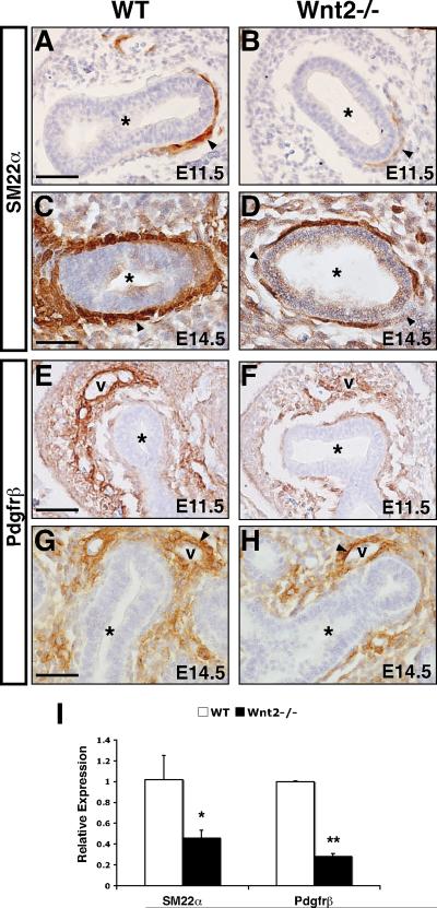
Smooth muscle in the lung is thought to derive from the developing lung mesenchyme. Smooth muscle formation relies upon coordination of both autocrine and paracrine signaling between the budding epithelium and adjacent mesenchyme to govern its proliferation and differentiation. However, the pathways initiating the earliest aspects of smooth muscle specification and differentiation in the lung are poorly understood. Here, we identify the Wnt2 ligand as a critical regulator of the earliest aspects of lung airway smooth muscle development. Using Wnt2 loss and gain of function models, we show that Wnt2 signaling is necessary and sufficient for activation of a transcriptional and signaling network critical for smooth muscle specification and differentiation including myocardin/Mrtf-B and the signaling factor Fgf10. These studies place Wnt2 high in a hierarchy of signaling molecules that promote the earliest aspects of lung airway smooth muscle development.


Figures

Figure 1
Figure 1. Wnt2−/− lungs exhibit airway smooth muscle (ASM) defects
SM22α immunostaining on cross-sections of E18.5 control wild type (A, C) and Wnt2−/− null mutant embryos (B, D) demonstrates reduced ASM in the distal airways of Wnt2−/− null lungs. Higher magnification (C, D) reveals the lack of a contiguous layer of smooth muscle surrounding the distal airway epithelium in Wnt2−/− null lungs (D, arrowheads). The smooth muscle layer surrounding adjacent blood vessels does not appear to be affected (arrows in A, B). Cross-sections of lacZ-stained E14.5 control BAT-GAL embryos shows lacZ expression in the developing epithelium (E, arrows) and primitive ASM cells (E, arrowheads). In Wnt2−/−:BAT-GAL mutant embryos there is decreased lacZ expression in primitive ASM cells (F, arrowhead), while some epithelial lacZ expression is retained (F, arrows). Axin2 immunostaining on E14.5 control wild type lung cross-sections demonstrates robust expression in the developing ASM (G, arrowhead), and this expression is reduced in Wnt2−/− mutant lungs (H, arrowhead). Q-PCR of Axin2 expression demonstrates a significant reduction in E14.5 Wnt2−/− lungs compared to control wild type lungs (I). * p <0.02 (Student's t test). N=5 lung buds. Airways denoted by (*) symbol on histological sections. Scale bars=200 μm (A, B, E, F), 100 μm (C, D, G, H).
Figure 2
Figure 2. Loss of Wnt2 disrupts development of the lung mesenchyme and immature smooth muscle
SM22α immunostaining (A–D) on cross-sections of control wild type (A and C) embryos shows expression in the primitive ASM layer surrounding the developing epithelium at E11.5 (A, arrowhead) and by E14.5 there is robust SM22α expression in the sub-epithelial smooth muscle layer (C, arrowhead). In contrast, Wnt2−/− null mutant lungs exhibit reduced SM22α expression in the primitive ASM layer at E11.5 (B, arrowhead), and this expression remains reduced at E14.5 (D, arrowheads). Pdgfrβ immunostaining on wild-type lungs shows expression throughout the developing lung mesenchyme surrounding the primitive airways and vessels (v) at E11.5 (E) and E14.5 (G). In Wnt2−/− null lungs Pdgfrβ expression is reduced at E11.5 (F) and E14.5 (H) in the mesenchyme underlying the airways. However, Pdgfrβ expression is retained in the mesenchyme condensing around primitive blood vessels (H, v). Q-PCR of SM22α and Pdgfrβ expression demonstrates a significant reduction in E11.5 Wnt2−/− lungs compared to control wild type lungs (I). *, p <0.02, ** p <0.001 (Student's t test). N=5 lung buds. Airways denoted by (*) symbol on histological sections. Scale bars=200 μm.
Figure 3
Figure 3. Wnt2 signaling is upstream of an early smooth muscle regulatory network
In situ hybridizations for myocardin expression on control wild type lung cross-sections (A and C) show expression in the primitive smooth muscle layer surrounding the developing airways at E11.5 (A, arrowheads) and later at E14.5 (C, arrowheads). In comparison, Wnt2−/− null lungs (B and D) show reduced myocardin expression in the primitive ASM layer at E11.5 (B, arrowhead) and E14.5 (D, arrowheads). Assessment of smooth muscle regulatory genes by Q-PCR demonstrates significantly reduced levels of myocardin and Mrtf-B expression in E11.5 Wnt2−/− null lung buds compared to control lung buds (E). *, p <0.02, (Student's t test). N=5 lung buds. Lung bud circumscribed with dashed line in panels A and B. Airways denoted by (*) symbol on histological sections. Scale bars=400 μm.
Figure 4
Figure 4. Assessment of endothelial differentiation in Wnt2−/− null lungs
Q-PCR indicates no significant change in expression levels of the endothelial marker gene Pecam in E12.5 Wnt2−/− null lung buds compared to control wild type lung buds (A). Assessment of the endothelial marker gene Flk1 reveals the expression level in Wnt2−/− null lung buds is significantly reduced to approximately 85% when normalized to control lung buds (B). Analysis of the Vegf family members indicates no significant change in expression levels between E12.5 control and Wnt2−/− null lung buds for VegfA (C) and VegfD (D). However, VegfC is reduced by approximately 30% in Wnt2−/− null lungs when normalized to control lungs (E). Expression of Vegfr3, a receptor for VegfC in the lung, is not significantly reduced in Wnt2−/− null lungs (F). Analysis of the lymphatic marker gene Lyve1 shows no change in expression level between control and Wnt2−/− null lung buds (G). *, p <0.05 (Student's t test). N=5 lung buds. N.S.= not statistically significant.
Figure 5
Figure 5. Evaluation of signaling factors important for smooth muscle development in Wnt2−/− null lungs
In situ hybridization on wild-type (A, D, G, J) and Wnt2−/− null mutant embryos (B, E, H, K) to detect expression of Wnt7b (A and B), Fgf10 (D and E), Shh (G and H), and Bmp4 (J and K) at E10.5 (A and B) and E11.5 (D,E,G,H,J,K). Q-PCR to detect expression of Wnt7b (C), Fgf10 (F), Shh (I), and Bmp4 (L) in wild type and Wnt2−/− null mutant lung buds at E11.5. *, p < 0.02, ** p <0.001 (Student's t test). N=5 lung buds. N.S.= not statistically significant. Lung bud circumscribed with dashed line in panels D, E, J, and K. Primitive airway denoted by (*) symbol on histological sections. Scale bars=100 μm (A–K).
Figure 6
Figure 6. Wnt2 signaling is sufficient to promote smooth muscle development in primary lung mesenchymal cells and the intact lung
Assessment of smooth muscle regulatory and marker gene expression by Q-PCR in 10T1/2 cells (A), wild type E11.5 lung bud explants (B, E), and isolated E11.5 primary lung mesenchyme (F) cultured in the absence (control) or presence of rWnt2 protein. Treating 10T1/2 cells for 48 hours with rWnt2 leads to increased expression of smooth muscle genes including SM22α, SMA, myocardin, Pdgfrα, Pdgfrβ, and Fgf10 (A). Treatment of wild type lung bud explants with rWnt2 leads to increased smooth muscle gene expression including increased expression of myocardin, Mrtf-B, and Fgf10 (B). Immunostaining on cross-sections of cultured lung bud explants shows increased expression of SM22α in the developing ASM in the presence of rWnt2 (D) as compared to control untreated explants (C). Expression of Wnt7b but not Bmp4 was increased by treatment with rWnt2 in lung explants (E). Treatment of isolated lung mesenchyme from E11.5 embryos with rWnt2 results in increased expression of Fgf10 as well as myocardin, Mrtf-B, Sm22α, Pdgfrα, and SMA (F). *, p < 0.05, ** p <0.01 (Student's t test). N=5 lung bud explants. Scale bars=200 μm.
Figure 7
Figure 7. Wnt2 signaling regulates smooth muscle development through activation of downstream Fgf10 signaling
Assessment of smooth muscle gene expression by Q-PCR in wild type lung bud explants treated with either PBS (control) or rFgf10 (A). Treating wild type lung bud explants with rFgf10 leads to increased expression of smooth muscle genes including SM22α, SMA, and Pdgfrα (A). SM22α immunostaining on cross-sections of lung bud explants treated with rFgf10 shows increased expression in the developing ASM as compared to control untreated lung bud explants (B and C). Assessment of smooth muscle gene expression by quantitative PCR in cultured E11.5 Wnt2+/+ wild type and Wnt2−/− null lung bud explants (D). Expression levels of SM22α, Pdgfrβ, and SMA expression levels were significantly upregulated in rFgf10-treated Wnt2−/− null lung bud explants as compared to untreated Wnt2−/− null lung bud explants while Wnt7b was increased to a non-significant level (D). Wnt2 expression levels were also measured to confirm the genotyping of lung bud explant populations (D). SM22α immunostaining on cross-sections of control Wnt2−/− null lung bud explants shows reduced expression in the primitive ASM as compared to Wnt2+/+ wild type explants (E and F). In the presence of rFgf10, there is increased SM22α expression in the developing ASM, similar to that in Wnt2+/+ wild type lung bud explants (compare E and G). *, p < 0.05 (Student's t test). N=8 lung bud explants. n.s.=not significant. Scale bars=100 μm.
Figure 8
Figure 8. Model of Wnt2 signaling in ASM development
Wnt2 signaling in the primitive lung mesenchyme promotes the proliferation and differentiation of the Pdgfrα/β-expressing multipotent mesenchyme into early-stage smooth muscle cells in part, by activating a myogenic transcriptional network including myocardin and Mrtf-B and activating expression of the pro-myogenic ligand Wnt7b in the adjacent epithelium. Wnt2 signaling also activates Fgf10 signaling, which further promotes amplification and differentiation of immature smooth muscle cells into a mature smooth muscle cells expressing ASM markers including SM22α, SMA, and Pdgfrα. Whether the amplification and differentiation of the smooth muscle program by Fgf10 is direct or indirect through intermediates (i.e. factor X) is unclear.

Similar articles

Cited by

References

    1. Badri KR, Zhou Y, Schuger L. Embryological origin of airway smooth muscle. Proc Am Thorac Soc. 2008;5:4–10. - PMC - PubMed
    1. Banerji S, Ni J, Wang SX, Clasper S, Su J, Tammi R, Jones M, Jackson DG. LYVE-1, a new homologue of the CD44 glycoprotein, is a lymph-specific receptor for hyaluronan. J Cell Biol. 1999;144:789–801. - PMC - PubMed
    1. Bellusci S, Furuta Y, Rush MG, Henderson R, Winnier G, Hogan BL. Involvement of Sonic hedgehog (Shh) in mouse embryonic lung growth and morphogenesis. Development. 1997a;124:53–63. - PubMed
    1. Bellusci S, Grindley J, Emoto H, Itoh N, Hogan BL. Fibroblast growth factor 10 (FGF10) and branching morphogenesis in the embryonic mouse lung. Development. 1997b;124:4867–4878. - PubMed
    1. Betsholtz C. Insight into the physiological functions of PDGF through genetic studies in mice. Cytokine Growth Factor Rev. 2004;15:215–228. - PubMed

Publication types

Substances