Review
. 2012 Feb;52(2):401-9.
doi: 10.1016/j.yjmcc.2011.06.007. Epub 2011 Jun 16.

Compartmentation of membrane processes and nucleotide dynamics in diffusion-restricted cardiac cell microenvironment

Affiliations
Review

Compartmentation of membrane processes and nucleotide dynamics in diffusion-restricted cardiac cell microenvironment

Alexey E Alekseev et al. J Mol Cell Cardiol. 2012 Feb.

Abstract

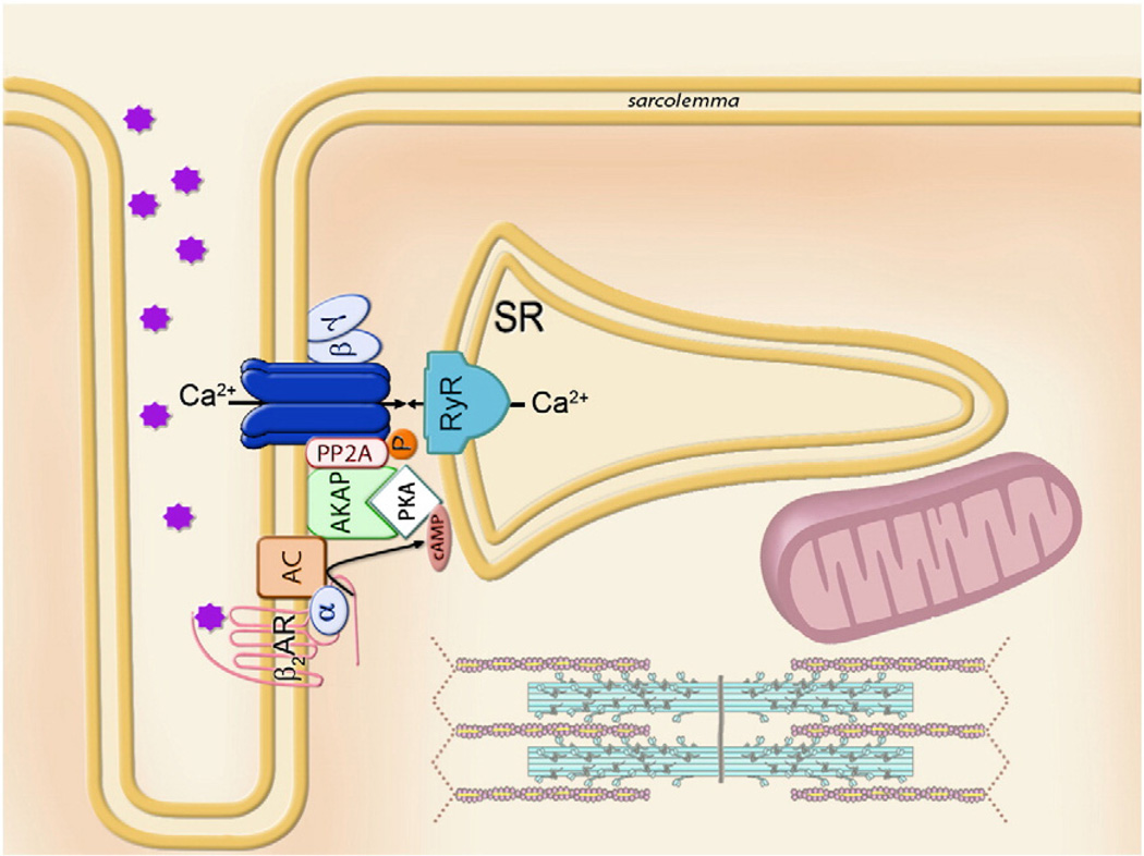
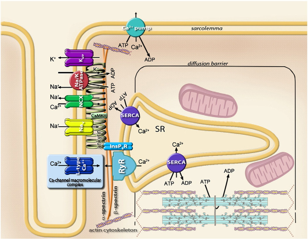
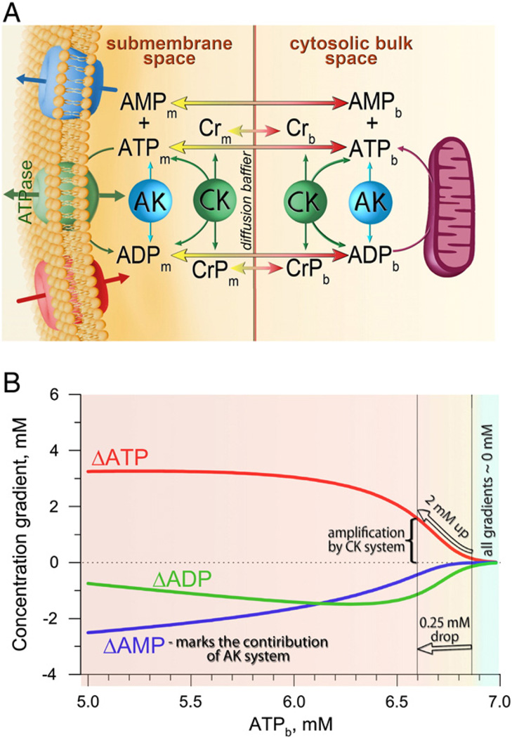
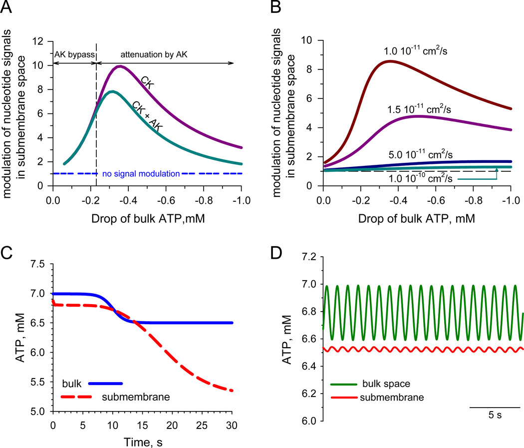
Orchestrated excitation-contraction coupling in heart muscle requires adequate spatial arrangement of systems responsible for ion movement and metabolite turnover. Co-localization of regulatory and transporting proteins into macromolecular complexes within an environment of microanatomical cell components raises intracellular diffusion barriers that hamper the mobility of metabolites and signaling molecules. Compared to substrate diffusion in the cytosol, diffusional restrictions underneath the sarcolemma are much larger and could impede ion and nucleotide movement by a factor of 10(3)-10(5). Diffusion barriers thus seclude metabolites within the submembrane space enabling rapid and vectorial effector targeting, yet hinder energy supply from the bulk cytosolic space implicating the necessity for a shunting transfer mechanism. Here, we address principles of membrane protein compartmentation, phosphotransfer enzyme-facilitated interdomain energy transfer, and nucleotide signal dynamics at the subsarcolemma-cytosol interface. This article is part of a Special Issue entitled "Local Signaling in Myocytes".


Figures

Fig. 1
Fig. 1
Model of a G-protein coupled signaling multiprotein module, which integrates β2-adrenoreceptor (β2AR) with the heteromeric G-protein (α and βγ subunits) and adenylate cyclase (AC), which by producing cAMP activates protein kinase A (PKA), anchored to the membrane through AKAP. Phosphorylation status and therefore operation of the final effector, L-type Ca2+ channel, is regulated by the ratio between local PKA and phosphatase (PP2A) activities. The Ca-channel macromolecular complex ensures rapid and vectorial activation of the specific effector and downstream pathways, exemplified here as Ca2+ release from sarcoplasmic reticulum (SR) through the ryanodine receptors (RyR).
Fig. 2
Fig. 2
Sarcolemmal metabolic unit comprised of the cardiac ankyrin-B membrane-associated protein complex, the Ca-channel macromolecular complex (see Fig. 1) and components of the sarcoplasmic reticulum. Ankyrin bound to β2-spectrin targets Na+/K+ ATPase, voltage-dependent Na+ (Nav) and ATP-sensitive K+ (KATP) channels, Na+–Ca2+ exchanger (NCX), IP3 receptor (IP3R), and, through interactions with obscurin (not shown), protein phosphatase 2A (PP2A). Coordination of calmodulin-dependent kinase (CaMKII) by β-spectrin in the proximity to Na+ channel is also indicated.
Fig. 3
Fig. 3
A: Remodeled ventricular myocyte from the heart of a TNFα-TG transgenic mouse manifests distorted cellular architecture (bottom) compared to the rod-shaped cell from control wildtype counterparts (top), visualized by scanning electron microscopy. B: In the open cell-attached mode of the patch-clamp technique, obtained by cell permeabilization with digitonin (Abraham et al. [55]), uncoupling of bulk mitochondria with 2,4-dinitrophenol (DNP), in the constant presence of inhibitory concentration of ATP, activated sarcolemmal KATP channel activity in cardiac myocytes from control hearts (top). Impeded by cellular remodeling, the communication between the cytosol and the sarcolemma resulted in a blunted KATP channel response to DNP-induced metabolic inhibition (bottom), despite intact intrinsic KATP channel gating properties (data not shown). C: Under chemical hypoxia, induced by DNP, APD90 (action potential duration measured at 90% of its amplitude) was markedly shortened in controlled (closed circlers), but not in TNFα-remodeled hearts (open circles) due to disturbed energy signals communication to sarcolemmal KATP channels (see Hodgson et al. [75] for further information).
Fig. 4
Fig. 4
A: Diffusion of adenine nucleotides between intracellular compartments facilitated by creatine (CK) and adenylate (AK) kinase systems, implying that delivery of ATP from one cellular compartment to another, in addition to passive diffusion flux, is accompanied by influx into submembrane space of high-energy phosphate equivalents (creatine phosphate, CrP) or efflux of AMP, which then are locally involved in CK- or AK-catalyzed ATP synthesis. Indexes ‘m’ and ‘b’ denote the metabolite concentrations in the submembrane and cytosolic bulk spaces, respectively. Influx and efflux of ions through sarcolemmal channels and concomitant ATPase flux are also indicated. B: Differences in ATP, ADP and AMP concentrations (ΔATP, ΔADP and ΔAMP) between the bulk and submembrane space at different bulk ATP concentrations (ATPb). Positive gradient values correspond to the drop of nucleotide concentrations directed from the diffusion barrier towards the sarcolemma. Nucleotide gradients were constructed by resolving equation 6 from Selivanov et al. [49], at JATPase = 4.7 × 10⁻⁶ µmol/cm2/s, D = 1.6 × 10⁻¹¹ cm2/s, 7 mM total nucleotide pool, 200 nm thickness of subsarcolemmal space and variable bulk ATP.
Fig. 5
Fig. 5
A: Effectiveness of modulation of nucleotide signals, i.e. changes of ATP in submembrane space (Δ[ATP]m) relative to a drop of ATP in bulk cytosolic levels (Δ[ATP]b), was calculated as d[ATP]m/d[ATP]b for creatine kinase (CK) alone and co-active CK and AK systems. The horizontal dotted line corresponds to a passive signal response (i.e. no signal modulation) in the absence of systems catalyzing phosphotransfer reactions. Note that higher changes in ATPb undergo a lower amplification, an effect enhanced by AK when a drop of ATPb exceeds the “AK bypass” threshold. B: Modulation of nucleotide signals under co-active CK and AK systems at different values of the apparent diffusion coefficient for nucleotides in submembrane space. C: Kinetic simulation of a nucleotide signal in submembrane space in response to a sustained drop of bulk ATP. Calculations were performed by resolving the system of differential equations (in the text) using JATPase = 4.7 × 10⁻⁶ µmol/cm2/s, D = 1.6 − 10−11 cm2/s, 200 nm thickness of subsarcolemmal space, 7 mM total nucleotide ([ANP]) and 40 mM Cr/CrP ([CrT]) pools. The nucleotide signal, generated in cytosol as 0.5 mM drop of ATP (blue solid line) is amplified in the membrane vicinity and reaches steady-state within >30 s delay (red dotted line). D: Same approach and values of parameters were used to simulate cytosolic ATP oscillations during contraction at 1 Hz frequency that were effectively filtered out in the vicinity of the sarcolemma. (For interpretation of the references to color in this figure legend, the reader is referred to the web version of this article.)

Similar articles

Cited by

References

    1. Alberts B, Johnson A, Lewis J, Raff R, Roberts K, Walter P. Molecular Biology of the Cell. 4th edition. New York: Garland Science; 2002. The compartmentalization of cells; pp. 659–669.
    1. Saks V, Monge C, Guzun R. Philosophical basis and some historical aspects of system biology: from Hegel to Noble—applications for bioenergetic research. Int J Mol Sci. 2009;10:1161–1192. - PMC - PubMed
    1. Martin W. Evolutionary origins of metabolic compartmentation in eukaryotes. Philos Trans R Soc Lond B Biol Sci. 2010;365:847–855. - PMC - PubMed
    1. Saks V, Beraud N, Wallimann T. Metabolic compartmentation—a system level property of muscle cells: real problems of diffusion in living cells. Int J Mol Sci. 2008;9:751–767. - PMC - PubMed
    1. Rockman HA, Koch WJ, Lefkowitz RJ. Seven-transmembrane-spanning receptors and heart function. Nature. 2002;415:206–212. - PubMed