. 2011 Sep;160(1-2):159-65.
doi: 10.1016/j.virusres.2011.06.005. Epub 2011 Jun 16.

VLPs of Leptopilina boulardi share biogenesis and overall stellate morphology with VLPs of the heterotoma clade

Affiliations

VLPs of Leptopilina boulardi share biogenesis and overall stellate morphology with VLPs of the heterotoma clade

Gwenaelle Gueguen et al. Virus Res. 2011 Sep.

Abstract

Viruses and virus-like particles (VLPs) of insect parasitoids modify host-parasite interactions. The Drosophila wasp, Leptopilina heterotoma, produce 300 nm spiked VLPs that bind to the host's blood cells via surface projections. L. heterotoma is a generalist wasp that attacks over a dozen Drosophila species. Oviposition introduces VLPs into the hemolymph of Drosophila larvae. VLPs lyse hemocytes and obliterate immune signaling in infected larval hosts. L. boulardi, a member of a distinct Leptopilina clade, is a specialist, whose host range is limited to the melanogaster group. As a step toward understanding a potential relationship between venom contents and host range in these wasps, we used electron microscopy to characterize VLPs from the virulent L. boulardi-17 (Lb-17) strain. While the Lb-17 VLPs can neither lyse blood cells nor suppress host defense, their biogenesis is surprisingly similar to that of L. heterotoma. Like L. heterotoma VLPs, L. boulardi VLPs are stellate; but they have fewer spikes, each spike being significantly longer than the spikes in L. heterotoma VLPs. The Lb-17 VLPs possess a dimple, making them clearly distinct from L. heterotoma VLPs. We discuss the significance of these cross-clade differences in VLP morphologies in relation to their biological activities and the host range of the wasp.


Conflict of interest statement

The authors declare no conflict of interest.

Figures
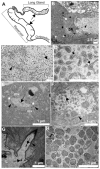
Figure 1
Figure 1. Transmission electron micrographs of sections of L. boulardi-17 venom gland
A. Schematic showing the organization of the long gland, reservoir and ovipositor complex of the wasp’s venom gland. Arrows point to the regions from where the ultrathin sections were prepared. (B–D) Contents of Lb-17 long gland. B. Fine structure of the long gland region with secretory cells (SC) and lumen (L). C. The lumen contains numerous long filamentous structures (arrowheads) and immature VLPs (arrows). D. Immature VLPs at higher magnification (arrows). (E–G) One cytoplasmic canal originates from each secretory cell and empties into the long gland lumen (see Ferrarese et al., 2009 for more details). E. Cross section from the rough canal portion with distinct microvilli (mv), proximal to the secretory cell. The rough canal has immature VLPs (arrow) in its lumen. The rough canal lumen is surrounded by a mesh-like matrix (white stars). Arrowheads indicate electron-dense particles between the microvilli and inside the matrix. F. Cross section of a smooth canal (no microvilli, distal to the secretory cell, and joins the lumen-see G.) with immature VLPs (arrow). Arrowhead points to the cuticle surrounding the canal. G. Smooth canal (cuticle-lined – arrowhead) ending into the gland lumen. VLPs (arrows) from the canal are dumped into the gland lumen (L) towards the reservoir. H. Contents of the venom gland reservoir. The reservoir contains VLPs ranging from 200–450 nm in diameter. VLPs at this stage of biogenesis contain electron-light regions.
Figure 2
Figure 2
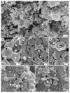
(A–D) Scanning electron micrographs of structures recovered from Lb-17 venom gland, or (E–F) from D. melanogaster hemolymph post-parasitization. A. Clusters of VLPs recovered from the wasp venom gland. (B–D) VLP morphologies in the venom gland. Stellate particles are found in both the venom gland (C) and in the host hemolymph compartments (E–F). Black stars indicate VLPs dimple, white stars indicate knobs at the end of the extensions and arrowheads point to spikes.
Figure 3
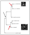
Figure 3. Phylogeny of Leptopilina heterotoma and boulardi clades (adapted from (Allemand et al., 2002)) superimposed with VLP morphologies
The first, more parsimonious, hypothesis is that wasps of the clade ancestral to the heterotoma and boulardi clades possessed stellate VLPs (black arrow). After the divergence of these clades, VLPs evolved independently acquiring distinct morphologies. In an alternative scenario, VLPs in both clades arose independently (red arrows). VLPs from L. longipes have not been reported yet. Studies of VLPs from additional species of the Leptopilina genus will help to distinguish these hypotheses. Panels to the right of the phylogeny allow comparison of surface morphologies of L. heterotoma (top) and Lb-17 (bottom) VLPs.

Similar articles

Cited by

References

    1. Allemand R, Lemaitre C, Frey F, Bouletreau M, Vavre F, Nordlander G, van Alphen J, Carton Y. Phylogeny of six African Leptopilina species (Hymenoptera : Cynipoidea, Figitidae), parasitoids of Drosophila, with description of three new species. Ann Soc Entomol Fr. 2002;38(4):319–332.
    1. Bezier A, Annaheim M, Herbiniere J, Wetterwald C, Gyapay G, Bernard-Samain S, Wincker P, Roditi I, Heller M, Belghazi M, Pfister-Wilhem R, Periquet G, Dupuy C, Huguet E, Volkoff AN, Lanzrein B, Drezen JM. Polydnaviruses of braconid wasps derive from an ancestral nudivirus. Science. 2009a;323(5916):926–930. - PubMed
    1. Bezier A, Herbiniere J, Lanzrein B, Drezen JM. Polydnavirus hidden face: the genes producing virus particles of parasitic wasps. J Invertebr Pathol. 2009b;101(3):194–203. - PubMed
    1. Chiu H, Govind S. Natural infection of D. melanogaster by virulent parasitic wasps induces apoptotic depletion of hematopoietic precursors. Cell Death Differ. 2002;9(12):1379–1381. - PubMed
    1. Chiu HL, Morales J, Govind S. Identification and immuno-electron microscopy localization of p40, a protein component of immunosuppressive virus-like particles from Leptopilina heterotoma, a virulent parasitoid wasp of Drosophila. J Gen Virol. 2006;87:461–470. - PMC - PubMed

Publication types