. 2011 Sep 8:190:207-27.
doi: 10.1016/j.neuroscience.2011.06.005. Epub 2011 Jun 12.

Caudal nuclei of the rat nucleus of the solitary tract differentially innervate respiratory compartments within the ventrolateral medulla

Affiliations

Caudal nuclei of the rat nucleus of the solitary tract differentially innervate respiratory compartments within the ventrolateral medulla

G F Alheid et al. Neuroscience. .

Abstract

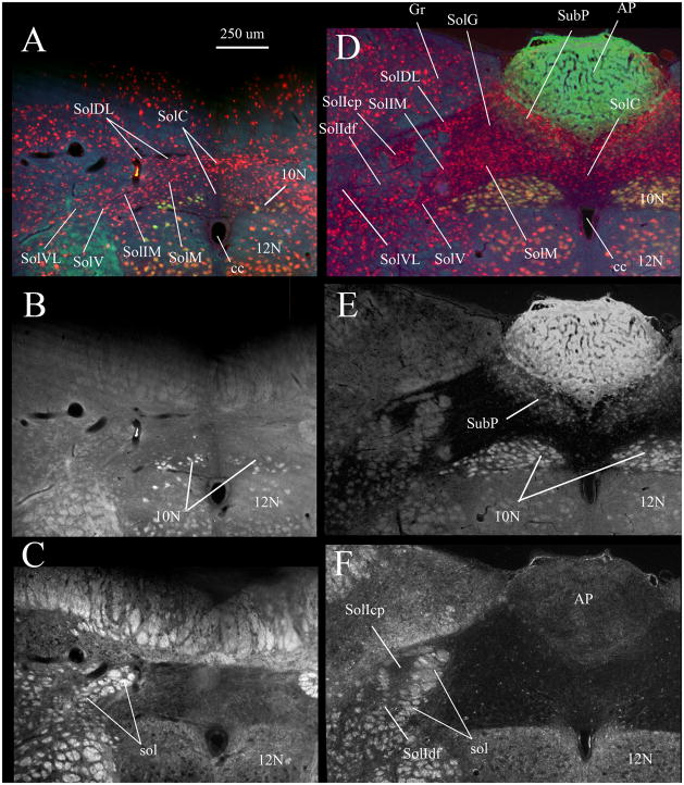
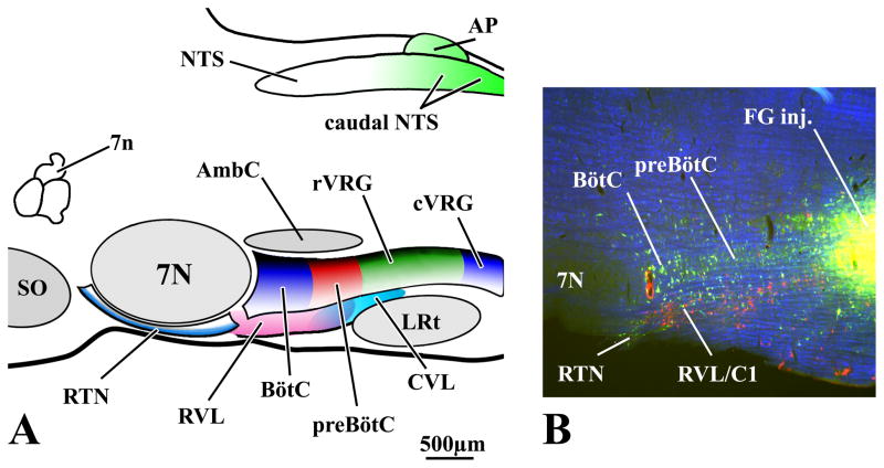
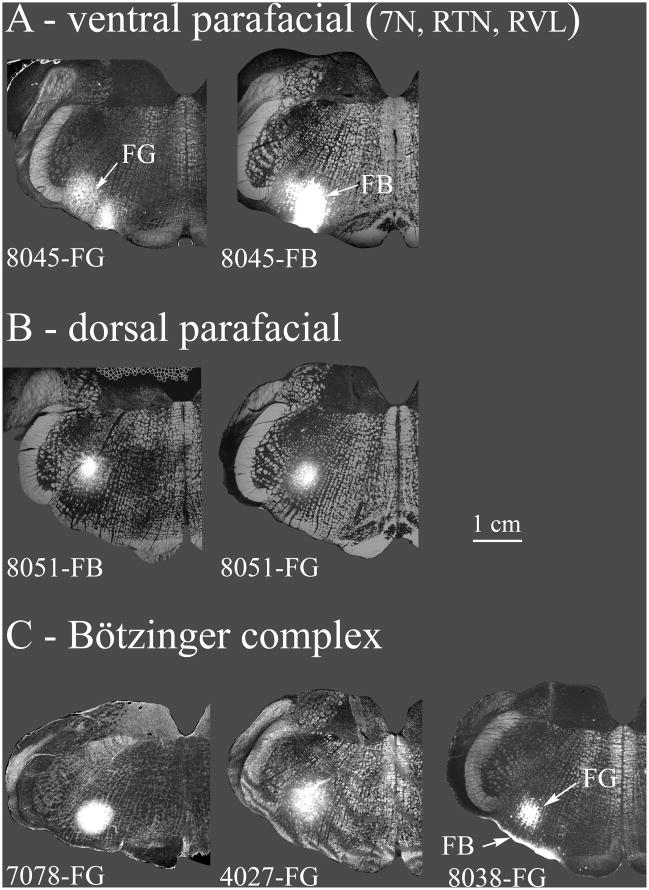
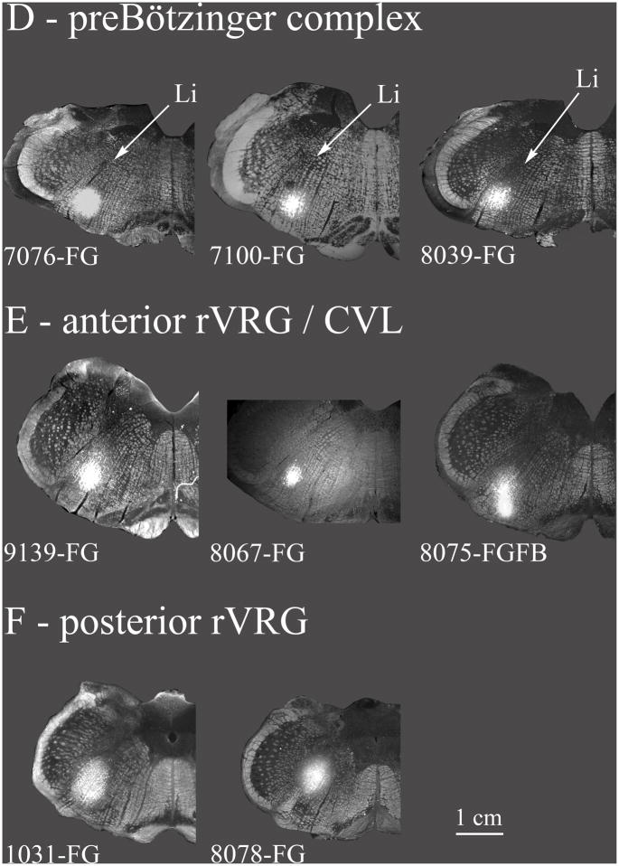
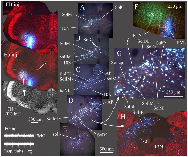
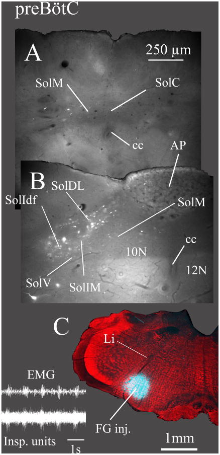
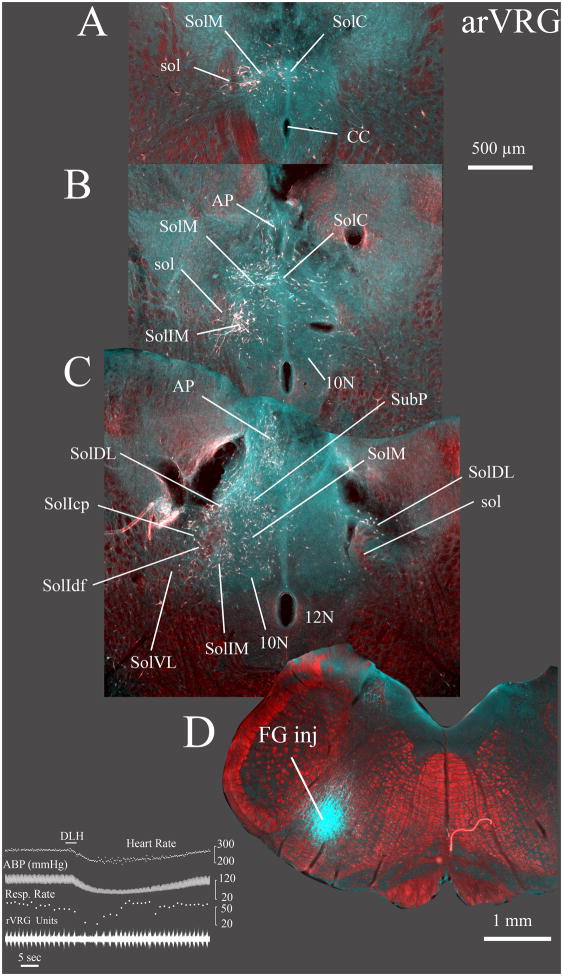
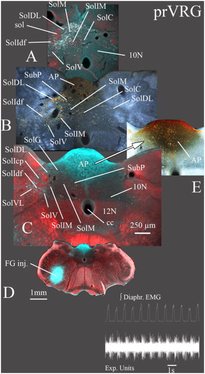
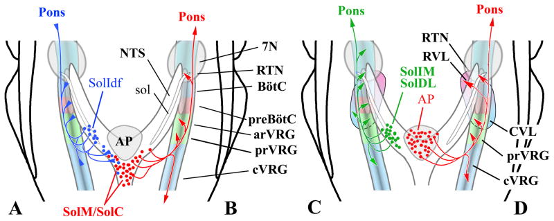
A substantial array of respiratory, cardiovascular, visceral and somatic afferents are relayed via the nucleus of the solitary tract (NTS) to the brainstem (and forebrain). Despite some degree of overlap within the NTS, specificity is maintained in central respiratory reflexes driven by second order afferent relay neurons in the NTS. While the topographic arrangement of respiratory-related afferents targeting the NTS has been extensively investigated, their higher order brainstem targets beyond the NTS has only rarely been defined with any precision. Nonetheless, the various brainstem circuits serving blood gas homeostasis and airway protective reflexes must clearly receive a differential innervation from the NTS in order to evoke stimulus appropriate behavioral responses. Accordingly, we have examined the question of which specific NTS nuclei project to particular compartments within the ventral respiratory column (VRC) of the ventrolateral medulla. Our analyses of NTS labeling after retrograde tracer injections in the VRC and the nearby neuronal groups controlling autonomic function indicate a significant distinction between projections to the Bötzinger complex and preBötzinger complex compared to the remainder of the VRC. Specifically, the caudomedial NTS, including caudal portions of the medial solitary nucleus and the commissural division of NTS project relatively densely to the region of the retrotrapezoid nucleus and rostral ventrolateral medullary nucleus as well as to the rostral ventral respiratory group while avoiding the intervening Bötzinger and preBötzinger complexes. Area postrema appears to demonstrate a pattern of projections similar to that of caudal medial and commissural NTS nuclei. Other, less pronounced differential projections of lateral NTS nuclei to the various VRC compartments are additionally noted.


Figures

Fig. 1
Fig. 1. Caudal NTS subdivisions viewed via NeuN immunostaining (A, D), FluoroGold labeling of vagal motoneurons and area postrema (B, E) and by darkfield illumination of solitary tract axons (C, F)
Nuclei composing the caudal NTS are indicated in A and D. A–C show the same section ~ 300 μm caudal to obex where the commissural nucleus of the solitary tract (SolC) is most prominent. D–F show a single section ~ 400 μm rostral to obex where the area postrema is near its maximal mediolateral extent. In A and D red neurons are immunofluorescent for neuron specific nuclear protein (NeuN). In A, B, D and E labeling in the dorsal motor nucleus of the vagus (10N), hypoglossal nucleus (12N), area postrema (AP) and subpostrema nucleus (SubP) resulted from a subcutaneous injection of FluoroGold. In C and F darkfield illumination identifies the myelinated fascicles of the solitary tract (sol). A and D are true color RGB images captured using a triple band-pass epifluorescence filter (uv/DAPI, FITC, Texas Red) and a RGB liquid crystal filter. Images in B and E were extracted from the green channel of the RGB images in A and D respectively.
Fig. 2
Fig. 2. Compartments of the Ventral Respiratory Column (VRC) of the Medulla
Compartments of the rat VRC (RTN, BötC, preBötC, rVRG, cVRG) are color-coded and projected onto a sagittal plane in A with local landmarks in gray. Approximate locations of the RVL and CVL are depicted ventral to the VRC. Overlapping aspects of RVL and CVL are depicted by the merged pink and cyan areas located beneath the preBötC. Caudal NTS regions (including the area postrema) providing input to the ventral medullary compartments are also indicated (green). B is a 50 μm thick parasagittal section at about the same scale and mediolateral location as the schematic in A. In B, respiratory-related neurons in the BötC and preBötC are retrogradely labeled from a FG injection in the rVRG. Tyrosine hydroxylase (TH) immunofluorescent neurons (red) ventrally adjacent to rostral compartments of the VRC are a component of the RVL. Note that TH neurons in RVL are not, in general, retrogradely labeled by retrograde tracers injected in the VRC. B is a pseudocolored image with monochrome images of TH neurons (red) and FG neurons (FITC-filter, green) and darkfield images (blue) inserted in the red, green and blue channels (respectively) of a digital image.
Fig. 3
Fig. 3. Retrograde tracer injection sites
The various retrograde tracer injections in the VRC and nearby areas (7N, RVL, CVL) are summarized. A: Ventral parafacial injections were located ventrally at the caudal end of the facial nucleus. These included the facial nucleus but also the RTN, and a rostral portion of RVL. These injections are depicted in greater detail in Fig. 4. B: Injections are located near the caudal facial nucleus but more dorsal than in A. The dorsal parafacial injections are depicted in greater detail in Fig. 5. C: Injections were classified as within the Bötzinger complex based on their location just caudal to the facial nucleus and by the types of neurons recorded proximal to the injections (see results). In case 8038 the bright band of labeling ventral to the FG injection resulted from a second injection tract where an FB injection was inadvertently made at the ventral surface of the medulla. Very few FB retrogradely labeled neurons were noted in the caudal NTS. Case 7078 is depicted in greater detail in Fig. 6. D: Injections were classified as within the preBötzinger complex on the basis of extracellular recordings near the tracer injection sites (see results) along with the location of the injection at the caudal end of the compact part of n. ambiguus, at approximately the same level as the folded portion of the linear nucleus of the medulla (Li) and just rostral to the lateral reticular formation. Case 7076 is depicted in greater detail in Fig. 7. E: Injections in the anterior part of rVRG (arVRG) were identified by the presence of inspiratory neurons at the injection site, the presence of the anterior portion of the lateral reticular nucleus and the absence of the area postrema; it was distinguished from posterior rVRG by the differential responses to DLH injection, bradypnea in anterior but not posterior rVRG. CVL neurons are generally located ventral to anterior rVRG (e.g. Figs. 2, 10) although functional overlap is indicated where DLH injections aimed at arVRG decrease blood pressure and heart rate (see results and case 9139 depicted in Fig. 8). F: Posterior rVRG injections, as with anterior rVRG, were localized by the presence of inspiratory neurons and the lateral reticular nucleus. In case 8075 FG and FB were differentially targeted at posterior VRG and CVL but overlapped due to the somewhat elongated shape of the FB injection. The presence of the area postrema helps discriminate the posterior rVRG from anterior rVRG as does the absence of significant changes in respiratory rhythm consequent to local DLH injections. Case 1031 is described in Fig. 9. Injection sites are depicted using a grayscale darkfield image of the relevant section as the base image overlaid by the injection site image using the “lighten” command in Photoshop®. Injections were saved as epifluorescent images for native FG/FB fluorescence, or as inverted brightfield images for DAB stained FG sections.
Fig. 4
Fig. 4. Most caudal NTS nuclei and area postrema project to the RTN/RVL region
In this example (case 8045), two retrograde tracers, FG and Fast Blue (FB) were injected at the caudal end of the facial nucleus. FG was injected where inspiratory unit activity was recorded (inset lower left) and FB was injected slightly caudal and ventral to the FG injection (100 μm caudal, 300 μm medial) but where no respiratory unit activity was evident. The FG injection overlapped the caudal facial nucleus (7N; in grayscale darkfield image from section with FG injection site). In addition to overlap of 7N, some dye extended to the superficial layers of the ventral medulla containing the RTN (F, detail of area marked on section with FG injection). FG and FB injections both overlapped TH immunolabeled neurons in RVL (red neurons in F). Sections A–E progress from caudal to rostral with A ~ 300 μm caudal and E ~ 900 μm rostral to obex. G is an enlarged view of the area in D. A–E and G are true color images taken with a liquid crystal RGB filter and uv excitation. Retrograde labeling from both the FG and FB injections was widespread, preferentially in ipsilateral caudal NTS and ipsilateral area postrema. H is at about the same level as D and illustrates the preferential ipsilateral retrograde labeling in NTS and AP. Labeled cells were found in every nucleus of the ipsilateral caudal NTS (A–E, and G) except SolCe. In most NTS nuclei, FG and FB labeled cells were coextensive, although not always double labeled (see detail in G). FB labeled cells generally appeared brighter than FG labeled cells in the aqueous media used to coverslip this material; double-labeled cells appeared as an intense blue-white color. EMG in inset is inspiratory motor activity recorded from the chest wall. Injection sites for FB and FG and the higher power image in H are depicted as pseudo-colored images composed by placing a monochrome darkfield image of the relevant section in the red channel of a blank RGB image, then combining this “red darkfield image” with a true color image of both injected dyes (blue FB and gold FG). The true color image was placed in a layer superficial to the darkfield image and combined with the latter using the “lighten” command in Photoshop CS3® which resulted in replacement of only the darker red areas by the brighter colors of the injection sites or, in H, by the retrograde labeled neurons. EMG in inset shows inspiratory motor activity. ECG activity was manually reduced on the EMG trace to facilitate visualization of the respiratory cycle.
Fig. 5
Fig. 5. Retrograde labeling of NTS nuclei from tracer injections dorsal to the facial nucleus
FG and FB were injected immediately dorsal to the facial nucleus with the FG injection located at the caudal end of the nucleus and FB located ~ 400 μm more rostrally (case 8051). Respiratory activity (inspiratory units, see inset) was recorded at the FG injection site but not observed at the FB site. Sections in A–D progress from caudal to rostral with A ~ 400 μm caudal and D ~ 900 μm rostral to obex. In contrast to the pattern of labeling following injections ventral to the facial nucleus (Fig. 4), there was only light retrograde labeling of most caudal NTS nuclei. An exception to this light labeling was the considerable FG labeling of cells in the central NTS nucleus (SolCe in D), particularly due to involvement of the AmbC by the FG injection (Cunningham and Sawchenko, 2000). Additionally, moderately dense retrograde labeling of the intermediate NTS nucleus (SolIM) was seen after the FG, but many fewer neurons after the FB injection. Significant FG and FB labeling of cells in the dorsal reticular formation is also evident in C and D, likely representing premotor neurons to the facial nucleus (Travers and Norgren, 1983). EMG in inset shows inspiratory motor activity. ECG activity was manually reduced on the EMG trace to facilitate visualization of the respiratory cycle.
Fig. 6
Fig. 6. Lateral and rostral NTS nuclei are retrogradely labeled from the Bötzinger complex (BötC), while few neurons in caudomedial NTS nuclei project to BötC
FG was injected caudal to the facial nucleus and below the compact part of nucleus ambiguus (C; case 7078). Section A is ~ 200 μm caudal and B is ~ 300 μm rostral to obex. Expiratory unit activity characteristic of BötC was recorded at the injection site (inset). Moderately dense retrograde labeling was apparent in only a few NTS nuclei, most prominently in the intermediate nucleus (SolIM). Relatively few neurons were labeled in the medial (SolM) or commissural (SolC) nuclei and no retrograde labeling was observed in area postrema (AP). EMG in inset is inspiratory motor activity. ECG activity was manually reduced on the EMG trace to facilitate visualization of the respiratory cycle.
Fig. 7
Fig. 7. Lateral and rostral NTS nuclei are retrogradely labeled from the preBötzinger complex (PreBötC) but very few neurons are labeled in caudomedial NTS nuclei or in area postrema (AP)
FG was injected at the level of the caudal end of the compact part of nucleus ambiguus (C; case 7076). Inspiratory unit activity was recorded at the injection site (inset). The FG injection was located at the level at which the dorsal part of the linear nucleus of the medulla (Li) bends ventromedially. Section A is ~ 350 μm caudal and B is ~ 300 μm rostral to obex. Retrograde labeling was moderately dense in the diffuse part of the interstitial (SolIdf), intermediate (SolIM) and dorsolateral (SolDL) nuclei but very sparse in the medial (SolM) and commissural (SolC) nuclei and in the area postrema. EMG in inset is inspiratory motor activity. ECG activity was manually reduced on the EMG trace to facilitate visualization of the respiratory cycle.
Fig. 8
Fig. 8. Widespread labeling is evident in the caudal NTS after injections into a region containing the anterior part of the rostral VRG (arVRG) and the caudal ventrolateral nucleus of the medulla (CVL)
FG was injected into the right arVRG (case 9139) at the site of respiratory unit activity (not shown) while a second electrode was used to record population respiratory activity in the left rVRG. Prior to injecting FG, injection of 3 nl of a 5 mM DLH solution (inset) in the right arVRG evoked a slowing of respiratory rate and decreases in heart rate and arterial pressure. In this instance, the central respiratory rhythm is represented by inspiratory unit activity recorded in the rVRG contralateral to the DLH and FG injections. Sections A–C progress from caudal to rostral with A ~ 500 μm caudal and C ~ 200 μm rostral to obex. Substantial retrograde labeling was evident throughout the caudal NTS, especially the commissural (SolC) and caudal part of the medial (SolM) nuclei. However, labeling in the ventrolateral NTS (SolVL) was relatively light particularly compared with FG injections more caudally in the posterior part of the rVRG (compare C with Fig. 9B). In the contralateral NTS, a significant number of retrogradely labeled neurons were evident in the dorsolateral nucleus (SolDL in C). Retrograde labeling is depicted as a pseudo-colored image composed of a monochrome darkfield image placed in the red channel of a blank RGB image, which was combined with inverted brightfield images of the DAB immunolabeled FG neurons. The inverted brightfield image was placed in a layer superficial to the darkfield image and combined with the latter using the lighten” command in Photoshop CS3®. The retrograde labeled neurons in the inverted images appear white on a dark background and therefore replace the (red) darkfield image where they occur. The resulting composite image provides white neurons against a red darkfield image. The FG injection site (D) is a pseudocolored image consisting of a low magnification darkfield image placed in the red channel of a blank RGB image. A grayscale inverted brightfield image of the same section was placed in both the green and blue channels. The FG injection consequently appears cyan against a red darkfield background image.
Fig. 9
Fig. 9. Widespread labeling is evident in the caudal NTS after injections in the posterior part of the rostral VRG (prVRG)
FG was injected into the rVRG immediately where respiratory neurons were recorded (inset; case 1031). Sections A–C progress from caudal to rostral with A ~ 100 μm caudal and C ~ 300 μm rostral to obex. Retrograde labeling was widespread in the ipsilateral caudal NTS (A – C) and similar to that following injections in the anterior rVRG (arVRG in Fig. 8). However, prVRG provided denser labeling in the ventrolateral nucleus (SolVL; B, C) and sparser labeling in the compact part of the interstitial nucleus (SolIcp; C). Retrograde labeling in the AP (B, E) had an ipsilateral predominance but with a greater bilateral representation compared to more rostral injections in the arVRG (Fig. 8). Sections B and E are normal darkfield images with the retrograde DAB labeled neurons appearing gold. Sections A, C are pseudo-colored images composed of the DAB immunolabeled FG neurons assembled in Photoshop® as described in the legend for Fig. 8. C is pseudocolored image of the FG injection site constructed similarly to that described for the section with the FG injection site in Fig. 8.
Fig. 10
Fig. 10. Schematic diagram of differential caudal NTS connections with ventrolateral medulla
The diagrams provide a synopsis of the differing patterns of innervation of the ventrolateral medulla suggested by the present report in concert with other reports that have examined this question (see Discussion). Arrows at the end of the schematic axons indicate likely excitatory terminations. Triangles at the end of axons indicate inhibitory projections (A). In A: the output of the lateral, caudal NTS is depicted (blue axons). This projection pattern is typified by 2nd order NTS neurons for slowly adapting pulmonary stretch receptors (Ezure et al., 2002). These neurons are mostly GABAergic, and, in the rat, are mainly located medially within SolIdf (Ezure and Tanaka, 2004; Takakura et al., 2007). B: illustrates the relatively selective projections from SolC and from caudal SolM (red axons) to rVRG and cVRG as well as to RTN, but not to the intervening BötC and preBötC. C: depicts projections from the lateral part of caudal NTS to ventrolateral medulla (green axons). This is typified by the projections from SolDL and from SolIM that project widely throughout the ventrolateral medulla with the exception that SolDL does not project to RVL. D: shows projections from the area postrema (red axons). Overall these are very similar to the projections originating from SolC and caudal SolM. Significant area postrema projections appear to target the RTN/RVL area as well as the region of rVRG/CVL. In contrast, no area postrema projections are seen to BötC and preBötC compartments of the VRC.

References

    1. Alheid GF. Efferents from medial and commissural nuclei (SolM, SolC) of the rat nucleus of the solitary tract (NTS) target the ventral respiratory group (VRG) and regions adjacent to the facial nucleus while avoiding the Bötzinger and preBötzinger complex. Society for Neuroscience. 2010 Abstracts #388.
    1. Alheid GF, Gray PA, Jiang MC, Feldman JL, McCrimmon DR. Parvalbumin in respiratory neurons of the ventrolateral medulla of the adult rat. J Neurocytol. 2002;31:693–717. - PubMed
    1. Alheid GF, McCrimmon DR. The chemical neuroanatomy of breathing. Respir Physiol Neurobiol. 2008;164:3–11. - PMC - PubMed
    1. Barraco IRA. Nucleus of the Solitary Tract. Boca Raton, FL: CRC Press; 1994.
    1. Bieger D, Hopkins DA. Viscerotopic representation of the upper alimentary tract in the medulla oblongata in the rat: the nucleus ambiguus. J Comp Neurol. 1987;262:546–562. - PubMed

Publication types