. 2012 Apr;8(3):291-8.
doi: 10.1016/j.nano.2011.06.012. Epub 2011 Jun 24.

Protective effect of the apoptosis-sensing nanoparticle AnxCLIO-Cy5.5

Affiliations

Protective effect of the apoptosis-sensing nanoparticle AnxCLIO-Cy5.5

Howard H Chen et al. Nanomedicine. 2012 Apr.

Abstract

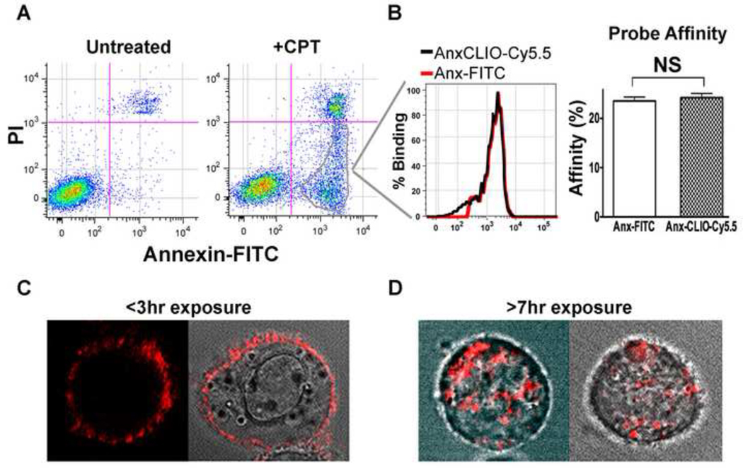
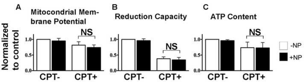
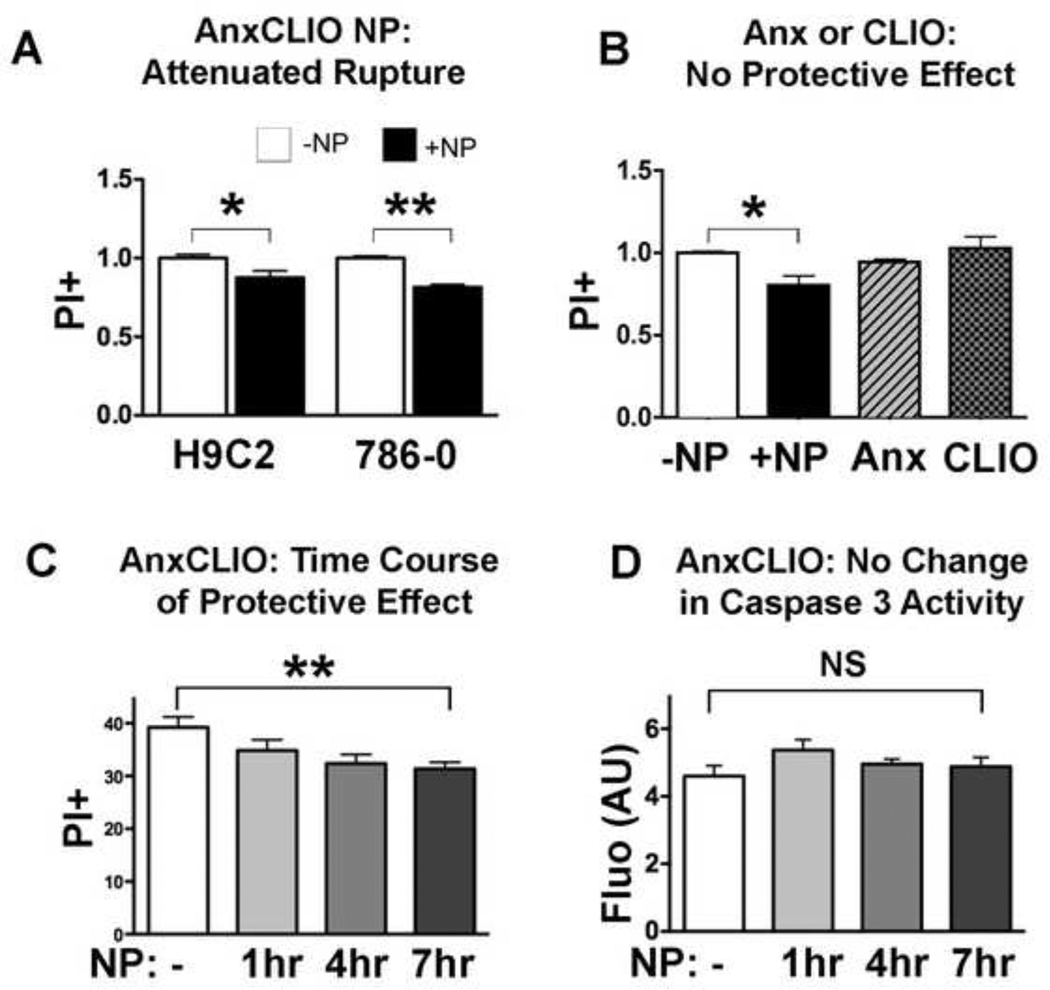
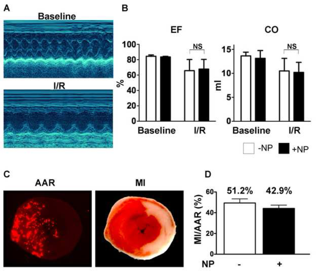
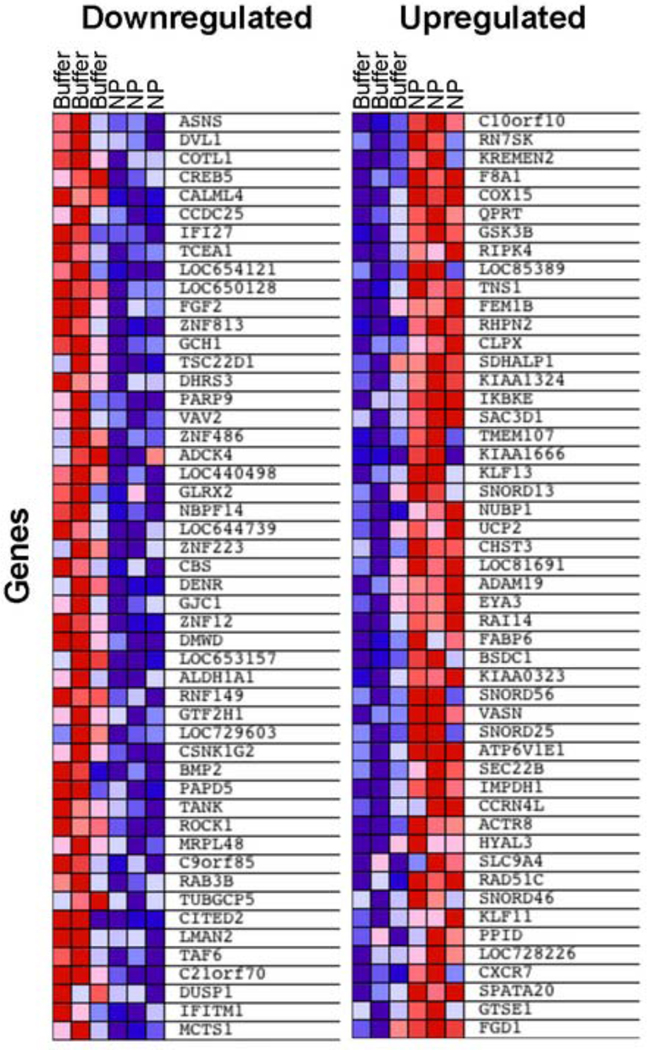
The diagnostic utility of the apoptosis-sensing nanoparticle (NP), AnxCLIO-Cy5.5, is well established. Here we sought to define the pathophysiological impact of the nanoparticle (NP) on apoptotic cells. Confocal microscopy showed that AnxCLIO-Cy5.5 remained bound to apoptotic cell membranes for 3 hours but by 7 hours had become completely internalized. AnxCLIO-Cy5.5 exposure did not impact energetics, metabolism or caspase-3 activity in apoptotic cells. Gene expression in cells exposed to AnxCLIO-Cy5.5 did not reveal upregulation of pro-inflammatory or cell-death pathways. Moreover, exposure to AnxCLIO-Cy5.5 decreased the frequency of membrane rupture of early apoptotic cells. Similarly, in mice exposed to 1 hour of ischemia -reperfusion, the injection of AnxCLIO-Cy5.5 at the onset of reperfusion reduced infarct size/area at risk by 16.2%. Our findings suggest that AnxCLIO-Cy5.5 may protect apoptotic cells by stabilizing their cell membranes and has the potential to become a theranostic agent, capable of both identifying and salvaging early apoptotic cells. From the Clinical Editor: This study demonstrates that AnxCLIO-Cy5.5 nanoparticles may protect apoptotic cells by cell membrane stabilization and have the potential to become a "theranostic agent" capable of identifying and salvaging early apoptotic cells.


Conflict of interest statement

Conflict-of-Interest: None

Figures

Figure 1
Figure 1
Flow Cytometry and Confocal Microscopy of apoptotic cells exposed to AnxCLIO-Cy5.5. (A) Camptothecin (CPT) treatment induces three cell populations: normal (left lower quadrant), early apoptotic (right lower quadrant), and late apoptotic/necrotic (right upper quadrant). (B) The percentage of early apoptotic cells labeled with AnxCLIO-Cy5.5 is almost identical to the number labeled with Anx-FITC. Likewise, the percentage of total cells labeled with both probes is highly similar, suggesting that AnxCLIO-Cy5.5 has an equal affinity for apoptotic cells to that of Anx-FITC. (C, D) Time-lapse confocal microscopy of apoptotic 786-0 cells exposed to AnxCLIO-Cy5.5. (C) Three hours after exposure to the probe, AnxCLIO-Cy5.5 is exclusively visualized on the cell membrane. (D) Within seven hours of exposure, however, AnxCLIO-Cy5.5 has become completely internalized into the cells. Two sets of different cells are shown in each panel. NS=not significant (p>0.05).
Figure 2
Figure 2
Impact of AnxCLIO-Cy5.5 on cell energetics and metabolism. (A) Mitochondrial membrane potential (JC-1), (B) cell reduction capacity (Resazurin), and (C) cell ATP content (Celltiter Glo) assays show significant differences between camptothecin treated (CPT+) and normal cells. However, no differences are seen between apoptotic (CPT+) cells exposed to the AnxCLIO-Cy5.5 nanoparticle (+NP) or buffer only (−NP). AnxCLIO-C5.5 does not perturb the energetics or metabolism of apoptotic cells. NS=not significant (p>0.05).
Figure 3
Figure 3
Impact of AnxCLIO-Cy5.5 exposure on the membrane stability of early apoptotic (PI negative) cells. White bars = no nanoparticle, Black bars = AnxCLIO-Cy5.5 nanoparticle. (A) Exposure to the AnxCLIO-Cy5.5 nanoparticle (+NP), reduced the proportion of apoptotic cells undergoing membrane rupture and becoming PI positive. (B) Exposure of apoptotic CHO cells to either the AnxCLIO-Cy5.5 nanoparticle, annexin alone or the cross-linked iron oxide nanoparticle (CLIO). A protective effect was seen only with the annexin-labeled nanoparticle. No effect was seen with either annexin alone or the unlabeled nanoparticle alone. (C) The protective effect of AnxCLIO-Cy5.5 on membrane stability was greater in those batches of cells exposed to the probe for longer periods of time. (D) No significant differences in caspase-3 activity were seen in cells exposed to AnxCLIO-Cy5.5. This suggests that the protective effect of AnxCLIO-Cy5.5 is due to a direct effect on the cell membrane and not on the apoptotic signaling cascade. *p<0.05; **p<0.01; NS=not significant (p>0.05).
Figure 4
Figure 4
Impact of AnxCLIO-Cy5.5 on mice exposed to ischemia-reperfusion injury (1 hour ligation of left coronary artery). The mice were injected intravenously at the onset of reperfusion with AnxCLIO-Cy5.5 (+NP) or PBS (−NP). (A) M-Mode echocardiography of a mouse heart before and 24 hours after surgery. The left ventricle is dilated and the anterior wall is severely hypocontractile 24 hours after surgery. (B) Ejection fraction (EF) and cardiac output (CO) were significantly lower after ischemia-reperfusion injury versus baseline, but there were no significant differences between those mice injected with AnxCLIO-Cy5.5 and PBS. (C) Measurement of infarct size (MI)/area at risk (AAR): The AAR in the section is defined by the absence of signal from fluorescent microspheres. The MI is defined by the pale area with TTC staining on the section. (D) MI/AAR was reduced by 16.2% in the mice injected with AnxCLIO-Cy5.5 (p=0.12).
Figure 5
Figure 5
Differentially expressed genes in apoptotic cells exposed to the AnxCLIO-Cy5.5 nanoparticle (NP) or buffer only. The most down- (left panel) and up-regulated (right panel) features (i.e. genes) in response to AnxCLIO-Cy5.5 (based on their signal-to-noise scores) are shown. Columns correspond to independent biological replicate samples. Heat map colors (red=high expression, blue=low expression) are normalized by row.

References

    1. Whelan RS, Kaplinskiy V, Kitsis RN. Cell death in the pathogenesis of heart disease: mechanisms and significance. Annual review of physiology. 2010;72:19–44. - PubMed
    1. Gottlieb RA, Burleson KO, Kloner RA, Babior BM, Engler RL. Reperfusion injury induces apoptosis in rabbit cardiomyocytes. The Journal of clinical investigation. 1994;94(4):1621–1628. - PMC - PubMed
    1. Korngold EC, Jaffer FA, Weissleder R, Sosnovik DE. Noninvasive imaging of apoptosis in cardiovascular disease. Heart failure reviews. 2008;13(2):163–173. - PMC - PubMed
    1. Dumont EA, Reutelingsperger CP, Smits JF, et al. Real-time imaging of apoptotic cell-membrane changes at the single-cell level in the beating murine heart. Nature medicine. 2001;7(12):1352–1355. - PubMed
    1. Hofstra L, Liem IH, Dumont EA, et al. Visualisation of cell death in vivo in patients with acute myocardial infarction. Lancet. 2000;356(9225):209–212. - PubMed

Publication types

MeSH terms