. 2011:102:105-25.
doi: 10.1016/B978-0-12-374912-3.00005-5.

Rise of the micromachines: microfluidics and the future of cytometry

Affiliations

Rise of the micromachines: microfluidics and the future of cytometry

Donald Wlodkowic et al. Methods Cell Biol. 2011.

Abstract

The past decade has brought many innovations to the field of flow and image-based cytometry. These advancements can be seen in the current miniaturization trends and simplification of analytical components found in the conventional flow cytometers. On the other hand, the maturation of multispectral imaging cytometry in flow imaging and the slide-based laser scanning cytometers offers great hopes for improved data quality and throughput while proving new vistas for the multiparameter, real-time analysis of cells and tissues. Importantly, however, cytometry remains a viable and very dynamic field of modern engineering. Technological milestones and innovations made over the last couple of years are bringing the next generation of cytometers out of centralized core facilities while making it much more affordable and user friendly. In this context, the development of microfluidic, lab-on-a-chip (LOC) technologies is one of the most innovative and cost-effective approaches toward the advancement of cytometry. LOC devices promise new functionalities that can overcome current limitations while at the same time promise greatly reduced costs, increased sensitivity, and ultra high throughputs. We can expect that the current pace in the development of novel microfabricated cytometric systems will open up groundbreaking vistas for the field of cytometry, lead to the renaissance of cytometric techniques and most importantly greatly support the wider availability of these enabling bioanalytical technologies.


Conflict of interest statement

The authors declare no conflicting financial interest.

Figures

Fig. 1
Fig. 1
The design of miniaturized LOC technologies is a promising avenue to address the inherent complexity of cellular systems with massive experimental parallelization, throughput, and analysis on a single cell level. (A) Microfluidics is an emerging field of engineering aimed at manipulating ultralow volumes of liquids in networks of microchannels with dimensions between 1 and 1000 μm. Fluid flow in microfluidic channels is dominated by viscous rather than inertial forces. Laminar flow describes the conditions where all fluid particles move in parallel to the flow direction. Note that during microfluidic circuitry is dominated only by a limited and local diffusion. Miniaturization of LOC promises greatly reduced equipment costs, simplified operation, increased sensitivity, and throughput by implementing many innovative and integrated on-chip analytical modules. (B) Microfabrication allows for design of new analytical functionalities. These for instance enable immobilization, manipulation, treatment, and analysis of single cells. An example of microcage (microjail) structure is shown that allows for micromechanical trapping and immobilization of single human cells.
Fig. 2
Fig. 2
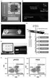
Innovative microfluidic flow cytometers (μFCM). (A) (B) Overview of the Agilent CellLab Chip technology. A glass chip with an etched network of microfluidic channels is mounted into the plastic cartridge providing the interface for the modified Agilent 2100 Bioanalyzer. Note the principles of microcytometry such as microhydrodynamic cell focusing using isobuoyant sheath buffer and double-point laser interrogation point (right panel). Systems permit for an automated, clog-free operation. No laser alignment or operator engagement is necessary for a sequential analysis of up to six samples on one chip. (C) Disposable microfluidic cartridge of the Fishman-R microfluidic flow cytometer with a two-way 2D hydrodynamic focusing of cells. (D) Optical configuration of Fishman-R microfluidic flow cytometers. Multiparameter detection capabilities are comparable to the conventional flow cytometers. Note forward scatter (FSC), side scatter (SSC), and four fluorescence detectors used in combination with spatially separated solid state 473 and 640 nm solid-state diode lasers. Side scatter detection is performed using innovative SLER technology (side scattered light detection using edge reflection) recently developed by On-chip Biotechnologies Co. (Tokyo, Japan). (E) Off-chip interface of the Fishman-R microfluidic flow cytometer housing the pneumatic and optical modules (data courtesy of Dr Kazuo Takeda, On-chip biotechnologies Co., Tokyo, Japan). (F) Multicolor immunophenotyping performed on the microfluidic Fishman-R flow cytometer as compared to conventional flow cytometers. Note that μFACS analysis requires merely 20 μl of blood and yields comparative multiparameter data to FACS (data courtesy of Dr Kazuo Takeda, On-chip Biotechnologies Co., Tokyo, Japan).
Fig. 3
Fig. 3
Gigasort™ Clinical Grade Cell Sorter (CytonomeST LLC, Boston, MA, USA) is the fastest fully sterile optical cell sorter ever produced and can be named as the most revolutionary advancement in FACS since its invention in the early 1960s. (A) A microfabricated glass die measuring 2 × 3 inches and containing 72 parallel microfluidic switches (microsorters). Using a massively parallel array of simultaneously operating microfluidic sorter cores, the technology achieves ultra-high sorting speeds of up to 1 billion events per hour. As such it can operate at over seven times the throughput of any conventional FACS. (B) Early prototype of Gigasort™ Clinical Grade Cell Sorter. (C) Principles of Gigasort operation. Each switch (microsorter) operates in sequential steps at a rate of 2000 cell sorting operations per second. In Step 1, cells pass through the microfluidic channel. In Step 2, laser beams illuminate the cells and optical emission occurs. In Step 3, emission is analyzed and accept/reject sort decisions are made by the off-chip hardware and software modules. In Step 4, microactuator pushes the diaphragm that moves the sorted cell into the upper portion of the laminar fluid stream. In Step 5, the desired cell (green) is moved into the keep area, whereas non-sorted cells (red) are directed to waste compartment. Data courtesy of Dr John C Sharpe, CytonomeST LLC (Boston, MA, USA). (See plate no. 7 in the color plate section.)
Fig. 4
Fig. 4
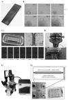
High-throughput drug screening on innovative microfabricated chips. (A) Picovitro microarray plates and slides as an example of a proprietary, microfabricated high-density cell array. Microwells 650 × 650 μm are fabricated by anodically bonding the silicon grid wafer to a 500 μm borofloat glass substrate (left panel). (B) Example of a long-term cell proliferation analysis on a Picovitro array. Long-term clone formation was started with a single K-562 cell FACS sorted to one well and cultured for up to 2 weeks. Data courtesy of Dr Sara Lindström, Picovitro AB (Stockholm, Sweden). (C) (D) CellTRAY®–a novel micro-etched live cell screening technology. Independently addressable regions of glass or plastic microwells allow for a multiplexed and time-resolved experimentation at a single cell level. Fully integrated and automated CellTRAY® system mounted on a microscope stage. On-microscope incubator and integrated microfluidics system allow for long-term experiments with automated, precise time-lapse imaging of live cells over the course of several days. Data courtesy of Dr Cathy Owen, Nanopoint Inc. (Honolulu, HI, USA). (E) (F) The BioFlux System (Fluxion Biosciences, San Francisco, CA, USA) that provides the state-of-the-art ability to emulate the physiological shear flow in an in vitro model. Shear force, flow rates, temperature, and compound addition, however, can be independently controlled and automated through the dedicated software. The BioFlux system leverages the advantages of microfluidics to create a network of laminar flow cells integrated into standard microtiterwell plates to ensure compatibility with common microscope stages making it thus compatible with bright field, fluorescence, confocal microscopy, and possibly also laser scanning cytometry. Data courtesy of Dr Mike Schwartz, Fluxion Biosciences (San Francisco, CA, USA).
Fig. 5
Fig. 5
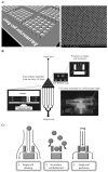
Living cell microarrays. (A) Single cell microarray platform fabricated in a glass substratum using femtosecond pulse laser μ-machining. Static cell microarray allows for a single cell docking into an array of microfabricated wells. Single wells are 25 μm in diameter. Cells passively sediment into a predefined pattern and are inaccessible to neighboring cells. This minimizes the influence of extrinsic factors, such as physical cell-to-cell contacts and paracrine signaling. This design facilitates real-time analysis at both single cell and population level. (B) Design of the microfluidic cell array (microfluidic array cytometer) with an active (hydrodynamic) cell docking into an array of microfabricated cell traps (microjails). Note the triangular chamber that contains a low-density cell positioning array. Array of microjails was fabricated in a biocompatible elastomer, polydimethylsiloxane (PDMS), and bonded to a glass substrate. The microfluidic array cytometer allows for a gentle trapping of single live cells for prolonged periods of time. (C) Principles of hydrodynamic cell docking and continuous microperfusion on a microfluidic array cytometer.

Similar articles

Cited by

References

    1. Adams JD, Kim U, Soh HT. Multitarget magnetic activated cell sorter. Proc Natl Acad Sci U S A. 2008;105:18165–18170. - PMC - PubMed
    1. Adams JD, Tom Soh H. Perspectives on utilizing unique features of microfluidics technology for particle and cell sorting. JALA Charlottesv Va. 2009;14:331–340. - PMC - PubMed
    1. Andersson H, van den Berg A. Microtechnologies and nanotechnologies for single-cell analysis. Curr Opin Biotechnol. 2004;15:44–49. - PubMed
    1. Arndt-Jovin DJ, Jovin TM. Computer-controlled multiparameter analysis and sorting of cells and particles. J Histochem Cytochem. 1974;22:622–625. - PubMed
    1. Baret JC, Miller OJ, Taly V, Ryckelynck M, El-Harrak A, Frenz L, Rick C, Samuels ML, Hutchison JB, Agresti JJ, Link DR, Weitz DA, Griffiths AD. Fluorescence-activated droplet sorting (FADS): efficient microfluidic cell sorting based on enzymatic activity. Lab Chip. 2009;9:1850–1858. - PubMed

Publication types

MeSH terms