. 2011 Jul;134(Pt 7):1959-71.
doi: 10.1093/brain/awr148.

Distinctive genetic and clinical features of CMT4J: a severe neuropathy caused by mutations in the PI(3,5)P₂ phosphatase FIG4

Affiliations

Distinctive genetic and clinical features of CMT4J: a severe neuropathy caused by mutations in the PI(3,5)P₂ phosphatase FIG4

Garth Nicholson et al. Brain. 2011 Jul.

Abstract

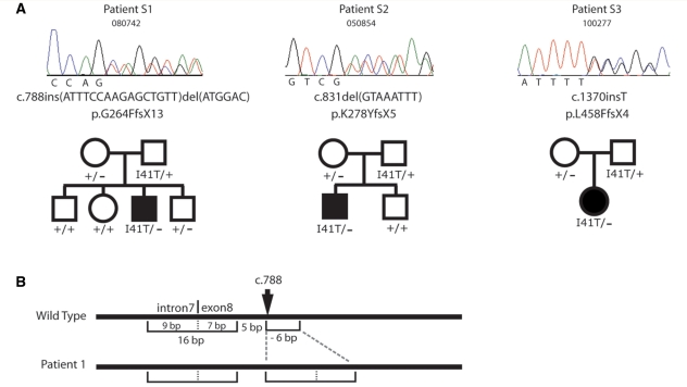
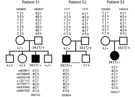
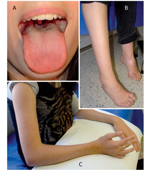
Charcot-Marie-Tooth disease is a genetically heterogeneous group of motor and sensory neuropathies associated with mutations in more than 30 genes. Charcot-Marie-Tooth disease type 4J (OMIM 611228) is a recessive, potentially severe form of the disease caused by mutations of the lipid phosphatase FIG4. We provide a more complete view of the features of this disorder by describing 11 previously unreported patients with Charcot-Marie-Tooth disease type 4J. Three patients were identified from a small cohort selected for screening because of their early onset disease and progressive proximal as well as distal weakness. Eight patients were identified by large-scale exon sequencing of an unselected group of 4000 patients with Charcot-Marie-Tooth disease. In addition, 34 new FIG4 variants were detected. Ten of the new CMT4J cases have the compound heterozygous genotype FIG4(I41T/null) described in the original four families, while one has the novel genotype FIG4(L17P/nul)(l). The population frequency of the I41T allele was found to be 0.001 by genotyping 5769 Northern European controls. Thirty four new variants of FIG4 were identified. The severity of Charcot-Marie-Tooth disease type 4J ranges from mild clinical signs to severe disability requiring the use of a wheelchair. Both mild and severe forms have been seen in patients with the same genotype. The results demonstrate that Charcot-Marie-Tooth disease type 4J is characterized by highly variable onset and severity, proximal as well as distal and asymmetric muscle weakness, electromyography demonstrating denervation in proximal and distal muscles, and frequent progression to severe amyotrophy. FIG4 mutations should be considered in Charcot-Marie-Tooth patients with these characteristics, especially if found in combination with sporadic or recessive inheritance, childhood onset and a phase of rapid progression.


Figures

Figure 1
Figure 1
Autosomal recessive inheritance of FIG4 mutations in three Australian families with CMT4J. (A) DNA chromatographs demonstrating the null mutations in three patients who also carry the FIG4-I41T allele. Autosomal recessive inheritance in evident in these families. (B) Structure of the mutant allele in Patient S1.
Figure 2
Figure 2
The FIG4-I41T allele is inherited on a shared chromosome 6 haplotype in three unrelated Australian families. The shared alleles are indicated in bold (lower left).
Figure 3
Figure 3
The missense mutation E302K is a functionally null allele of FIG4. (A) Residue E302 is evolutionarily conserved in yeast, invertebrates and mammals. Dots represent identity to the amino acid in the human sequence. (B and C) The enlarged vacuole in FIG4 null yeast is rescued by the wild-type (WT) gene but not by the E302K mutant. (D) Location of the E302K mutation between the protein interaction domain and the catalytic domain of FIG4 in the FIG4 protein (courtesy of Yuxin Mao).
Figure 4
Figure 4
CMT4J patient A7 is a compound heterozygote carrying the I41T mutation in combination with a deletion of FIG4 exon 2. (A) Sequencing of exon 2 from genomic DNA of Patient A7 detected only the c.122C nucleotide encoding threonine 41, and did not detect the reference T allele, indicating that the patient does not carry a wild-type allele. (B and C) Quantitative polymerase chain reaction using intronic primers flanking exon 2 demonstrates reduced copy number for exon 2 compared with diploid controls. The data indicate that the wild-type copy of exon 2 is deleted in this patient. (D) Maximal size of the deletion that includes exon 2. Genotyping polymorphic single nucleotide polymorphisms flanking FIG4 on chromosome 6 identified a region of 0.94 Mb, including the 100 kb FIG4 gene, in which none of the tested markers were heterozygous. This interval, bounded by the heterozygous single nucleotide polymorphisms rs11153138 and rs4245540, defines the maximal size of the deletion. Each symbol represents one genotyped marker. The identities and locations of the single nucleotide polymorphisms and microsatellites are presented in Supplementary Table 1 (www.genome.ucsc.edu, human genome build 38).
Figure 5
Figure 5
Proximal and distal muscle weakness in a patient with CMT4J. At the age of 19 years, Patient S3 demonstrated several features of proximal muscle wasting. (A) Wasted tongue with ‘triple furrow’ sign and (B) foot deformity and cocked up toes. The difference in leg length follows a left femoral fracture. (C) Proximal and distal wasting of the upper limb.
Figure 6
Figure 6
Abnormal sural nerve myelination in early stage disease in a patient with CMT4J. Right sural nerve biopsy at 7 years of age. A later biopsy of the left sural nerve, at the age of 41 years, revealed further loss of axons (data not shown). (A) Cross section of a sural nerve fascicle with extensive loss of large myelinated fibres. There is relative sparing of moderate to small diameter fibres, which range from thinly myelinated (arrowheads) to absent myelin (arrows). Stained with toluidine blue; ×20 magnification. (B) Electron microscopy demonstrating one naked axon with myelin breakdown products (arrowhead) and one thinly myelinated axon with surrounding onion bulb remnants and no clear Schwann cell processes (arrow). (C) Thinly myelinated axon with multiple thin myelin bands within the surrounding onion bulb (arrow). Also visible, a dysmyelinated axon (arrowhead) and a thinly myelinated axon (curving arrow). (D) Electron microscopy demonstrating a minimally myelinated axon with myelin breakdown products. The double basal lamina of a remnant Schwann cell process is visible (arrows).

References

    1. Barisic N, Claeys KG, Sirotkovic-Skerlev M, Lofgren A, Nelis E, De Jonghe P, et al. Charcot-Marie-Tooth disease: a clinico-genetic confrontation. Ann Hum Genet. 2008;72:416–41. - PubMed
    1. Biesecker LG, Mullikin JC, Facio FM, Turner C, Cherukuri PF, Blakesley RW, et al. The ClinSeq Project: piloting large-scale genome sequencing for research in genomic medicine. Genome Res. 2009;19:1665–74. - PMC - PubMed
    1. Chow CY, Landers JE, Bergren SK, Sapp PC, Grant AE, Jones JM, et al. Deleterious variants of FIG4, a phosphoinositide phosphatase, in patients with ALS. Am J Hum Genet. 2009;84:85–88. - PMC - PubMed
    1. Chow CY, Zhang Y, Dowling JJ, Jin N, Adamska M, Shiga K, et al. Mutation of FIG4 causes neurodegeneration in the pale tremor mouse and patients with CMT4J. Nature. 2007;448:68–72. - PMC - PubMed
    1. Davis EE, Zhang Q, Liu Q, Diplas BH, Davey LM, Hartley J, et al. TTC21B contributes both causal and modifying alleles across the ciliopathy spectrum. Nat Genet. 2011;43:189–96. - PMC - PubMed

Publication types

MeSH terms