. 2011 Aug 1;187(3):1496-505.
doi: 10.4049/jimmunol.1002910. Epub 2011 Jun 24.

Overexpression of Ste20-related proline/alanine-rich kinase exacerbates experimental colitis in mice

Affiliations

Overexpression of Ste20-related proline/alanine-rich kinase exacerbates experimental colitis in mice

Yutao Yan et al. J Immunol. .

Abstract

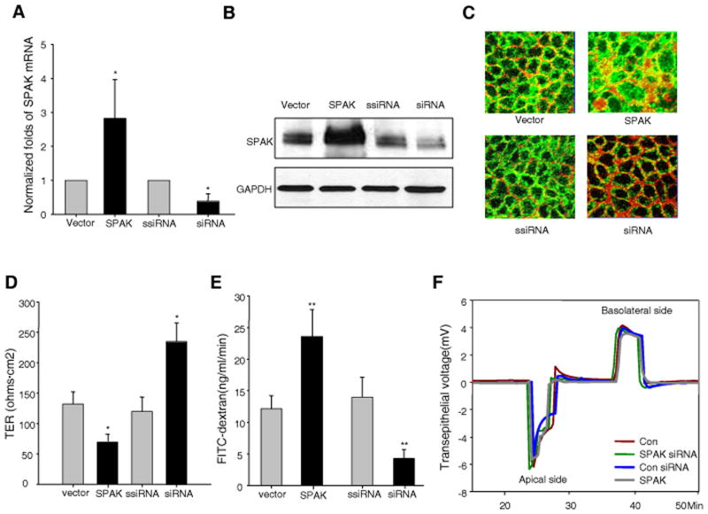
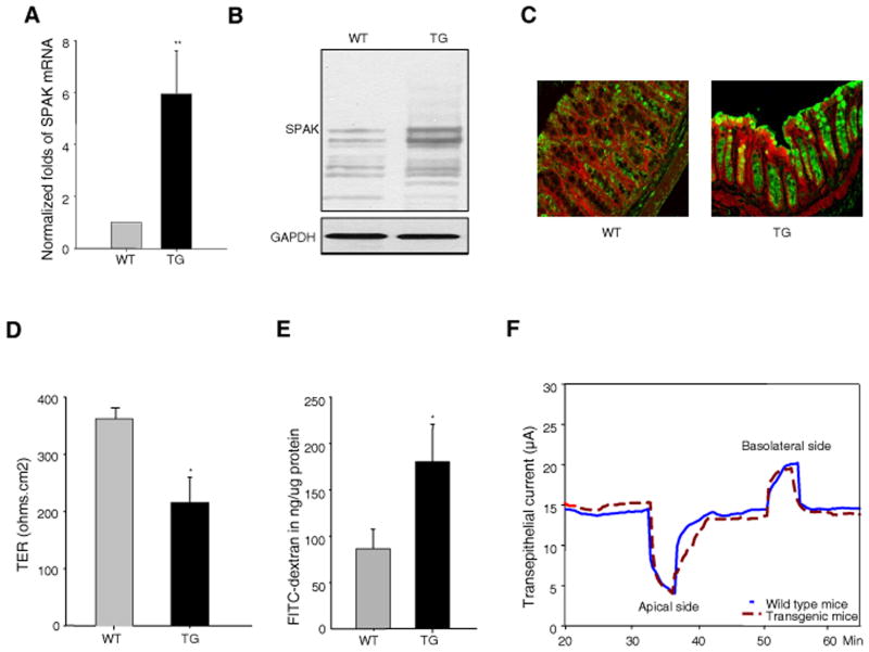
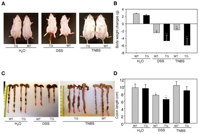
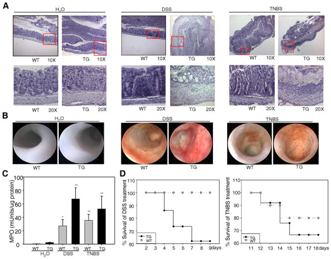
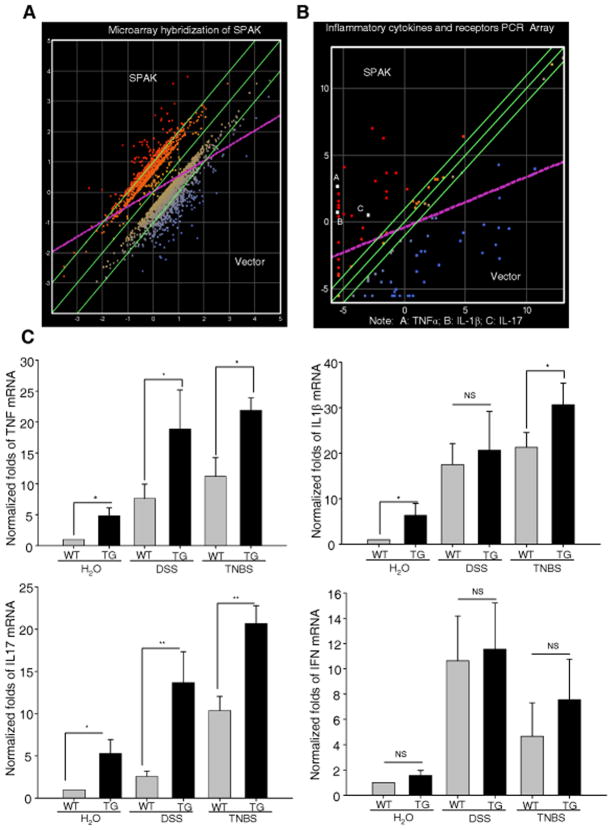
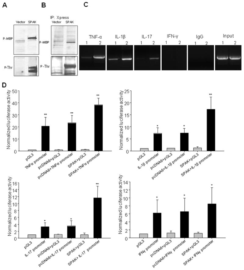
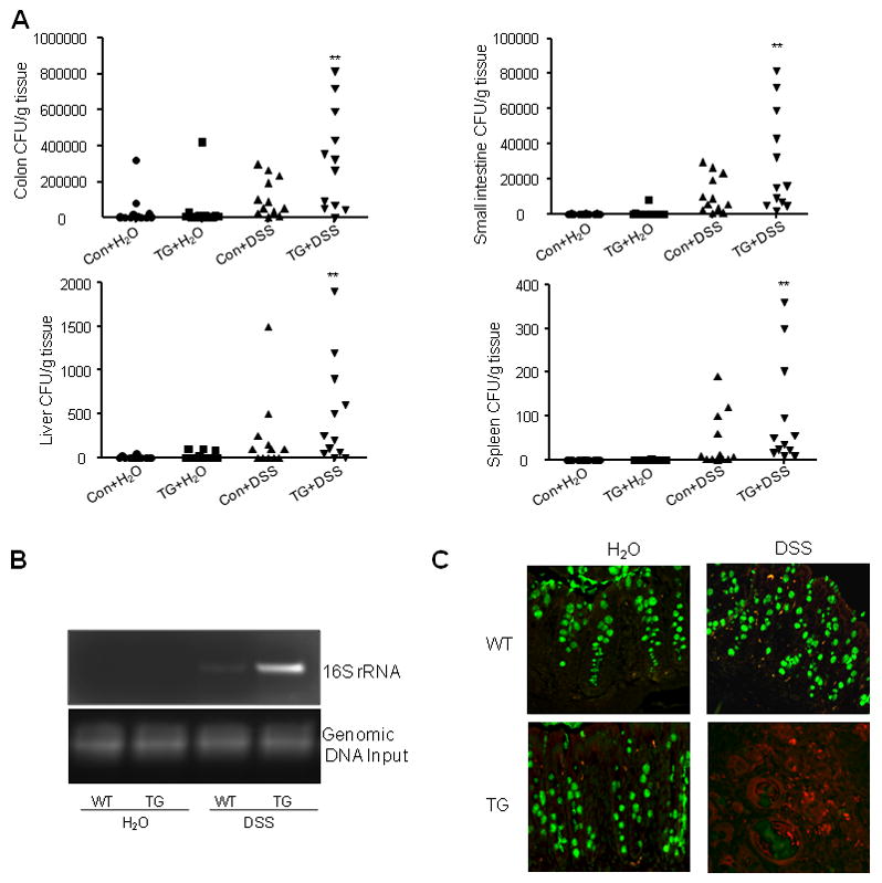
Inflammatory bowel disease, mainly Crohn's disease and ulcerative colitis, are characterized by epithelial barrier disruption and altered immune regulation. Colonic Ste20-like proline/alanine-rich kinase (SPAK) plays a role in intestinal inflammation, but its underlying mechanisms need to be defined. Both SPAK-transfected Caco2-BBE cells and villin-SPAK transgenic (TG) FVB/6 mice exhibited loss of intestinal barrier function. Further studies demonstrated that SPAK significantly increased paracellular intestinal permeability to FITC-dextran. In vivo studies using the mouse models of colitis induced by dextran sulfate sodium (DSS) and trinitrobenzene sulfonic acid showed that TG FVB/6 mice were more susceptible to DSS and trinitrobenzene sulfonic acid treatment than wild-type FVB/6 mice, as demonstrated by clinical and histological characteristics and enzymatic activities. Consistent with this notion, we found that SPAK increased intestinal epithelial permeability, which likely facilitated the production of inflammatory cytokines in vitro and in vivo, aggravated bacterial translocation in TG mice under DSS treatment, and consequently established a context favorable for the triggering of intestinal inflammation cascades. In conclusion, overexpression of SPAK inhibits maintenance of intestinal mucosal innate immune homeostasis, which makes regulation of SPAK important to attenuate pathological responses in inflammatory bowel disease.


Figures

Figure 1
Figure 1. SPAK is involved in the regulation of barrier function in IECs in vitro
SPAK expression is modulated in Caco2-BBE cells by SPAK/pcDNA6 and SPAK siRNA transient transfection compared with vector pcDNA6 and scramble siRNA (ssiRNA) transient transfection by (A) real time PCR, (B) western blot and (C) immunoflurescrence (SPAK-green, beta-actin-red). (D) Transepithelial resistance (TER) assay with Ussing chamber in Caco2-BBE monolayer: over-expression of SPAK decreases TER, while knock down of SPAK expression by siRNA increases TER. (E) FITC-dextran (4 kDa) was added to the apical side of polarized monolayers of Caco2-BBE cells at 10 mg/ml, and the basolateral reservoir was sampled at 2 h after the addition of FITC-dextran to the apical side. Histograms show mean ± SEM of ng/ml/min FITC-dextran translocation to the basolateral reservoir. (F) The dilution potential was determined by the change of transepithelial voltage upon switching from symmetrical bathing solutions (apical and basolateral side) to a 2:1 NaCl concentration gradient in Ussing chamber. Data are representative of three independent experiments. Error bars represent the means ± SEM. P-values were determined by Student’s t test. *: P<0.05, **: P<0.01.
Figure 2
Figure 2. SPAK transgenic mice display loss of intestinal barrier function
SPAK expression is enhanced in colonic mucosa in TG mice by (A) real time PCR, (B) Western blot and (C) (SPAK-green, beta-actin-red). (D) Transepithelial resistance (TER) assay with Ussing chamber in mouse colonic mucosa, TG mice have lower TER compared to WT mice in ex vivo experiments. (E) WT and TG mice were starved for 4 h and then gavaged with FITC-dextran (4 kDa). Serum was collected retro-orbitally 4 hours after gavage; fluorescence intensity of each sample was measured; and FITC-dextran concentrations were determined from standard curves generated by serial dilution of FITC-dextran. (F) In 2:1 NaCl short-circuit current assay in mouse colonic mucosa, the 128 mM NaCl solution was replaced with 52 mM NaCl in Kreb’s solution, and osmolarity was maintained with mannitol. Transepithelial current was monitored and recorded at 10 sec intervals, with the voltage continuously clamped at zero. Data are expressed as means ± SEM (n = 9 mice per group). Statistical differences of TG versus WT mice are reported. Data are pooled from three independent experiments. *: P<.05, **: P<.01.
Figure 3
Figure 3. SPAK TG mice were more susceptible to DSS- and TNBS-induced mouse colitis
(A) Phenotypic assay of WT and TG mice with 3.5 % DSS treatment for 10 days or with 150 mg/kg body weight of TNBS treatment for 48 hours. (B) Body weight measurement in WT and TG mice with 3.5 % DSS treatment for 10 days or with 150 mg/kg body weight of TNBS treatment for 48 hours. (C) Colonic phenotypic assay in WT and TG mice with 3.5 % DSS treatment for 10 days or with 150 mg/kg body weight of TNBS treatment for 48 hours. (D) Measurement of mouse colon length in WT and TG mice with 3.5 % DSS treatment for 10 days or with 150 mg/kg body weight of TNBS treatment for 48 hours. Data are expressed as means ± SEM (n = 4 mice per group). Statistical differences of TG versus WT mice are reported. Data are pooled from three independent experiments. *: P<0.05, **: P<0.01.
Figure 4
Figure 4. SPAK TG mice exhibit aggravated inflammation
(A) Representative photomicrographs of paraffin-embedded, hematoxylin-stained sections of the distal colon. Original magnification 10 × (upper panels) and 20 × (lower panels). (B) Intestinal inflammation was evaluated macroscopically in vivo using a murine miniature endoscope. Representative images of 6 different mice are shown. (C) WT and TG mice were given water or 3.5 % DSS for 10 days or 150 mg/kg body weight of TNBS for 48 hours as described in Figure 3, distal colon tissue was collected and subjected for MPO activity measurement. Data are expressed as means ± SEM (n = 9 mice per group). Statistical analysis was performed using an unpaired two-tailed Student’s t test. *: P<0.05, **: P<0.01. (D) After 10 days of 3.5 % DSS or 48 hours of TNBS, mice were given tap water and followed for mortality during recovery phase.
Figure 5
Figure 5. SPAK facilitates the production of proinflammatory cytokines
(A) Microarray hybridizations using the Affymetrix GeneChip exhibited alteration of expression of hundreds of different genes in human IECs. The mean of three independent experiments for each gene was calculated and used for data clustering. Only genes that showed significant change (over 2-fold difference) were selected for further characterization. DNASTAR ArrayStar 2 analytic software packages were used for Scatter plotting. (B) PCR array of cytokines demonstrated increase of proinflammatory cytokines, including TNF-α, IL-1β and IL-17 in human IECs. (C) Real time PCR was performed in WT and TG mice for proinflammatory cytokines, including TNF-α, IL-1β and IL-17, with or without DSS treatment. IFN-γ acted as a negative control. Data are expressed as means ± SEM (n = 9 mice per group). Statistical differences of TG versus WT mice are reported *: P<0.05, **: P<0.01.
Figure 6
Figure 6. Mechanism assays of SPAK involvement in the production of proinflammatory cytokines
(A) In vitro kinase assay of SPAK N-terminus expressed by TNT Quick Coupled Transcription/Translation System. MBP acts as substrate for kinase assay. The kinase assay complex was subjected to Western blot with anti-phospho MBP and threonine antibody. (B) In vivo kinase assay of N-terminus of SPAK, Immunoprecipitate of Caco2-BBE nuclear protein with Xpress antibody. MBP acted as a substrate for the kinase assay. The kinase assay complex was subjected to Western blot with anti-phospho MPB antibody and threonine antibody. (C) ChIP assay exhibited association of SPAK and cytokine genes, (1) Vector-transfected-Caco2-BBE cells; (2) SPAK-transfected-Caco2-BBE cells. (D) Transient transfection of different constructs into Caco2-BBE cells and transactivation assays of SPAK and the genes related to cytokines TNF-α, IL-1β and IL-17. The results are representative of three independent experiments performed in triplicate, and error bars represent standard deviation analyzed by Student’s t test by InStat v3.06 (GraphPad) software. *: P<0.05, **: P<0.01.
Figure 7
Figure 7. SPAK aggravates the luminal bacteria burden and translocation
(A) Colon, small intestine, spleen and liver tissue from SPAK TG mice or WT littermates were homogenized and cultured on nonselective media. CFUs were counted as described in Material & Methods. Data are expressed as means ± SEM (n = 9 mice per group). Statistical differences of TG versus WT mice with or without DSS treatment are reported. Data are pooled from three independent experiments. *: P<0.05, **: P<0.01. (B) PCR of bacterial 16S gene using DNA isolated from equal surface areas of colonic mucosa. Amplification was performed for 20 cycles. (C) SPAK facilitates the translocation of luminal bacteria inside the crypt of colon. FISH with the probe of Alexa Fluor 555-conjugated EUB and mucus staining in each slide were performed. There is very sparse presence of bacteria (in red) in untreated WT and TG mice. After DSS treatment, there is a dramatic increase of translocation of bacteria in both WT and TG mice; further, significantly more bacterial colonies were observed in TG mice.

Similar articles

Cited by

References

    1. Clayburgh DR, Shen L, Turner JR. A porous defense: the leaky epithelial barrier in intestinal disease. Lab Invest. 2004;84:282–291. - PubMed
    1. Turner JR. Intestinal mucosal barrier function in health and disease. Nat Rev Immunol. 2009;9:799–809. - PubMed
    1. Schmitz H, Barmeyer C, Fromm M, Runkel N, Foss HD, Bentzel CJ, Riecken EO, Schulzke JD. Altered tight junction structure contributes to the impaired epithelial barrier function in ulcerative colitis. Gastroenterology. 1999;116:301–309. - PubMed
    1. Swidsinski A, Loening-Baucke V, Theissig F, Engelhardt H, Bengmark S, Koch S, Lochs H, Dorffel Y. Comparative study of the intestinal mucus barrier in normal and inflamed colon. Gut. 2007;56:343–350. - PMC - PubMed
    1. Zeissig S, Burgel N, Gunzel D, Richter J, Mankertz J, Wahnschaffe U, Kroesen AJ, Zeitz M, Fromm M, Schulzke JD. Changes in expression and distribution of claudin 2, 5 and 8 lead to discontinuous tight junctions and barrier dysfunction in active Crohn’s disease. Gut. 2007;56:61–72. - PMC - PubMed

Publication types

MeSH terms

Substances