

ML1419c peptide immunization induces Mycobacterium leprae-specific HLA-A*0201-restricted CTL in vivo with potential to kill live mycobacteria
- PMID: 21705623
- PMCID: PMC3140574
- DOI: 10.4049/jimmunol.1100980
ML1419c peptide immunization induces Mycobacterium leprae-specific HLA-A*0201-restricted CTL in vivo with potential to kill live mycobacteria
Abstract
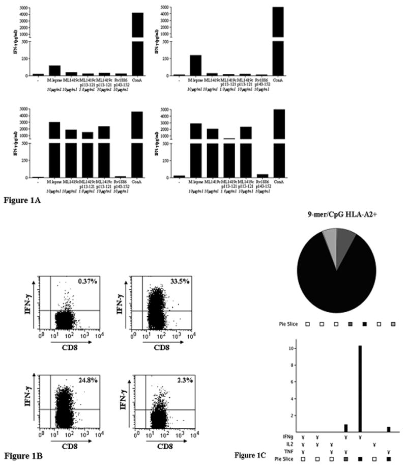
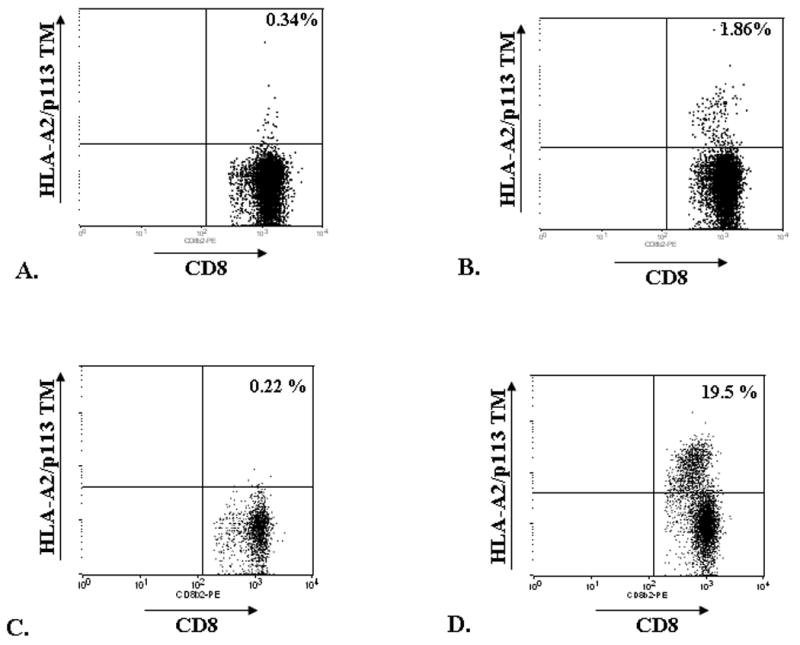
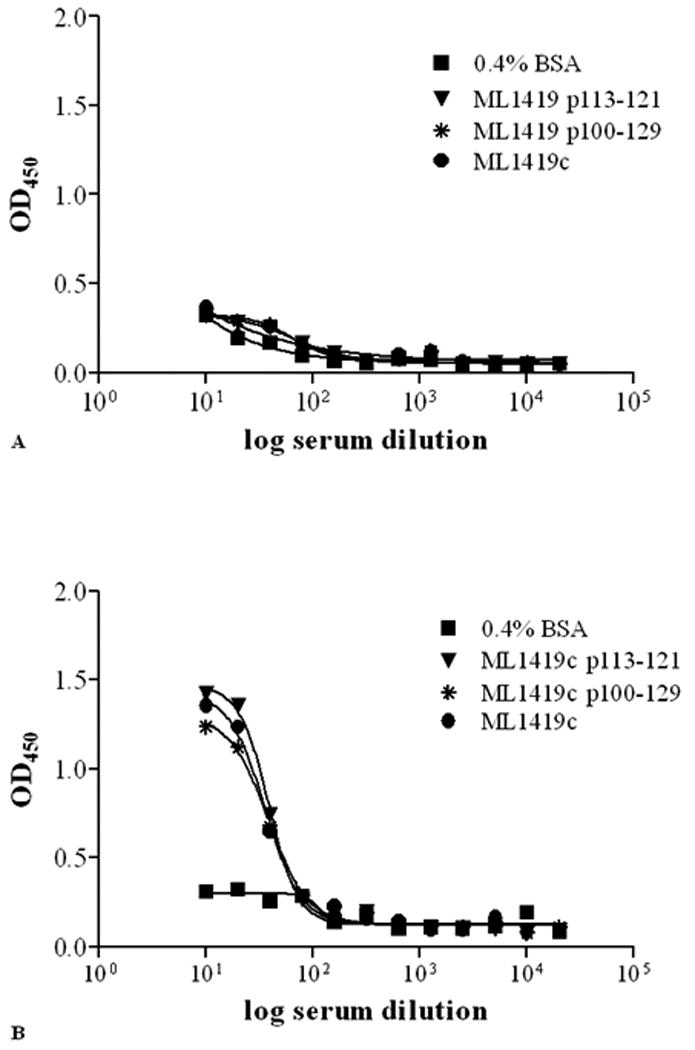
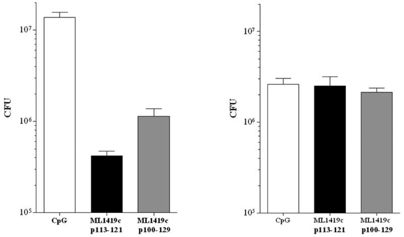
MHC class I-restricted CD8(+) T cells play an important role in protective immunity against mycobacteria. Previously, we showed that p113-121, derived from Mycobacterium leprae protein ML1419c, induced significant IFN-γ production by CD8(+) T cells in 90% of paucibacillary leprosy patients and in 80% of multibacillary patients' contacts, demonstrating induction of M. leprae-specific CD8(+) T cell immunity. In this work, we studied the in vivo role and functional profile of ML1419c p113-121-induced T cells in HLA-A*0201 transgenic mice. Immunization with 9mer or 30mer covering the p113-121 sequence combined with TLR9 agonist CpG induced HLA-A*0201-restricted, M. leprae-specific CD8(+) T cells as visualized by p113-121/HLA-A*0201 tetramers. Most CD8(+) T cells produced IFN-γ, but distinct IFN-γ(+)/TNF-α(+) populations were detected simultaneously with significant secretion of CXCL10/IFN-γ-induced protein 10, CXCL9/MIG, and VEGF. Strikingly, peptide immunization also induced high ML1419c-specific IgG levels, strongly suggesting that peptide-specific CD8(+) T cells provide help to B cells in vivo, as CD4(+) T cells were undetectable. An additional important characteristic of p113-121-specific CD8(+) T cells was their capacity for in vivo killing of p113-121-labeled, HLA-A*0201(+) splenocytes. The cytotoxic function of p113-121/HLA-A*0201-specific CD8(+) T cells extended into direct killing of splenocytes infected with live Mycobacterium smegmatis expressing ML1419c: both 9mer and 30mer induced CD8(+) T cells that reduced the number of ML1419c-expressing mycobacteria by 95%, whereas no reduction occurred using wild-type M. smegmatis. These data, combined with previous observations in Brazilian cohorts, show that ML1419c p113-121 induces potent CD8(+) T cells that provide protective immunity against M. leprae and B cell help for induction of specific IgG, suggesting its potential use in diagnostics and as a subunit (vaccine) for M. leprae infection.
Figures
Similar articles
-
Identification of major epitopes of Mycobacterium tuberculosis AG85B that are recognized by HLA-A*0201-restricted CD8+ T cells in HLA-transgenic mice and humans.J Immunol. 2000 Dec 1;165(11):6463-71. doi: 10.4049/jimmunol.165.11.6463. J Immunol. 2000. PMID: 11086086
-
A CD8+ T cell heptaepitope minigene vaccine induces protective immunity against Chlamydia pneumoniae.J Immunol. 2005 May 1;174(9):5729-39. doi: 10.4049/jimmunol.174.9.5729. J Immunol. 2005. PMID: 15843575
-
Interferon-gamma (IFN-gamma) and tumour necrosis factor-alpha (TNF-alpha) are necessary in the early stages of induction of CD4 and CD8 cytotoxic T cells by Mycobacterium leprae heat shock protein (hsp) 65 kD.Clin Exp Immunol. 1998 Nov;114(2):196-203. doi: 10.1046/j.1365-2249.1998.00702.x. Clin Exp Immunol. 1998. PMID: 9822276 Free PMC article.
-
HLA and leprosy in the pre and postgenomic eras.Hum Immunol. 2006 Jun;67(6):439-45. doi: 10.1016/j.humimm.2006.03.009. Epub 2006 Apr 3. Hum Immunol. 2006. PMID: 16728267 Review.
-
Derivation of HLA-B*0702 transgenic mice: functional CTL repertoire and recognition of human B*0702-restricted CTL epitopes.Hum Immunol. 2003 Feb;64(2):211-23. doi: 10.1016/s0198-8859(02)00786-3. Hum Immunol. 2003. PMID: 12559623
Cited by
-
Synthetic Long Peptide Derived from Mycobacterium tuberculosis Latency Antigen Rv1733c Protects against Tuberculosis.Clin Vaccine Immunol. 2015 Sep;22(9):1060-9. doi: 10.1128/CVI.00271-15. Epub 2015 Jul 22. Clin Vaccine Immunol. 2015. PMID: 26202436 Free PMC article.
-
Diguanylate cyclase activity of the Mycobacterium leprae T cell antigen ML1419c.Microbiology (Reading). 2016 Sep;162(9):1651-1661. doi: 10.1099/mic.0.000339. Epub 2016 Jul 22. Microbiology (Reading). 2016. PMID: 27450520 Free PMC article.
-
Vaccines for Leprosy and Tuberculosis: Opportunities for Shared Research, Development, and Application.Front Immunol. 2018 Feb 26;9:308. doi: 10.3389/fimmu.2018.00308. eCollection 2018. Front Immunol. 2018. PMID: 29535713 Free PMC article. Review.
-
Immunogenicity evaluation of a rationally designed polytope construct encoding HLA-A*0201 restricted epitopes derived from Leishmania major related proteins in HLA-A2/DR1 transgenic mice: steps toward polytope vaccine.PLoS One. 2014 Oct 13;9(10):e108848. doi: 10.1371/journal.pone.0108848. eCollection 2014. PLoS One. 2014. PMID: 25310094 Free PMC article.
References
-
- Kaufmann SH. CD8+ T lymphocytes in intracellular microbial infections. Immunol Today. 1988;9:168–174. - PubMed
-
- van Pinxteren LA, Cassidy JP, Smedegaard BH, Agger EM, Andersen P. Control of latent Mycobacterium tuberculosis infection is dependent on CD8 T cells. Eur J Immunol. 2000;30:3689–3698. - PubMed
-
- Ottenhoff TH, T Mutis. Role of cytotoxic cells in the protective immunity against and immunopathology of intracellular infections. Eur J Clin Invest. 1995;25:371–377. - PubMed
Publication types
MeSH terms
Substances
Grants and funding
LinkOut - more resources
Full Text Sources
Molecular Biology Databases
Research Materials
