. 2011 Sep;59(9):864-74.
doi: 10.1369/0022155411413929. Epub 2011 Jun 24.

Expression of focal adhesion proteins in the developing rat kidney

Affiliations

Expression of focal adhesion proteins in the developing rat kidney

Sato Matsuura et al. J Histochem Cytochem. 2011 Sep.

Abstract

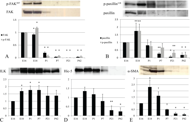
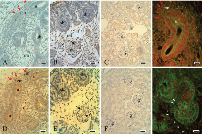
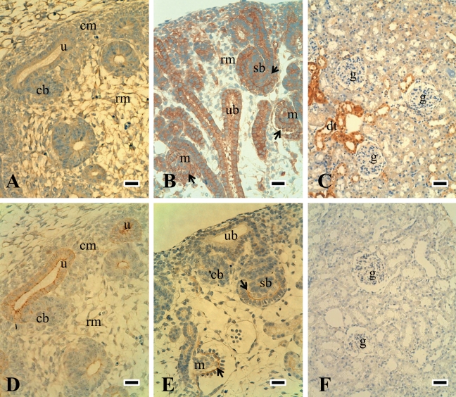
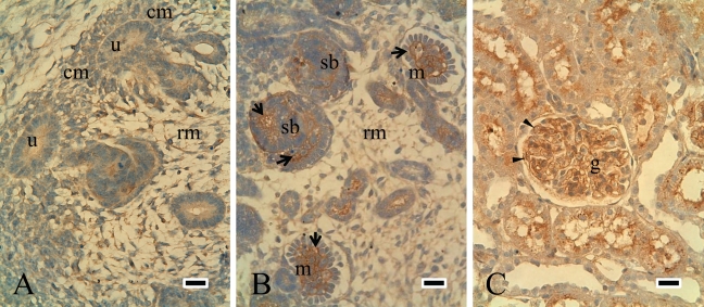
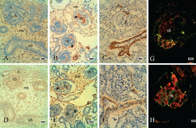
Focal adhesions play a critical role as centers that transduce signals by cell-matrix interactions and regulate fundamental processes such as proliferation, migration, and differentiation. Focal adhesion kinase (FAK), paxillin, integrin-linked kinase (ILK), and hydrogen peroxide-inducible clone-5 (Hic-5) are major proteins that contribute to these events. In this study, we investigated the expression of focal adhesion proteins in the developing rat kidney. Western blotting analysis revealed that the protein levels of FAK, p-FAK(397), paxillin, p-paxillin(118), and Hic-5 were high in embryonic kidneys, while ILK expression persisted from the embryonic to the mature stage. Immunohistochemistry revealed that FAK, p-FAK(397), paxillin, and p-paxillin(118) were strongly expressed in condensed mesenchymal cells and the ureteric bud. They were detected in elongating tubules and immature glomerular cells in the nephrogenic zone. Hic-5 was predominantly expressed in mesenchymal cells as well as immature glomerular endothelial and mesangial cells, suggesting that Hic-5 might be involved in mesenchymal cell development. ILK expression was similar to that of FAK in the developmental stages. Interestingly, ILK was strongly expressed in podocytes in mature glomeruli. ILK might play a role in epithelial cell differentiation as well as kidney growth and morphogenesis. In conclusion, the temporospatially regulated expression of focal adhesion proteins during kidney development might play a role in morphogenesis and cell differentiation.


Conflict of interest statement

The author(s) declared no potential conflicts of interest with respect to the authorship and publication of this article.

Figures

Figure 1.
Figure 1.
FAK, paxillin, ILK, Hic-5, and α-SMA expression during the development of rat kidneys. FAK, p-FAK397, paxillin, and p-paxillin118 were highly expressed in embryonic kidneys (A and B). ILK expression was detected in embryonic, postnatal, and mature kidneys (C). Hic-5 and α-SMA were highly expressed in E16 to P1 during development (D and E). Results are shown as means ± standard deviations from at least three independent experiments. Significant differences versus E16: p < 0.05; p < 0.01; **p < 0.001; *p < 0.0001.
Figure 2.
Figure 2.
Expression of FAK and p-FAK397 during the development of rat kidneys. Expression of FAK and p-FAK397 in E16 was intense at the cell membrane of the ureteric bud and mesenchymal cells, especially in the condensed mesenchyme (red arrows) (A and D). Around the nephrogenic zone in E18, FAK and p-FAK397 were expressed in S-shaped bodies (sb) as well as in immature glomerular endothelial and mesangial cells (black arrows in B and E, which are the serial sections). FAK expression was decreased and localized in tubules in P42 (C), while p-FAK397 was not detected in mature glomeruli of P42 (F). Double-staining experiments were performed to compare the expression of FAK (green, FITC) and PCNA-positive cells (red, TRITC) (G and H). FAK was expressed on the cell membrane of the ureteric bud and condensed mesenchymal cells (red arrows), where the PCNA was positive in E16 (G). In E18, FAK expression was also positive in cleft-invading endothelial and mesangial cells (arrow head) and mesenchymal cells (*), where the PCNA expression was weak (H). rm = renal mesenchymal cells; red arrows and “cm” = condensed mesenchymal cells; ub = ureteric bud; u = ureteric bud tip; sb = S-shaped body; m = immature glomerulus; black arrows in B and E = immature endothelial and mesangial cells; g = glomerulus. Scale bars (A, B, D, and F) = 25 µm; scale bars (C and F) = 50 µm; scale bars (G and H) = 100 µm.
Figure 3.
Figure 3.
Expression of paxillin and p-paxillin118 during the development of rat kidneys. Paxillin and p-paxillin118 expression were similar to those of FAK and were detected at the cell membrane of the ureteric bud in E16 (A and D are the serial sections). Both were also observed in immature endothelial (black arrow) and mesangial cells in comma- and S-shaped bodies in the nephrogenic zone of E18 (B and E). Paxillin was decreased and was limited to distal tubules in P42 (C), while p-paxillin118 expression on tubules was very weak (F). rm = renal mesenchymal cells; cm = condensed mesenchymal cells; ub = ureteric bud; u = ureteric bud tips; sb = S-shaped body; m = immature glomerulus; black arrows in B and E = immature endothelial and mesangial cells; dt = distal tubules; g = glomerulus. Scale bars (A, B, D, and F) = 25 µm; scale bars (C and F) = 50 µm.
Figure 4.
Figure 4.
Expression of ILK during the development of rat kidneys. ILK expression was similar to that of FAK in developing stages. ILK was expressed in the ureteric bud and mesenchymal cells in E15 (A). In E18, ILK was detected in S-shaped bodies as well as in immature endothelial and mesangial cells (B). ILK was strongly expressed in podocytes and epithelial cells in P42 (C). rm = renal mesenchymal cells; cm = condensed mesenchymal cells; u = ureteric bud tips; sb = S-shaped body; m = immature glomerulus; arrow = immature endothelial and mesangial cells; arrowhead = podocytes; g = glomerulus. Scale bars = 25 µm.
Figure 5.
Figure 5.
Expression of Hic-5 and α-SMA–positive cells during the development of rat kidneys. Hic-5 was mainly expressed in mesenchymal cells in E16 but was not detected in the ureteric bud or epithelial cells (A). α-SMA–positive cells were detected in mesenchymal cells in E16 (D). Distributions of Hic-5–positive cells were similar to α-SMA–positive cells in E16. In E18, Hic-5 was detected in immature glomerular endothelial and mesangial cells and also in mesenchymal cells (black arrows in B). Most Hic-5 expression was very similar to the distribution of α-SMA–positive cells in E18 (B and E are the serial sections). In mature kidneys, both expressions were not observed in glomeruli and were limited to vascular smooth muscle cells (C and F). Double-staining experiments showed that Hic-5 (green, FITC) and CD31 (red, TRITC) were partially co-localized in immature glomeruli of E18 (yellow arrowhead in G and H). rm = renal mesenchymal cells; u = ureteric bud tips; ub = ureteric bud; sb = S-shaped body; m = immature glomerulus; black arrows in B and E = immature endothelial and mesangial cells; g = glomerulus; v = vascular smooth muscle cells; red in G and H = CD31; green in G and H = Hic-5. Scale bars (A-F) = 25 µm; scale bars (G and H) = 75 µm.

References

    1. Abrahamson DR. 2009. Development of kidney glomerular endothelial cells and their role in basement membrane assembly. Organogenesis. 5:275–287 - PMC - PubMed
    1. Bonadio JF, Sage H, Cheng F, Bernstein J, Striker GE. 1984. Localization of collagen types IV and V, laminin, and heparan sulfate proteoglycan to the basal lamina of kidney epithelial cells in transfilter metanephric culture. Am J Pathol. 116:289–296 - PMC - PubMed
    1. Brunskill EW, Witte DP, Yutzey KE, Potter SS. 2001. Novel cell lines promote the discovery of genes involved in early heart development. Dev Biol. 235:507–520 - PubMed
    1. Cai G, Huang H, Shapiro E, Zhou H, Yeh S, Melamed J, Greco MA, Lee P. 2005. Expression of androgen receptor associated protein 55 (ARA55) in the developing human fetal prostate. J Urol. 173:2190–2193 - PubMed
    1. Carey AV, Carey RM, Gomez RA. 1992. Expression of alpha-smooth muscle actin in the developing kidney vasculature. Hypertension. 19:II168–II175 - PubMed

Publication types

MeSH terms