. 2011 Jul 12;108(28):11393-8.
doi: 10.1073/pnas.1104922108. Epub 2011 Jun 24.

Five phosphonate operon gene products as components of a multi-subunit complex of the carbon-phosphorus lyase pathway

Affiliations

Five phosphonate operon gene products as components of a multi-subunit complex of the carbon-phosphorus lyase pathway

Bjarne Jochimsen et al. Proc Natl Acad Sci U S A. .

Abstract

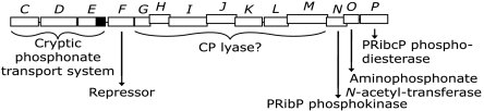
Organophosphonate utilization by Escherichia coli requires the 14 cistrons of the phnCDEFGHIJKLMNOP operon, of which the carbon-phosphorus lyase has been postulated to consist of the seven polypeptides specified by phnG to phnM. A 5,660-bp DNA fragment encompassing phnGHIJKLM is cloned, followed by expression in E. coli and purification of Phn-polypeptides. PhnG, PhnH, PhnI, PhnJ, and PhnK copurify as a protein complex by ion-exchange, size-exclusion, and affinity chromatography. The five polypeptides also comigrate in native-PAGE. Cross-linking of the purified protein complex reveals a close proximity of PhnG, PhnI, PhnJ, and PhnK, as these subunits disappear concomitant with the formation of large cross-linked protein complexes. Two molecular forms are identified, a major form of molecular mass of approximately 260 kDa, a minor form of approximately 640 kDa. The stoichiometry of the protein complex is suggested to be PhnG(4)H(2)I(2)J(2)K. Deletion of individual phn genes reveals that a strain harboring plasmid-borne phnGHIJ produces a protein complex consisting of PhnG, PhnH, PhnI, and PhnJ, whereas a strain harboring plasmid-borne phnGIJK produces a protein complex consisting of PhnG and PhnI. We conclude that phnGHIJK specify a soluble multisubunit protein complex essential for organophosphonate utilization.


Conflict of interest statement

The authors declare no conflict of interest.

Figures

Fig. 1.
Fig. 1.
Organization and gene products of the 14-cistron phnCDEFGHIJKLMNOP operon of E. coli K-12 (3, 10). Transcription occurs from left to right. Each phn cistron is shown as a rectangle above which the cistron is designated in italics. Displacement of a cistron from the previous cistron indicates translational coupling. The function of each gene product is shown below the gene organization. Assignment of phnG to phnM as coding for CP lyase is presumptive (see text for details). The phnE cistron of E. coli K-12 is cryptic due to the presence of an 8-bp duplication, which causes premature termination of translation. The untranslated part of the phnE cistron is shown as a black box. Abbreviations: PRibP, ribosyl 1,5-bisphosphate; PRibcP, 5-phosphoribosyl 1,2-cyclic phosphate.
Fig. 2.
Fig. 2.
Characterization of PhnGHIJK protein complex. Position of molecular mass standards are indicated by Roman numerals: i, 14.4; ii, 20.1; iii, 30; iv, 45; v, 66; and vi, 97 kDa. (A) SDS-PAGE of polypeptides specified by pMN1 (phnGHIJKLM). Lane 1, pellet obtained by sonication and centrifugation of cells of strain HO2735/pMN1 followed by dissolving in 6 M urea. The polypeptides of the bands labeled 1 to 8 were identified by MS/MS (Table 1); lane 2, molecular mass standard. (B) SDS-PAGE of selected fractions after elution of PhnGHIJK protein complex by size-exclusion chromatography in Sephacryl S300. Lanes 1 to 7: fraction 18, 20, 22, 24, 26, 28, and 30, respectively. Protein was concentrated by precipitation with trichloroacetic acid; lane 8: molecular mass standard. (C) SDS-PAGE of purified PhnGHIJK protein complex after size-exclusion chromatography and concentration. Lane 1, side fraction; lane 2, peak fraction; lane 3, molecular mass standard. Polypeptides of bands labeled 1 to 7 were identified by MS/MS (with Mowse scores given in parentheses): band 1, PhnI (748); band 2, PhnJ (1281); band 3, PhnJ (1175); band 4, PhnJ (1237); band 5, PhnK (1077); band 6, PhnH (520) and band 7, PhnG (871). (D) SDS-PAGE of purified PhnGHIJK protein complex. Lanes 1 and 2, protein complex purified by ion-exchange and size-exclusion chromatography from strain HO2735 (Δphn)/pMN1 (phnGHIJKLM) and HO2735/pHO571 (phnGHIJK), respectively; lane 3, histidine-tailed protein complex purified by Ni-chelate affinity chromatography from HO2735/pHO575 (phnGHIJK). Protein was concentrated by centrifugation through a Vivaspin column prior to analysis; lane 4, molecular mass standard. (E) Native-PAGE of purified PhnGHIJK protein complex. Polypeptides of the bands labeled 1 to 5 were identified by MS/MS (with Mowse scores given in parenthesis): band 1, PhnI (931), PhnJ (846), PhnK (602), PhnH (330), PhnG (623); band 2, PhnI (1089), PhnJ (1069), PhnK (783), PhnH (338), PhnG (532); band 3, PhnI (977), PhnJ (845), PhnK (698), PhnH (323), PhnG (668); band 4, PhnI (1010), PhnJ (921), PhnK (307), PhnH (286), PhnG (592); band 5, PhnI (1022), PhnJ (927), PhnH (287), PhnG (639). (F) Cross-linking of PhnGHIJK protein complex analyzed by SDS-PAGE followed by silver staining (27). Lanes 1 to 5, protein complex incubated with glutaraldehyde for 0, 1, 3.5, 7, and 24 h, respectively. Phn polypeptides are labeled I, J, K, H, and G; cross-linking products are labeled a, b, c, d, and e.
Fig. 3.
Fig. 3.
Cross-linking of PhnH analyzed by SDS-PAGE followed by silver staining. Lanes 1 to 4, PhnH incubated with glutaraldehyde for 0, 1, 3, and 8 h, respectively; lane 5, same as lane 4, but threefold more loaded; lane 6, molecular mass standard labeled as in Fig. 2; lane 7, untreated PhnH. PhnH monomer is labeled H, whereas cross-linking products are labeled a, b, and c.
Fig. 4.
Fig. 4.
Characterization of Phn protein complex specified by phn deletions. Molecular mass standard is labeled as in Fig. 2. (A) Synthesis of polypeptides specified by plasmids containing various phn deletions. After sonication of plasmid-harboring cells of strain HO2735 and centrifugation, pellet was dissolved in 6 M urea and analyzed by SDS-PAGE. Cells contained: lane 1, pFM31 (phnΔGHIJK); lane 2, pFM32 (phnGΔHIJK); lane 3, pFM33 (phnGHΔIJK); lane 4, pFM34 (phnGHIΔJK); lane 5, pHO572 (phnGHIJΔK); lane 6, pHO571 (phnGHIJK); lane 7, pUHE23-2; i.e., empty vector. The position of each Phn polypeptide is indicated in lane 6. (B) PhnGHIJ and PhnGI protein complexes of phnK and phnH deletions analyzed by SDS-PAGE. Lane 1, PhnGHIJK protein complex purified from strain HO2735 (Δphn)/pHO571 (phnGHIJK); lane 2, PhnGI protein complex purified from HO2735/pFM32 (phnGΔHIJK); lane 3, PhnGHIJ protein complex purified from HO2735/pHO572 (phnGHIJ); lane 4, molecular mass standard. (C) Cross-linking of PhnGI protein complex analyzed by SDS-PAGE and silver staining. Lane 1, molecular mass standard; lanes 2 to 7, protein complex incubated with glutaraldehyde for 0, 2, 5, 30, 60, and 180 min, respectively. Phn polypeptides are indicated by I and G, cross-linking product is indicated by a. (D) Native-PAGE of PhnGHIJ and PhnGI protein complexes. Lane 1, PhnGHIJK protein complex identical to that shown in Fig. 2E; lane 2, PhnGHIJ protein complex; lane 3, PhnGI protein complex. Polypeptides of bands labeled 1–5 were identified by MS/MS (with Mowse scores given in parenthesis): band 1, PhnI (935), PhnJ (971), PhnH (289), PhnG (610); band 2, PhnI (940), PhnJ (947), PhnH (273), PhnG (574); band 3, PhnI (928), PhnJ (807), PhnH (275), PhnG (649); band 4, elongation factor G (1681); band 5, PhnI (1105), PhnG (639).
Fig. 5.
Fig. 5.
Proposed reactions for mehylphosphonic acid catabolism by the CP lyase pathway. Compounds: a, 5-phosphoribosyl 1-methylphosphonate; b, 5-phosphoribosyl 1,2-cyclic phosphate; c, ribosyl 1,5-bisphosphate; d, a proposed, unknown intermediate. The existence of intermediates b and c, as well as a dephosphorylated derivative of intermediate a have been confirmed (15). Reactions: i, CP lyase, presumably identical the PhnGHIJK protein complex; ii, phnP specified phosphoribosyl cyclic phosphodiesterase. Reactions iii and iv have not yet been defined. Both of these may be exchange reactions involving an unidentified cosubstrate, x. Omitted from the scheme is the reaction catalyzed by phnN-specified ribosyl 1,5-bisphosphate phosphokinase, which may serve to initiate the reaction cycle by supplying ribosyl 1,5-bisphosphate (intermediate c).

References

    1. Hsieh YJ, Wanner BL. Global regulation by the seven-component Pi signaling system. Curr Opin Microbiol. 2010;13:198–203. - PMC - PubMed
    1. VanBogelen RA, Olson ER, Wanner BL, Neidhardt FC. Global analysis of proteins synthesized during phosphorus restriction in Escherichia coli. J Bacteriol. 1996;178:4344–4366. - PMC - PubMed
    1. Chen CM, Ye QZ, Zhu ZM, Wanner BL, Walsh CT. Molecular biology of carbon-phosphorus bond cleavage. Cloning and sequencing of the phn (psiD) genes involved in alkylphosphonate uptake and C-P lyase activity in Escherichia coli B. J Biol Chem. 1990;265:4461–4471. - PubMed
    1. Metcalf WW, Wanner BL. Mutational analysis of an Escherichia coli fourteen-gene operon for phosphonate degradation, using Tn phoA' elements. J Bacteriol. 1993;175:3430–3442. - PMC - PubMed
    1. Rizk SS, Cuneo MJ, Hellinga HW. Identification of cognate ligands for the Escherichia coli phn D protein product and engineering of a reagentless fluorescent biosensor for phosphonates. Protein Sci. 2006;15:1745–1751. - PMC - PubMed

Publication types

MeSH terms

LinkOut - more resources