. 2011 Jun 26;43(8):811-4.
doi: 10.1038/ng.864.

Dynamic CpG island methylation landscape in oocytes and preimplantation embryos

Affiliations

Dynamic CpG island methylation landscape in oocytes and preimplantation embryos

Sébastien A Smallwood et al. Nat Genet. .

Abstract

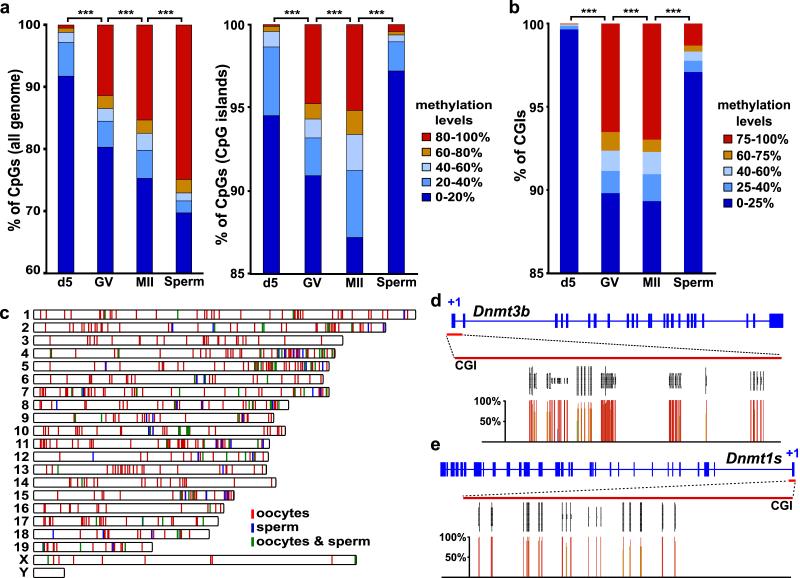
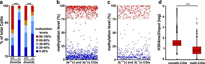
Elucidating how and to what extent CpG islands (CGIs) are methylated in germ cells is essential to understand genomic imprinting and epigenetic reprogramming. Here we present, to our knowledge, the first integrated epigenomic analysis of mammalian oocytes, identifying over a thousand CGIs methylated in mature oocytes. We show that these CGIs depend on DNMT3A and DNMT3L but are not distinct at the sequence level, including in CpG periodicity. They are preferentially located within active transcription units and are relatively depleted in H3K4me3, supporting a general transcription-dependent mechanism of methylation. Very few methylated CGIs are fully protected from post-fertilization reprogramming but, notably, the majority show incomplete demethylation in embryonic day (E) 3.5 blastocysts. Our study shows that CGI methylation in gametes is not entirely related to genomic imprinting but is a strong factor in determining methylation status in preimplantation embryos, suggesting a need to reassess mechanisms of post-fertilization demethylation.


Figures

Figure 1
Figure 1. DNA methylation landscape in oocytes and sperm determined by RRBS
a-b, Distribution of CpG methylation levels across the genome (a, left), within CGIs (a, right), and CGI methylation (b), in immature (d5), mature (GV and MII) oocytes and sperm (***: p<0.001, χ2 test). The number of CpGs and CGIs analysed is indicated in Suppl. Fig 1b. c, Chromosome distribution of the 1062 CGIs methylated in oocytes and 185 CGIs methylated in sperm (100 CGIs in both). d, CpG methylation levels (percentage of all cytosines called methylated) at the Dnmt3b and Dnmt1s promoter CGIs in GV oocytes. The grey vertical lines represent the sequencing read depth of individual cytosines; below, percentage methylation of the corresponding CpGs is represented by coloured vertical lines.
Figure 2
Figure 2. Mechanism of DNA methylation establishment in oocytes
a, Distribution of CpG methylation levels across the genome in Dnmt3a−/− and Dnmt3L−/− oocytes and their wild-type counterparts (+/+); the number of CpGs analysed is indicated in Suppl. Fig 1b (***: p<0.001, χ2 test). b-c, Methylation levels of CGIs in Dnmt3a−/− and Dnmt3L−/− oocytes; only those CGIs for which methylation was ≥75% in the corresponding wild-type oocytes are displayed. d, Overall correlation between H3K4me3 enrichment determined in d15 oocytes by ChIP-seq and methylation status of CGIs (all CGIs irrespective of genomic location; ***: p<0.001, Mann-Whitney U test).
Figure 3
Figure 3. Biological significance and fate of CGI methylation in oocytes
a, mRNA expression levels in d10 and GV oocytes of the genes associated with methylated CGIs, either promoter (red, n=410) or intragenic (blue, n=555). b, Methylation levels in blastocysts of the CGIs identified as methylated in mature oocytes; twelve known germline DMRs with informative coverage are displayed in red (range 45.2%-58.7%). c, Range of methylation in blastocysts of the CGIs methylated specifically in oocytes (n=803) or sperm (n=51), methylated in both oocytes and sperm (n=86) and unmethylated in gametes (n=11512). d, Bisulphite sequencing in GV oocytes, sperm and C57BL/6JxCAST/Ei hybrid E3.5 blastocysts of the Syt2 CGI. Bisulphite sequence profiles from the maternal (mat) and paternal (pat) alleles in blastocysts were discriminated by polymorphisms between C57BL/6J and CAST/Ei. Open circles represent unmethylated CpGs and filled circles methylated CpGs.

References

    1. Bartolomei MS. Genomic imprinting: employing and avoiding epigenetic processes. Genes & Development. 2009;23:2124–2133. - PMC - PubMed
    1. Morgan HD, Santos F, Green K, Dean W, Reik W. Epigenetic reprogramming in mammals. Human Molecular Genetics. 2005;14:R47–R58. - PubMed
    1. Sasaki H, Matsui Y. Epigenetic events in mammalian germ-cell development: reprogramming and beyond. Nat Rev Genet. 2008;9:129–140. - PubMed
    1. Bourc’his D, Xu G-L, Lin C-S, Bollman B, Bestor TH. Dnmt3L and the Establishment of Maternal Genomic Imprints. Science. 2001;294:2536–2539. - PubMed
    1. Kaneda M, et al. Essential role for de novo DNA methyltransferase Dnmt3a in paternal and maternal imprinting. Nature. 2004;429:900–903. - PubMed

Methods References

    1. Hemberger M, Dean W, Reik W. Epigenetic dynamics of stem cells and cell lineage commitment: digging Waddington’s canal. Nat Rev Mol Cell Biol. 2009;10:526–537. - PubMed
    1. Krueger F, Andrews SR. Bismark: A flexible aligner and methylation caller for Bisulfite-Seq applications. Bioinformatics. 2011 doi:10.1093/bioinformatics/btr167. - PMC - PubMed
    1. Dahl JA, Collas P. A rapid micro chromatin immunoprecipitation assay (ChIP) Nat. Protocols. 2008;3:1032–1045. - PubMed
    1. Creyghton MP, et al. Histone H3K27ac separates active from poised enhancers and predicts developmental state. Proceedings of the National Academy of Sciences. 2010;107:21931–21936. - PMC - PubMed
    1. Tomizawa S, et al. Dynamic stage-specific changes in imprinted differentially methylated regions during early mammalian development and prevalence of non-CpG methylation in oocytes. Development. 2011;138:811–820. - PMC - PubMed

Publication types

MeSH terms