. 2011 Jun 26;14(8):1009-16.
doi: 10.1038/nn.2855.

Axin2 as regulatory and therapeutic target in newborn brain injury and remyelination

Affiliations

Axin2 as regulatory and therapeutic target in newborn brain injury and remyelination

Stephen P J Fancy et al. Nat Neurosci. .

Abstract

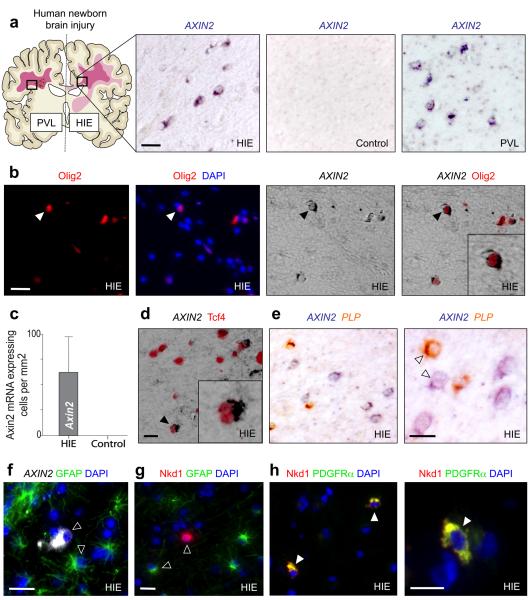
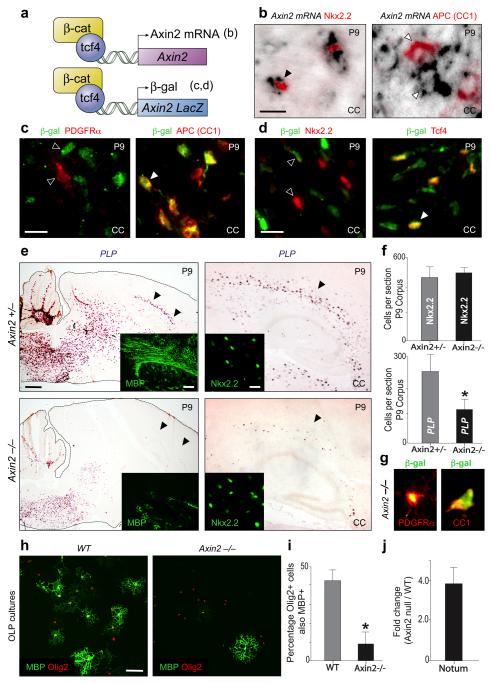
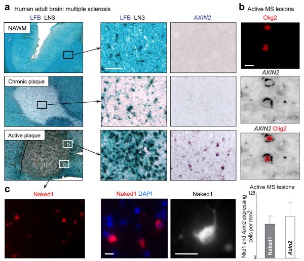
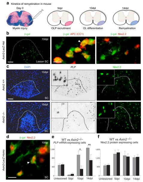
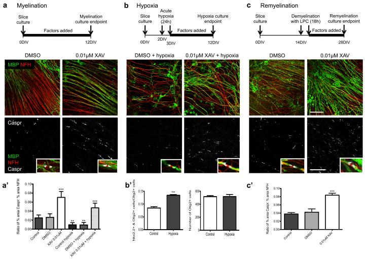
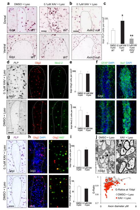
Permanent damage to white matter tracts, comprising axons and myelinating oligodendrocytes, is an important component of brain injuries of the newborn that cause cerebral palsy and cognitive disabilities, as well as multiple sclerosis in adults. However, regulatory factors relevant in human developmental myelin disorders and in myelin regeneration are unclear. We found that AXIN2 was expressed in immature oligodendrocyte progenitor cells (OLPs) in white matter lesions of human newborns with neonatal hypoxic-ischemic and gliotic brain damage, as well as in active multiple sclerosis lesions in adults. Axin2 is a target of Wnt transcriptional activation that negatively feeds back on the pathway, promoting β-catenin degradation. We found that Axin2 function was essential for normal kinetics of remyelination. The small molecule inhibitor XAV939, which targets the enzymatic activity of tankyrase, acted to stabilize Axin2 levels in OLPs from brain and spinal cord and accelerated their differentiation and myelination after hypoxic and demyelinating injury. Together, these findings indicate that Axin2 is an essential regulator of remyelination and that it might serve as a pharmacological checkpoint in this process.


Figures

Figure 1
Figure 1. AXIN2 mRNA expression identifies Wnt pathway activation in immature oligodendrocytes within neonatal human white matter injury
(a) AXIN2 mRNA is expressed in areas of affected subcortical white matter in human pediatric cases of Hypoxic ischemic encephalopathy (HIE) and also adjacent to the cystic core of a periventricular lesion of Periventricular Leukomalacia (PVL), but is not seen in age-matched controls. (b) AXIN2 mRNA is expressed solely in Olig2-positive cells within affected white matter in neonatal HIE. (c) Quantification of the number of AXIN2 mRNA expressing cells in areas of white matter from HIE cases and control subjects. (d) In the HIE cases, AXIN2 mRNA was expressed in a subset of the Tcf4 positive cells, which (e) segregated from the mature OL marker PLP in situ. (f) AXIN2 mRNA expression in HIE separates completely from cells expressing GFAP proteins. (g)(h) The independent Wnt activated target Naked1 (Nkd1) is upregulated in neonatal HIE; proteins are expressed cytoplasmically in OLP expressing PDGFRα(h), but separate completely from cells expressing GFAP proteins (g). Scale bar in all = 10μm.
Figure 2
Figure 2. Axin2 functions as a negative regulator of Wnt signaling in OLPs and promotes differentiation
(a) Schematic for Axin2 mRNA expression in WT mice and β-catenin/Tcf4-driven reporter expression in heterozygous Axin2-lacZ mice. (b) During developmental myelination of corpus callosum (CC), Axin2 mRNA is confined to earlier stage immature OLP that express Nkx2.2 protein, but separates from mature OL expressing APC (CC1) protein. (c, d) Using Axin2-lacZ heterozygous mice during developmental myelination, β-galactosidase proteins were first detectable at a stage of the OL lineage in P9 corpus callosum that express mature marker APC (CC1), but separated from OLP markers PDGFRα (c) and Nkx2.2 (d), suggesting that kinetics of reporter expression lags behind that of Axin2 mRNA during developmental myelination. Tcf4 expression was observed in only a subset of β-gal+ cells (d). Scale bar in b, c, d = 10μm. (e) Axin2-lacZ homozygous null animals demonstrated a significant reduction in mature OL expressing PLP mRNA (and MBP protein, inset) at P9 during developmental myelination of the corpus callosum, despite normal numbers of OLP expressing Nkx2.2 protein. Scale bar = 600μm (100μm in MBP inset; 15μm in Nkx2.2 inset). (f) Quantification during developmental myelination of the reduced (t test p=0.007) mature PLP-expressing OL in P9 Axin2 null corpus callosum (black bars) compared to heterozygous littermates (grey bars), despite normal numbers of Nkx2.2-expressing OLP. (g) Whilst β-gal expression separates from OLP markers in Axin2-lacZ heterozygotes (Fig. 2c, d), overlap of β-gal with PDGFRα is observed in Axin2−/− animals as OLP are delayed in their differentiation. (h, i) There is a marked impairment of Axin2−/− OL differentiation in vitro as evidenced by a significant (t test p < 0.005) reduction in the percentage of Olig2+ cells expressing MBP at 60 hours post differentiation in culture. Scale bar in h = 25μm. (j) There is a strong activation of the independent Wnt target Notum mRNA in Axin2−/− OLP cultures compared to WT, demonstrating that loss of Axin2 leads to an increase in Wnt pathway activity within these cultures.
Figure 3
Figure 3. AXIN2 is expressed in OLP in active MS lesions
(a) MS lesions were characterized according to Lock et al. Nat. Med. 8: 500-508 (2002), using Luxol Fast Blue (LFB) to assess demyelination and LN3 immunohistochemistry to assess inflammatory cell activity. AXIN2 mRNA is expressed in cells in active multiple sclerosis (MS) lesions, but not within normal appearing white matter (NAWM) or chronic plaques. Scale bar in a = 100μm. (b) AXIN2 mRNA expression within active MS lesions is specific to OL lineage, where it co-localizes with Olig2 proteins. (c) The independent Wnt activated target Naked1 (Nkd1) is also upregulated in active MS lesions (Suppl. Fig. 5). Nkd1 proteins are expressed in the cytoplasm of OLP with characteristic simple bipolar morphology. The density of cells expressing Nkd1 proteins in active MS lesions is similar to the density of cells expressing AXIN2 mRNAs. Scale bar in b, c = 10μm.
Figure 4
Figure 4. Axin2 function is essential for timely myelin repair
(a) Schematic showing use of adult murine lysolecithin injury for investigating remyelination kinetics. Such lesions in spinal cord show OLP recruitment (5 days post lesion (dpl)), differentiation (10 dpl) and myelination (14 dpl) with stereotyped timing in young adult animals, allowing precise assessment of remyelination kinetics. (b) Following demyelination of adult Axin2-lacZ heterozygote animals with lysolecithin in the spinal cord, β-gal proteins are observed within the lesion in mature OL (co-expressing APC) at 10 days post-lesioning (dpl), but separate from OLP marker Nkx2.2. Scale bar in b low power lesion = 80μm; in high power = 10μm. (c) Axin2−/− mice showed delayed repair compared to WT littermates. This was due to a reduced (t test p=0.03 at 10dpl, p=0.02 at 14dpl) OLP differentiation into mature OL expressing PLP mRNA in lesions at 10dpl, despite a normal recruitment of Nkx2.2-expressing OLP into lesions. Scale bar in c = 80μm. (d) OLP with dysregulated Wnt signaling in Axin2 null (Axin2-lacZ homozygote) mice at 10dpl following demyelination showed abnormal kinetics of mature marker acquisition, β-gal proteins co-localizing with OLP marker Nkx2.2, in contrast to Axin2-lacZ heterozygote mice above (b). Scale bar in d = 10μm. (e, f) Quantification of (e) mature OL (expressing PLP mRNA) and (f) OLP (expressing Nkx2.2 protein) in unlesioned and 5dpl, 10dpl and 14dpl demyelinated spinal cord of Axin2 null animals (black bars) and WT littermates (grey bars).
Figure 5
Figure 5. Axin protein stabilization through small molecule Tankyrase inhibition promotes OLP differentiation in vitro
(a) Tankyrase proteins are detected in the cytoplasm of mouse Olig2+ cells, separating from the PDGFRα+ (OLP) stage, at P9 during developmental myelination in the spinal cord (SC). (b) The onset of Tankyrase expression coincided with expression of β-galactosidase in Axin2-lacZ heterozygous reporter mice, at approximately the CC1+ stage of OL development. (c) Tankyrase is also expressed within OL lineage at 10dpl following demyelination with lysolecithin in the adult spinal cord white matter of the mouse, co-localizing with mature oligodendrocyte marker cytoplasmic Olig1 (inset). Scale bar in a, b, c = 10μm. (d) XAV939 treatment of mouse OLPs in vitro with 0.01 or 0.1 μM for 24 hours produced marked increases in the protein levels of both Axin2 and Axin1 versus vehicle controls, leading to an increased activity of the β-catenin degradation complex, evidenced by an increase in degraded phospho-β-catenin protein levels. (e) At 0.01 μM, XAV939 treatment effectively inhibited the Wnt pathway in OLP in vitro after 96 hours, evidenced by a reduction in mRNA levels of the Wnt target Axin2. (f, h) In vitro OLP differentiation assays demonstrate a significant increase in the proportion of Olig2+ cells expressing mature OL marker MBP in the presence of either 0.1 or 0.01 μM XAV939 at both 48 and 60 hours post-differentiation compared to vehicle control treatment. Scale bar in f = 40μm. (g) At 60 hours post differentiation of OLP in vitro in the presence of 0.01 μM XAV939 there is a significant increase in the quantity of MBP protein harvested from the culture compared to vehicle control. (i) Tankyrase protein is expressed within OL lineage in human pediatric HIE white matter injury, where it is co-expressed within Olig2 positive cells, with NOGO-A+ and cytoplasmic Olig1+ cells, but separated from Iba1+ macrophages/microglia and GFAP+ astrocytes. Scale bar in i = 10μm.
Figure 6
Figure 6. XAV939 treatment increases myelination, myelination following hypoxia, and remyelination in ex vivo mouse cerebellar slice cultures
(a) XAV939 promotes developmental myelination. Axons (NFH) are shown in red, myelin (MBP) shown in green, and paranodes showing compacted myelin sheaths (Caspr) are shown in white. (a’) Quantification of myelination using a ratio of percent area stained for Caspr to percent area stained for NFH. Values shown are mean + SD, and the data were analyzed by one-way ANOVA with Dunnett’s multiple comparison test, and significant differences (**p<0.01, ***p<0.001) are shown. Three independent experiments were conducted. (b) XAV939 promotes myelination and recovery following acute hypoxic insult. Axons (NFH) are shown in red, myelin (MBP) shown in green, and paranodes showing compacted myelin sheaths (Caspr) are shown in white. (b’) Acute hypoxia impedes differentiation of OLP in cerebellar slice cultures. Values shown are mean + SD, and the data were analyzed by unpaired t-test, and the significant difference (***p<0.001) is shown. (c) XAV939 promotes remyelination following demyelination by lysolecithin. Axons (NFH) are shown in red, myelin (MBP) shown in green, and paranodes showing compacted myelin sheaths (Caspr) are shown in white. Scale bar: 50μm MBP/NFH and 25μm in Caspr panels. (c’) Quantification of remyelination using a ratio of percent area stained for Caspr to percent area stained for NFH. Values shown are mean +/− SD, and the data were analyzed by one-way ANOVA with Dunnett’s multiple comparison test, and the significant difference (***p<0.001) is shown. Three independent experiments were conducted per condition tested and 5-10 separate slices were counted per experiment.
Figure 7
Figure 7. XAV939 treatment dramatically accelerates OLP differentiation and myelin regeneration during remyelination in vivo
(a) Injection of 0.1μM XAV939 into demyelinated lesions in young adult mouse spinal cord at the time of lysolecithin injection produces significant increases in the appearance of PLP mRNA expressing mature OL at 6dpl in dorsal or ventral funicular lesions compared to vehicle treated controls. (b) XAV939 effects are Axin2-dependent, as Axin2 null mice show significantly less OLP differentiation at 6dpl following XAV+lysolecithin treatment versus controls. (c) Quantification of (a) and (b) above at 6dpl during remyelination showing number of PLP mRNA expressing cells per mm2 after respective treatments. (d, e) Whilst total Olig2+ OL lineage numbers were similar at 6dpl in XAV939 treated lesions and controls, XAV939 produced a shift in the proportion of mature PLP+ OL versus immature Nkx2.2+ OLP, with a significant increase (t test p=0.002, n=4) in mature OL. (f) XAV939 treatment did not affect the astrocyte (GFAP) or macrophage (Iba1) infiltration into lesions at 6dpl. (g) XAV939 treatment in lesions was not cytoprotective to existing mature OL, as there were no PLP-expressing cells at 3dpl within lesions of either group. (h, i) The OLP recruitment into lesions and proliferation phase at 3dpl were unaffected by XAV939 treatment, evidenced by similar numbers of Olig2+ cells in the lesion, a similar proportion of which were Ki67+. Scale bar in a, b, d, f, g, h = 50μm. (j) The accelerated OLP differentiation produced by XAV939 treatment in lesions leads to an accelerated myelin regeneration at 10dpl, evidenced by significant increases in the thickness of restored myelin sheaths. G ratios were significantly different (t test p<0.0001) between control group (G ratio mean= 0.94, SEM 0.003) and XAV939-treated (G ratio mean= 0.90, SEM 0.004). Scale bar in j = 2μm.

Comment in

References

    1. Back SA, et al. Selective vulnerability of late oligodendrocyte progenitors to hypoxia-ischemia. J Neurosci. 2002;22:455–463. - PMC - PubMed
    1. Khwaja O, Volpe JJ. Pathogenesis of cerebral white matter injury of prematurity. Arch Dis Child Fetal Neonatal Ed. 2008;93:F153–161. - PMC - PubMed
    1. Woodward LJ, Anderson PJ, Austin NC, Howard K, Inder TE. Neonatal MRI to predict neurodevelopmental outcomes in preterm infants. N Engl J Med. 2006;355:685–694. - PubMed
    1. Compston A, Coles A. Multiple sclerosis. Lancet. 2008;372:1502–1517. - PubMed
    1. Chang A, Tourtellotte WW, Rudick R, Trapp BD. Premyelinating oligodendrocytes in chronic lesions of multiple sclerosis. N Engl J Med. 2002;346:165–173. - PubMed

Publication types

MeSH terms

Associated data