. 2011 Jun 26;475(7355):217-21.
doi: 10.1038/nature10177.

In vivo genome editing restores haemostasis in a mouse model of haemophilia

Affiliations

In vivo genome editing restores haemostasis in a mouse model of haemophilia

Hojun Li et al. Nature. .

Abstract

Editing of the human genome to correct disease-causing mutations is a promising approach for the treatment of genetic disorders. Genome editing improves on simple gene-replacement strategies by effecting in situ correction of a mutant gene, thus restoring normal gene function under the control of endogenous regulatory elements and reducing risks associated with random insertion into the genome. Gene-specific targeting has historically been limited to mouse embryonic stem cells. The development of zinc finger nucleases (ZFNs) has permitted efficient genome editing in transformed and primary cells that were previously thought to be intractable to such genetic manipulation. In vitro, ZFNs have been shown to promote efficient genome editing via homology-directed repair by inducing a site-specific double-strand break (DSB) at a target locus, but it is unclear whether ZFNs can induce DSBs and stimulate genome editing at a clinically meaningful level in vivo. Here we show that ZFNs are able to induce DSBs efficiently when delivered directly to mouse liver and that, when co-delivered with an appropriately designed gene-targeting vector, they can stimulate gene replacement through both homology-directed and homology-independent targeted gene insertion at the ZFN-specified locus. The level of gene targeting achieved was sufficient to correct the prolonged clotting times in a mouse model of haemophilia B, and remained persistent after induced liver regeneration. Thus, ZFN-driven gene correction can be achieved in vivo, raising the possibility of genome editing as a viable strategy for the treatment of genetic disease.


Figures

Figure 1
Figure 1. F9 ZFNs cleave human F9 intron 1 and induce homology-directed repair in vitro
a, F9 ZFNs target intron 1 of the human F9 gene, allowing for homology-directed repair upstream of 95% of F9 mutations. b, ZFN expression constructs (400 ng) were transfected (right lane) or not (left lane) into K562 cells and genomic DNA was harvested 3 days post-transfection. The Cel-I assay was used to determine the frequency of ZFN-induced insertions and deletions (indels) in both samples, indicated as the % Indels at the base of the right lane. ZFN (FLAG-tagged) expression is confirmed by α-FLAG immunoblotting, and αNFκB-p65 serves as a loading control. c, Schematic of RFLP assay detailing ZFN-mediated targeting of a NheI restriction site tag to the human F9 gene. d, Co-transfection of 400 ng of ZFN expression plasmid with increasing amounts of NheI donor plasmid (0–4 µg) results in increasing levels of homology-directed repair (HDR) at days 3 and 10 post-transfection, whereas transfection of the NheI donor alone (4 µg) does not result in detectable HDR. Black arrows denote NheI-sensitive cleavage products resulting from HDR. PCR performed using 32P-labeled nucleotides, followed by PAGE and band intensity quantification by autoradiography. Lanes with no quantification had no detectable HDR.
Figure 2
Figure 2. AAV8-mediated delivery of F9 ZFNs to hF9mut mouse liver results in cleavage of hF9mut intron 1 in vivo
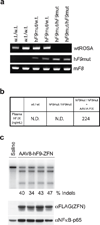
a, PCR genotyping of parental strain (w.t./w.t.), mouse heterozygous for human F9 mutant (hF9mut) construct knocked into the ROSA 26 locus (hF9mut/w.t.), and mouse homozygous for hF9mut knocked into the ROSA 26 locus (hF9mut/hF9mut). The murine Factor VIII (mF8) PCR product indicates no inhibition of PCR. b, Plasma hF.IX levels, assayed by hF.IX ELISA, in w.t. mice, homozygous hF9mut mice, and hF9mut mice injected with a viral vector expressing hF.IX (1e9 v.g. AAV-hF.IX injected via tail vein). N.D.= none detected. c, Tail vein injection of 1e11 v.g. AAV8-ZFN expression vector into hF9mut mice results in cleavage of intron 1. The Cel-I assay was performed on liver DNA isolated at day 7 post-injection to determine the frequency of ZFN-induced insertions and deletions (indels), indicated as the % Indels at the base of each lane, resulting from cleavage of the hF9mut intron. Lane with no quantification had no detectable cleavage products. Each lane represents an individual mouse. ZFN (FLAG-tagged) expression confirmed by α-FLAG immunoblotting of whole liver lysate.
Figure 3
Figure 3. F9 ZFNs promote AAV-mediated targeting of wild-type F9 exons 2–8 to hF9mut intron 1 in vivo
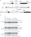
a, The hF9mut gene mutation (truncation of exons 7&8) can be bypassed by targeted integration of hF9 exons 2–8 into intron 1. Targeted and untargeted hF9mut alleles can be differentiated through PCR using primers P1, P2, and P3. Location of methionine start codon, and premature stop mutation indicated by arrows. The left arm of homology spans from the beginning of exon 1 to the ZFN target site. (Deletion of exon 1 from left homology arm does not alter results, Supplementary Figure 13). The right arm of homology spans intronic sequence 3’ of the ZFN target site. b, PCR analysis with primer pairs P1/P3 (upper panel) and P1/P2 (middle panel) demonstrating successful gene targeting by HDR upon I.P. co-injection of 5e10 v.g. AAV8-ZFN and 2.5e11 v.g. AAV8-Donor in hF9mut/HB mice at day 2 of life (n=5), but not with injection of 5e10 v.g. AAV8-ZFN alone (n=1), or co-injection of 5e10 v.g. AAV8-Mock and 2.5e11 v.g. AAV8-Donor (n=5). Mock vector replaces F9 ZFN coding sequences with Renilla Luciferase. PCR was performed using 32P-labeled nucleotides, followed by PAGE and product band intensity quantification by autoradiography to evaluate targeting frequency. Targeting frequencies are rounded down to the nearest whole number. Lower panel. IP injection of AAV8-ZFN expression vector into hF9mut mice results in cleavage of intron 1. The Cel-I assay was performed on liver DNA to determine the frequency of ZFN-induced insertions and deletions (indels), indicated as the % Indels at the base of each lane, resulting from cleavage of the hF9mut intron. Lanes with no quantification had no detectable HDR or indels. Each lane represents an individual mouse.
Figure 4
Figure 4. In vivo hF9mut gene correction results in stable circulating F.IX
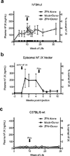
a, Plasma hF.IX levels in hF9mut mice following I.P. injection at day 2 of life with either 5e10 v.g. AAV8-ZFN alone (n=7), 5e10 v.g. AAV8-ZFN and 2.5e11 v.g. AAV8-Donor (n=7), or 5e10 v.g. AAV8-Mock and 2.5e11 v.g. AAV8-Donor (n=6). Partial hepatectomy (PHx) performed at time indicated by arrow. Plasma hF.IX assayed by ELISA. Error bars denote standard error. b, Plasma hF.IX levels in wild-type mice (n=3) following tail vein injection of 1e9 v.g. AAV-hF.IX (predominantly episomal) with subsequent PHx. Plasma hF.IX assayed by ELISA. Error bars denote standard error. c, Plasma hF.IX levels in w.t. C57BL/6J mice following I.P. injection at day 2 of life with either 5e10 v.g. AAV8-ZFN alone (n=8 pre-PHx, n=4 post-PHx), 5e10 v.g. AAV8-ZFN and 2.5e11 v.g. AAV8-Donor (n=9 pre-PHx, n=5 post-PHx), or 5e10 v.g. AAV8-Mock and 2.5e11 v.g. AAV8-Donor (n=6 pre-PHx, n=5 post-PHx). Plasma hF.IX assayed by ELISA. Error bars denote standard error.
Figure 5
Figure 5. Hepatic hF9mut gene correction results in phenotypic correction of hemophilia B
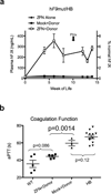
a, Plasma hF.IX levels in hF9mut/HB mice following I.P. injection at day 2 of life with either 5e10 v.g. AAV8-ZFN alone (n=10 pre-PHx, n=1 post-PHx), 5e10 v.g. AAV8-ZFN and 2.5e11 v.g. AAV8-Donor (n=9 pre-PHx, n=5 post-PHx), or 5e10 v.g. AAV8-Mock and 2.5e11 v.g. AAV8-Donor (n=9 pre-PHx, n=3 post-PHx). Plasma hF.IX assayed by ELISA. Error bars denote standard error. b, Test of clot formation by aPTT at week 14 of life of mice receiving I.P. injection at day 2 of life with 5e10 v.g. AAV8-ZFN and 2.5e11 v.g. AAV8-Donor (n=5), or 5e10 v.g. AAV8-Mock and 2.5e11 v.g. AAV8-Donor (n=3). aPTTs of wild-type (WT, n=5) and hemophilia B (HB, n=12) mice are shown for comparison (p-values from 2-tailed Student’s T-test of WT vs. ZFN+Donor, ZFN+Donor vs. Mock+Donor, and Mock+Donor vs. HB). Error bars denote standard error.

Similar articles

Cited by

References

    1. Urnov FD, Rebar EJ, Holmes MC, Zhang HS, Gregory PD. Genome editing with engineered zinc finger nucleases. Nat Rev Genet. 2010;11(9):636–646. - PubMed
    1. Urnov FD, et al. Highly efficient endogenous human gene correction using designed zinc-finger nucleases. Nature. 2005;435(7042):646–651. - PubMed
    1. Porteus MH, Baltimore D. Chimeric nucleases stimulate gene targeting in human cells. Science. 2003;300(5620):763. - PubMed
    1. Bibikova M, et al. Stimulation of homologous recombination through targeted cleavage by chimeric nucleases. Mol Cell Biol. 2001;21(1):289–297. - PMC - PubMed
    1. Cartier N, et al. Hematopoietic stem cell gene therapy with a lentiviral vector in X-linked adrenoleukodystrophy. Science. 2009;326(5954):818–823. - PubMed

Publication types

MeSH terms