. 2012 Jan 26;31(4):527-34.
doi: 10.1038/onc.2011.252. Epub 2011 Jun 27.

RUNX3 acts as a tumor suppressor in breast cancer by targeting estrogen receptor α

Affiliations

RUNX3 acts as a tumor suppressor in breast cancer by targeting estrogen receptor α

B Huang et al. Oncogene. .

Abstract

Transcription factor RUNX3 is inactivated in a number of malignancies, including breast cancer, and is suggested to function as a tumor suppressor. How RUNX3 functions as a tumor suppressor in breast cancer remains undefined. Here, we show that about 20% of female Runx3(+/-) mice spontaneously developed ductal carcinoma at an average age of 14.5 months. Additionally, RUNX3 inhibits the estrogen-dependent proliferation and transformation potential of ERα-positive MCF-7 breast cancer cells in liquid culture and in soft agar and suppresses the tumorigenicity of MCF-7 cells in severe combined immunodeficiency mice. Furthermore, RUNX3 inhibits ERα-dependent transactivation by reducing the stability of ERα. Consistent with its ability to regulate the levels of ERα, expression of RUNX3 inversely correlates with the expression of ERα in breast cancer cell lines, human breast cancer tissues and Runx3(+/-) mouse mammary tumors. By destabilizing ERα, RUNX3 acts as a novel tumor suppressor in breast cancer.


Conflict of interest statement

Conflict of interest: The authors declare no conflict of interest

Figures

Figure 1
Figure 1
Histopathology of mammary tumors from female Runx3+/− mice. (a) Incidence of mammary tumors in Runx3+/− mice. The generation of Runx3+/− BALB/c mice was described previously (Ito et al., 2008). The mice were maintained in pathogen-free conditions and monitored daily. All procedures involving animals were performed in accordance with the guidelines of Nagasaki University Animal Care and Use Committee. (b) Mammary gland specimens from wild-type and Runx3+/− BALB/c mice were fixed with 4% paraformaldehyde in PBS, embedded in paraffin, and sectioned at 4 μm. Sections were stained with hematoxylin and eosin (HE). HE sections were evaluated by a pathologist (M.S.-T.). Representative HE staining and RUNX3-immunohistochemical staining of a normal mammary gland from a WT mouse (left 4 panels, A-D) and an adenocarcinoma of mammary gland from a Runx3+/− mouse (right 4 panels, E-H) are shown. Boxed regions are enlarged to the right of each image. Scale bars: 100 (lower magnification) and 50 (higher magnification) μm. (c) (d) Immunohistochemical staining of Ki-67 (c) and ERα (d) in serial sections from (b). For the detection of RUNX3, Ki-67 and ERα protein expression, rehydrated specimens were treated for 40 min at 96°C with an antigen retrieval solution (S1700; DAKO). The specimens were incubated with antibodies against RUNX3 (R3-3F12 as previously described (Ito et al., 2009)), Ki-67 (M7249; DAKO) or ERα (SC-542; Santa Cruz Biotech). The EnVision+ system (DAKO) was used for visualization of signals in (b), (c) and (d).
Figure 2
Figure 2
RUNX3 suppresses the proliferation and tumorigenesis of MCF-7 cells. (a) Both vector-MCF-7 and RUNX3-MCF-7 cells were plated in 96-well plates in 0.1 ml phenol red-free MEM with double-stripped calf serum and cultured for three days. Cells were then stimulated with 100 pM E2 and cultured for up to 4 days. Cell proliferation was measured by OD 490 nm using the CellTiter 96® AQueous One Solution Cell Proliferation Assay (MTS) (Promega) and converted to cell number according to the standard curve. Data represent the average of three independent experiments +/−SD. (b) A total of 5000 cells from vector-MCF-7 or RUNX3-MCF-7 were suspended in DMEM containing 0.35% SELECT Agar® (Invitrogen) and then plated in 6-well plates coated with an initial underlay of 0.5% SELECT Agar® (Invitrogen) in culture medium. Colony growth was scored after 14 days of cell incubation at the normal condition. Representative photographs were taken at day 1 and day 14 to show colonies. All the colony formation assays presented in this study were repeated in at least 3 independent experiments. (c) Six-week old female severe combined immunodeficient (SCID) mice (C.B-17/IcrCrl-scidBR) (Charles River Laboratories) were subcutaneously implanted with a slow release pellet of 25 μg E2. Four days later, the mice were injected in the inguinal mammary fat pads with vector-MCF-7 or RUNX3-MCF-7 stable cell lines (5×10⁶). The recipient mice were monitored daily by palpation, and killed and dissected for tumor evaluation 30 days post-injection. Pictures were taken at day 30 after cell inoculation. Mouse injection was performed essentially as previously described (Yan et al., 2009). All experiments involving mice were approved by the Institutional Animal Care and Use Committee (IACUC). (d) Summary of the average weight of tumors from (c). Data represent mean ± SEM (n = 3). * P < 0.05 (paired t test).
Figure 3
Figure 3
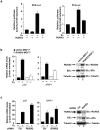
RUNX3 represses transcriptional activity of ERα. (a) MCF-7 cells were cultured in phenol red-free MEM with 10% double-stripped calf serum for three days followed by transfection with 4×ERE- (left panel) or PI-9-luciferase (right panel) reporter and RUNX3 plasmid DNA. Twenty-four hours after transfection, cells were stimulated with vehicle or E2 (100 nM) for 24 hr. Luciferase activity was measured and expressed as fold induction after normalization with Renilla luciferase. Results represent the average of three independent experiments +/− SD. (b) MCF-7 cells stably expressing control vector or RUNX3 were stimulated with E2 (100 nM) for 24 hr. The expression levels of pS2 and NRIP1 mRNA were measured with quantitative RT-PCR using Qiagen SYBR green PCR kit by 7300 real-time PCR system (ABI). The primers for pS2, NRIP1, and actin were purchased from Qiagen. Expression levels of RUNX3 and ERα protein in the MCF-7 stable cell lines are shown in the right panels. Intensity of each band was measured and analyzed by ImageJ 1.40g. Mean of three measurements is indicated beneath each band image. Data are representative of three independent experiments. (c) MDA-MB-361 cells were transfected with control or RUNX3 siRNA using Lipofectamine 2000 (Invitrogen). Sixty hours post-transfection, cells were left untreated or stimulated with E2 (100 nM) for 24 hr. The expression levels of pS2 and NRIP1 mRNA were measured by quantitative real-time PCR as in (b). Efficiency of siRNA knock-down as well as ERα expression levels are shown in the right panels. The predesigned siRNA targeting RUNX3 was purchased from Santa Cruz Biotech (SC-37679).
Figure 4
Figure 4
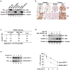
Expression of RUNX3 inversely correlated with ERα expression. (a) Expression levels of RUNX3 and ERα in various normal mammary and breast cancer cells. (b) Representative immunohistochemical staining of RUNX3 and ERα in normal and tumor breast tissues. Boxed regions are enlarged below. The detection of the expression of RUNX3 and ERα of the human tissue microarray was performed as previously described (Zhang et al., 2003). (d) Tissue sections of 80 breast cancer samples were immunostained with anti-RUNX3 or anti-ERα antibodies for the expression of RUNX3 and ERα and their correlation was analyzed by Sperman rank correlation test (p = 0.005). RUNX3 staining intensity was graded as previously described with a score 0 to 3 (Subramaniam et al., 2009a). Samples with a score 0 or 1 were graded as Low, samples with a score 2 or 3 were graded as High. For ERα, staining occurred in more than 50% of tumor cells was graded as High whereas staining occurred in less than 50% of tumor cells was graded as Low (Zhang et al., 2003). (d) MCF-7 cells were transfected with increasing amounts of Myc-RUNX3 as indicated. Forty-two hours post-transfection, cells were treated with vehicle or MG-132 (20 μM) for 6 hrs, and whole cell lysates were immunoblotted as indicated. (e) Vector-MCF-7 and RUNX3-MCF-7 cells were plated in 6 cm dishes. Twenty-four hours later, cells were incubated with DMEM lacking methionine and cysteine (Invitrogen) for 1hr, and then labeled with 35S-methionine (PerkinElmer) in a final concentration of 20 μCi/ml for 1 hr. The media was replaced with DMEM and cells were cultured (chased) for indicated time points. ERα was immunoprecipitated using an anti-ERα antibody and separated on SDS-page gel. 35S-labelled ERα was detected by autoradiography. Representative gels are shown in the upper panel. Quantification of the results from three independent experiments is shown in the lower panel.

Similar articles

Cited by

References

    1. Anderson E. The role of oestrogen and progesterone receptors in human mammary development and tumorigenesis. Breast Cancer Res. 2002;4:197–201. - PMC - PubMed
    1. Cheskis BJ, Greger JG, Nagpal S, Freedman LP. Signaling by estrogens. J Cell Physiol. 2007;213:610–7. - PubMed
    1. Chi XZ, Yang JO, Lee KY, Ito K, Sakakura C, Li QL, et al. RUNX3 suppresses gastric epithelial cell growth by inducing p21(WAF1/Cip1) expression in cooperation with transforming growth factor {beta}-activated SMAD. Mol Cell Biol. 2005;25:8097–107. - PMC - PubMed
    1. Duong V, Boulle N, Daujat S, Chauvet J, Bonnet S, Neel H, et al. Differential regulation of estrogen receptor alpha turnover and transactivation by Mdm2 and stress-inducing agents. Cancer Res. 2007;67:5513–21. - PubMed
    1. Ekena K, Weis KE, Katzenellenbogen JA, Katzenellenbogen BS. Identification of amino acids in the hormone binding domain of the human estrogen receptor important in estrogen binding. J Biol Chem. 1996;271:20053–9. - PubMed

Publication types

MeSH terms