. 2012 Jan 12;31(2):251-64.
doi: 10.1038/onc.2011.229. Epub 2011 Jun 27.

NF-κB mediates radio-sensitization by the PARP-1 inhibitor, AG-014699

Affiliations

NF-κB mediates radio-sensitization by the PARP-1 inhibitor, AG-014699

J E Hunter et al. Oncogene. .

Erratum in

Abstract

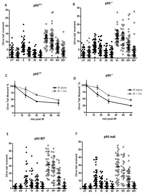
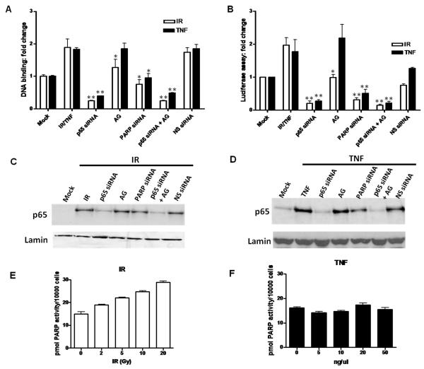
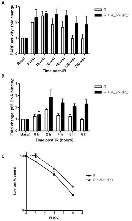
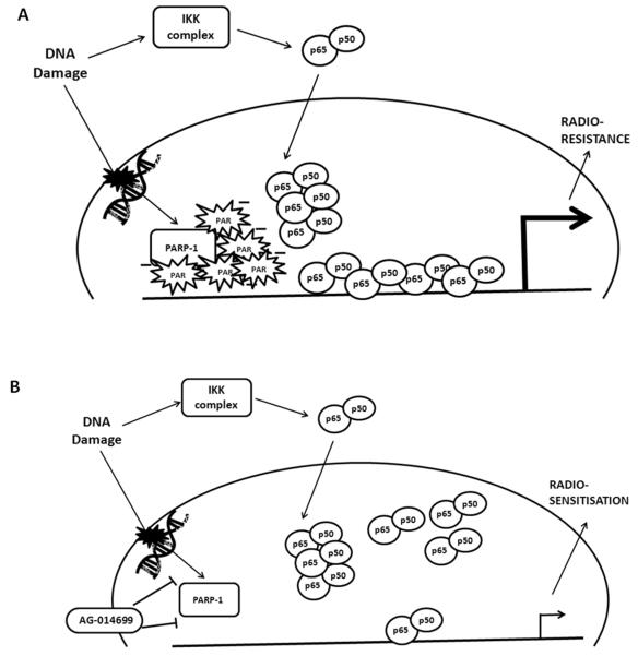
The stress-inducible transcription factor, nuclear factor (NF)-κB induces genes involved in proliferation and apoptosis. Aberrant NF-κB activity is common in cancer and contributes to therapeutic-resistance. Poly(ADP-ribose) polymerase-1 (PARP-1) is activated during DNA strand break repair and is a known transcriptional co-regulator. Here, we investigated the role of PARP-1 function during NF-κB activation using p65 small interfering RNA (siRNA), PARP siRNA or the potent PARP-1 inhibitor, AG-014699. Survival and apoptosis assays showed that NF-κB p65(-/-) cells were more sensitive to ionizing radiation (IR) than p65(+/+) cells. Co-incubation with p65 siRNA, PARP siRNA or AG-014699 radio-sensitized p65(+/+), but not p65(-/-) cells, demonstrating that PARP-1 mediates its effects on survival via NF-κB. Single-strand break (SSB) repair kinetics, and the effect SSB repair inhibition by AG-014699 were similar in p65(+/+) and p65(-/-) cells. As preventing SSB repair did not radio-sensitize p65(-/-) cells, we conclude that radio-sensitization by AG-014699 is due to downstream inhibition of NF-κB activation, and independent of SSB repair inhibition. PARP-1 catalytic activity was essential for IR-induced p65 DNA binding and NF-κB-dependent gene transcription, whereas for tumor necrosis factor (TNF)-α-treated cells, PARP-1 protein alone was sufficient. We hypothesize that this stimulus-dependent differential is mediated via stimulation of the poly(ADP-ribose) polymer, which was induced following IR, not TNF-α. Targeting DNA damage-activated NF-κB using AG-014699 may therefore overcome toxicity observed with classical NF-κB inhibitors without compromising other vital inflammatory functions. These data highlight the potential of PARP-1 inhibitors to overcome NF-κB-mediated therapeutic resistance and widens the spectrum of cancers in which these agents may be utilized.


Figures

Figure 1
Figure 1. Characterisation of cell lines and siRNA knockdown
(A) Western blots of whole cell extracts of p65+/+ and p65−/− MEFs at 48 h after transfection with vehicle alone (Mock), non-specific (NS) siRNA or p65 siRNA. (B) Western blots of whole cell extracts of p65+/+ and p65−/− at 48 h after transfection with vehicle alone (Mock), non-specific (NS) siRNA or PARP-1 siRNA. (C) Histogram showing PARP activity, measured using a validated immunoblot assay which quantifies PAR polymer formation, in p65+/+ MEFs after transfection with vehicle alone (mock), p65 siRNA, AG-014699, PARP-1 siRNA,a combination of p65 siRNA + AG-014699 or NS siRNA. (D) PARP activity measured using a validated immunoblot assay which quantifies PAR polymer formation in p65−/− MEFs following transfection with vehicle alone (mock), p65 siRNA, AG-014699, PARP-1 siRNA,a combination of p65 siRNA + AG-014699 or NS siRNA 10H antibody was used measure PAR formation Data are represented as the mean ±sem of three independent experiments. *Significance relative to mock treated control was p<0.05 using unpaired Student’s t-test.
Figure 2
Figure 2. Radio-sensitization by p65 knockdown, PARP-1 knockdown or AG-014699 is associated with the induction of apoptosis
The effects of increasing doses of IR either alone, or co-incubated with p65 siRNA, AG-014699, PARP-1 siRNA or a combination of p65 siRNA and AG-014699, on cell survival were assessed using the clonogenic survival assay. Cells were treated with relevant siRNA, (or vehicle control), left for 48 h, pre-treated with AG-014699, or DMSO control 1 h prior to IR then re-plated after a further 24 h and allowed to form colonies for 7-21 days. (Survival curve (A) shows p65+/+ MEFs, (B) p65−/− MEFs, (C) PARP+/+ MEFs and (D) PARP−/− MEFs) E The effect of IR alone (mock, white bar) ± p65 siRNA (black bar) ± AG-104699 (AG alone, light grey bar, AG + p65 siRNA, dark grey bar) on the induction of apoptosis in p65+/+ MEFs. Cells were treated with relevant siRNA, or control left for 48 h, pre-treated with AG-014699, or control 1 h prior to IR then allowed to repair for a further 24 h before harvesting and assessment of the induction of apoptosis by Annexin V FACs analysis. Results shown are normalised to untreated controls. F The effect of IR (mock, white bar) ± p65 siRNA (black bar) ± AG-104699 (AG alone, hatched bar, AG + p65 siRNA, striped bar) on the induction of apoptosis in p65−/− MEFs. Cells were treated with relevant siRNA, or control left for 48 h, pre-treated with AG-014699, or control 1 h prior to IR then allowed to repair for a further 24 h before harvesting and assessment of the induction of apoptosis by Annexin V FACs analysis. Results shown are normalised to untreated controls. All data shown are represented as the mean ±sem of three independent experiments. *Significance relative to mock treated control was p<0.05 using unpaired Student’s t-test.
Figure 3
Figure 3. AG-014699 inhibits Single strand break (SSB) repair to a similar extent regardless of cellular NF-κB status
Scatter diagrams showing the extent of single strand breaks (SSBs) in (A) p65+/+ MEFs and (B) p65−/− MEFs treated with 10 Gy IR ± AG-014699 (AG, denoted by +) and allowed to repair (0 min, 15 min, 30 min and 60 min). SSB repair was measured using the alkaline COMET assay and Olive Tail moment (shown here) is used as a measure of both the smallest detectable size of migrating DNA (reflected in the comet tail length) and the number of relaxed / broken pieces (represented by the intensity of DNA in the tail). All data were tested for Gaussian distribution and the lines shown indicate the mean of the data plotted. Line graph showing the kinetics of single strand break repair in p65+/+ (C) and p65−/− MEFs (D). Data was normalised to relevant controls and in both cases solid black lines represent repair over time of cells treated with 10 Gy IR, and dotted lines represent repair over time of cells treated with 10 Gy IR + AG-014699. Scatter diagrams showing the extent of single strand breaks (SSBs) in (E) p65−/− MEFs reconstituted with wild-type (WT) p65 and (F) p65−/− (control) MEFs treated with 10 Gy IR ± AG-014699 (AG, denoted by +) and allowed to repair (0 min, 15 min, 30 min and 60 min). SSB repair was measured using the alkaline COMET assay and Olive Tail moment is shown here. All data were tested for Gaussian distribution and the lines shown indicate the mean of the distribution. All results shown are the mean of three independent experiments ± SEM
Figure 4
Figure 4. PARP activity is essential for NF-κB activation following IR, not TNF-α
Histogram showing the effect of IR or TNF-α ± p65 siRNA ± AG-014699 (AG) ± PARP-1 siRNA ± (p65 siRNA + AG-014699) ±Non-specific (NS) siRNA control on NF-κB DNA binding activity, measured using an ELISA-based assay (A) and (B) NF-κB-dependent transcriptional activation, measured using a luciferase reporter assay, in p65+/+ MEFs. Open bars IR, black bars TNF-α. All results are the mean of three independent experiments with SEM. **Significance relative to mock treated control was p<0.01 using unpaired Student’s t-test. *Significance relative to mock treated control was p<0.05 using unpaired Student’s t-test. Western blotting data showing nuclear translocation of p65 following (C) IR or TNF-α (D) ± p65 siRNA ± AG-014699 (AG) ± PARP-1 siRNA ± (p65 siRNA + AG-014699) ± NS siRNA control, in p65+/+ MEFs. Cells were treated with relevant siRNA, or control, left for 48 h, pre-treated with AG-014699, or control 1 h prior to IR and harvested 1 h post-IR. Loading was normalised to lamin nuclear loading control in all cases. Histogram showing PARP activity, measured using a validated immunoblot assay which quantifies PAR polymer formation, in p65+/+ MEFs after increasing doses of IR (E) and TNF-α (F)
Figure 5
Figure 5. Persistence of NF-κB DNA binding following PARG inhibition
(A) Histogram showing PARP activity timecourse, measured using a validated immunoblot assay which quantifies PAR polymer formation, in p65+/+ MEFs following 10 Gy IR ± ADP-HPD (open bars IR, black bars IR + ADP-HPD). (B) Histogram showing NF-κB DNA binding timecourse measured using an ELISA-based method following IR ± ADP-HPD (open bars IR, black bars IR + ADP-HPD). (C) The effects of increasing doses of IR on cell survival, either alone (solid line) or co-incubated with ADP-HPD (dashed line) were assessed using the clonogenic survival in p65+/+ MEFs. Cells were pre-treated with ADP-HPD, or control 1 h prior to IR then re-plated after a further 24 h and allowed to form colonies for 7-21 days. All results are the mean of three independent experiments with SEM.
Figure 6
Figure 6. AG-014699 radio-sensitizes U251 glioblastoma cells via inhibition of NF-κB
(A)Western blots of whole cell extracts of U251 cells 48 h post transfection with vehicle alone (Mock), non-specific (NS) siRNA or p65 siRNA. (B) Western blots of whole cell extracts of U251 cells at 48 h after transfection with vehicle alone (Mock), non-specific (NS) siRNA or PARP-1 siRNA. (C)Histogram showing the effect of IR ± p65 siRNA ± AG-014699 (AG) ± PARP-1 siRNA ± (p65 siRNA + AG-014699) ±Non-specific (NS) siRNA control on NF-κB DNA binding activity, measured using an ELISA-based assay. All results are the mean of three independent experiments with SEM. **Significance relative to mock treated control was p<0.01 using unpaired Student’s t-test. (D)The effects of increasing doses of IR either alone, or co-incubated with p65 siRNA, AG-014699, PARP-1 siRNA or a combination of p65 siRNA and AG-014699, on U251 cell survival was assessed using the clonogenic survival assay. Cells were treated with relevant siRNA, (or vehicle control), left for 48 h, pre-treated with AG-014699, or DMSO control, 1 h prior to IR then re-plated after a further 24 h and allowed to form colonies for 7-21 days. (E) Scatter diagrams showing the extent of single strand breaks (SSBs) in U251 cells treated with 10 Gy IR ± AG-014699 (denoted by +) and allowed to repair (0 min, 15 min and 30 min). SSB repair was measured using the alkaline COMET assay and Olive Tail moment (described above) is shown here. All data were tested for Gaussian distribution and the lines shown indicate the mean of the data plotted. (F) Scatter diagrams showing the extent of single strand breaks (SSBs) in U251 cells treated with 10 Gy IR + p65 siRNA ± AG-014699 (denoted by +) and allowed to repair (0 min, 15 min and 30 min). SSB repair was measured using the alkaline COMET assay and Olive Tail moment (described above) is shown here. All data were tested for Gaussian distribution and the lines shown indicate the mean of the data plotted.
Figure 7
Figure 7. PAR is essential for DNA damage activated NF-κB
(A) DNA damage, such as IR, activates PARP-1, recruiting it to the site of damaged DNA. This results in formation of the negatively charged PAR polymer. The NF-κB p65-p50 heterodimer is also translocated to the nucleus following DNA damage via activation of the canonical pathway of NF-κB activation. When in proximity to regions with overall negative charge, such as that of the PAR polymer, a conformational change in p65 can be induced, exposing the positively charged DNA binding interface of p65. Thus, we propose that it is this negative charge on the PAR polymer that is attracting p65 to DNA bind, (as PARP-1 and the polymer are recruited to sites of damaged DNA), up-regulating transcription of NF-κB-dependent genes, protecting against apoptosis and conferring radio-resistance. (B) When the PARP inhibitor, AG-014699 is present, PARP-1 is no longer active and cannot form the polymer. Hence in this case, we see reduced DNA binding and transcriptional activation following IR, with induction of apoptosis and ultimately radio-sensitisation.

Similar articles

Cited by

References

    1. Althaus FR, Hofferer L, Kleczkowska HE, Malanga M, Naegeli H, Panzeter PL, et al. Histone shuttling by poly ADP-ribosylation. Mol Cell Biochem. 1994;138:53–9. - PubMed
    1. Barkett M, Gilmore T. Control of apoptosis by Rel/NF-kappaB transcription factors. Oncogene. 1999;18:6910–24. - PubMed
    1. Basseres DS, Baldwin AS. Nuclear factor-kappaB and inhibitor of kappaB kinase pathways in oncogenic initiation and progression. Oncogene. 2006;25:6817–6830. - PubMed
    1. Biswas DK, Shi Q, Baily S, Strickland I, Ghosh S, Pardee AB, et al. NF-kappa B activation in human breast cancer specimens and its role in cell proliferation and apoptosis. PNAS. 2004;101:10137–42. - PMC - PubMed
    1. Bonicalzi ME, Haince JF, Droit A, Poirier GG. Regulation of poly(ADP-ribose) metabolism by poly(ADP-ribose) glycohydrolase: where and when? Cell Mol Life Sci. 2005;62:739–50. - PMC - PubMed

Publication types