

The crystal structure of Streptococcus pyogenes uridine phosphorylase reveals a distinct subfamily of nucleoside phosphorylases
- PMID: 21707079
- PMCID: PMC3144492
- DOI: 10.1021/bi200707z
The crystal structure of Streptococcus pyogenes uridine phosphorylase reveals a distinct subfamily of nucleoside phosphorylases
Abstract
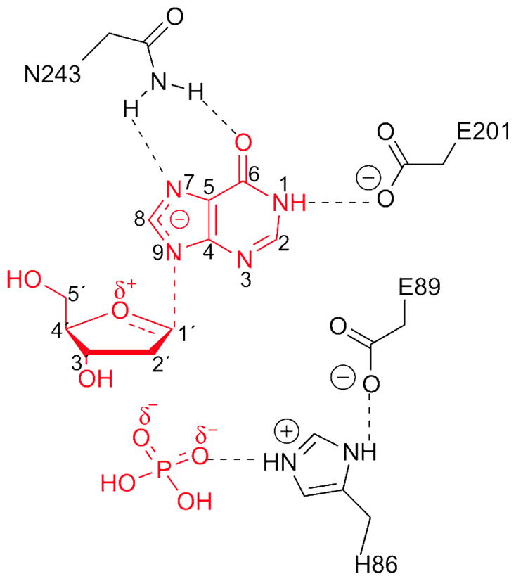
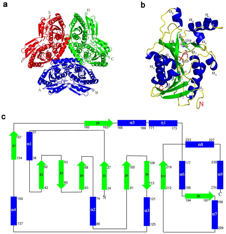
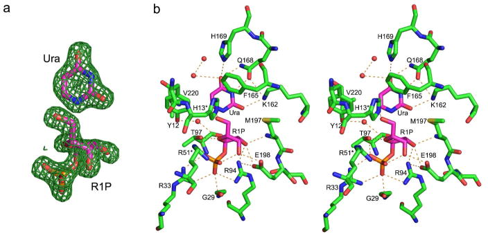
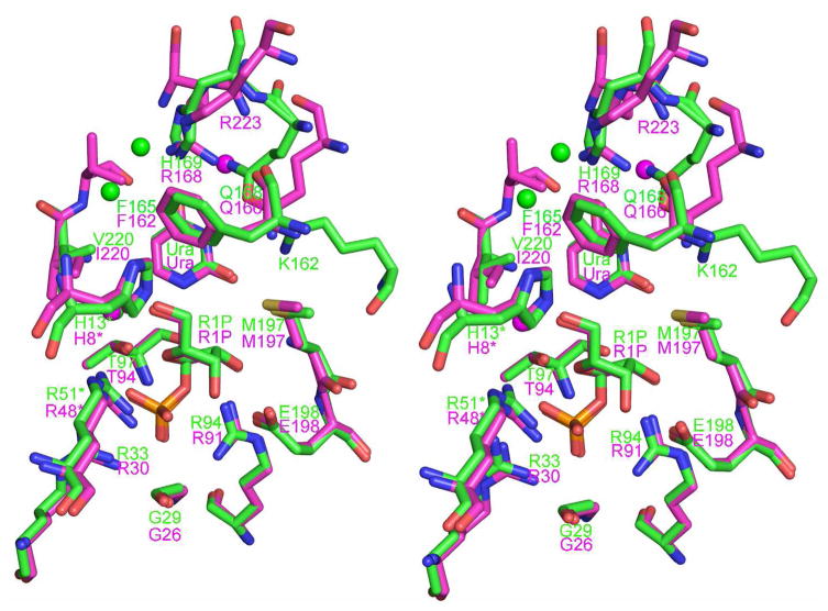
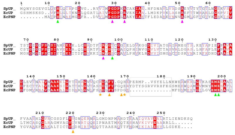
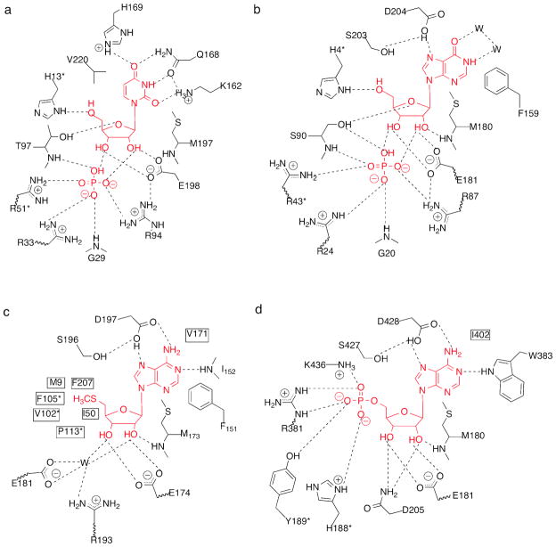
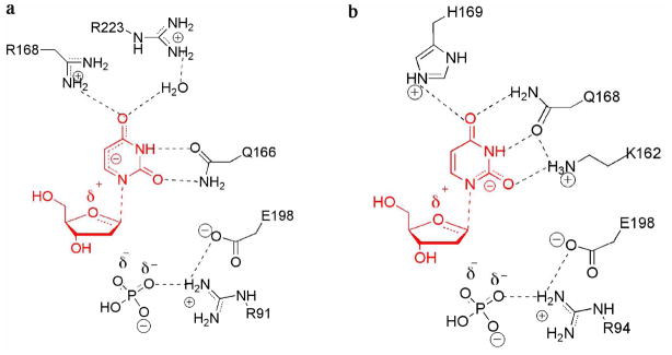
Uridine phosphorylase (UP), a key enzyme in the pyrimidine salvage pathway, catalyzes the reversible phosphorolysis of uridine or 2'-deoxyuridine to uracil and ribose 1-phosphate or 2'-deoxyribose 1-phosphate. This enzyme belongs to the nucleoside phosphorylase I superfamily whose members show diverse specificity for nucleoside substrates. Phylogenetic analysis shows Streptococcus pyogenes uridine phosphorylase (SpUP) is found in a distinct branch of the pyrimidine subfamily of nucleoside phosphorylases. To further characterize SpUP, we determined the crystal structure in complex with the products, ribose 1-phosphate and uracil, at 1.8 Å resolution. Like Escherichia coli UP (EcUP), the biological unit of SpUP is a hexamer with an α/β monomeric fold. A novel feature of the active site is the presence of His169, which structurally aligns with Arg168 of the EcUP structure. A second active site residue, Lys162, is not present in previously determined UP structures and interacts with O2 of uracil. Biochemical studies of wild-type SpUP showed that its substrate specificity is similar to that of EcUP, while EcUP is ∼7-fold more efficient than SpUP. Biochemical studies of SpUP mutants showed that mutations of His169 reduced activity, while mutation of Lys162 abolished all activity, suggesting that the negative charge in the transition state resides mostly on uracil O2. This is in contrast to EcUP for which transition state stabilization occurs mostly at O4.
Figures
Similar articles
-
Structural and catalytic analysis of two diverse uridine phosphorylases in Phytophthora capsici.Sci Rep. 2020 Jun 3;10(1):9051. doi: 10.1038/s41598-020-65935-9. Sci Rep. 2020. PMID: 32493959 Free PMC article.
-
Crystal structures of Escherichia coli uridine phosphorylase in two native and three complexed forms reveal basis of substrate specificity, induced conformational changes and influence of potassium.J Mol Biol. 2004 Mar 19;337(2):337-54. doi: 10.1016/j.jmb.2004.01.039. J Mol Biol. 2004. PMID: 15003451
-
Concerted action of two subunits of the functional dimer of Shewanella oneidensis MR-1 uridine phosphorylase derived from a comparison of the C212S mutant and the wild-type enzyme.Acta Crystallogr D Struct Biol. 2016 Feb;72(Pt 2):203-10. doi: 10.1107/S2059798315024353. Epub 2016 Jan 22. Acta Crystallogr D Struct Biol. 2016. PMID: 26894668
-
Structural analyses reveal two distinct families of nucleoside phosphorylases.Biochem J. 2002 Jan 1;361(Pt 1):1-25. doi: 10.1042/0264-6021:3610001. Biochem J. 2002. PMID: 11743878 Free PMC article. Review.
-
Pentose phosphates in nucleoside interconversion and catabolism.FEBS J. 2006 Mar;273(6):1089-101. doi: 10.1111/j.1742-4658.2006.05155.x. FEBS J. 2006. PMID: 16519676 Review.
Cited by
-
Enzymatic synthesis and phosphorolysis of 4(2)-thioxo- and 6(5)-azapyrimidine nucleosides by E. coli nucleoside phosphorylases.Beilstein J Org Chem. 2016 Dec 1;12:2588-2601. doi: 10.3762/bjoc.12.254. eCollection 2016. Beilstein J Org Chem. 2016. PMID: 28144328 Free PMC article.
-
Structural and catalytic analysis of two diverse uridine phosphorylases in Phytophthora capsici.Sci Rep. 2020 Jun 3;10(1):9051. doi: 10.1038/s41598-020-65935-9. Sci Rep. 2020. PMID: 32493959 Free PMC article.
-
Uridine phosphorylase from Trypanosoma cruzi: kinetic and chemical mechanisms.Biochemistry. 2011 Oct 25;50(42):9158-66. doi: 10.1021/bi2013382. Epub 2011 Sep 27. Biochemistry. 2011. PMID: 21932786 Free PMC article.
-
Crystallization of uridine phosphorylase from Shewanella oneidensis MR-1 in the laboratory and under microgravity and preliminary X-ray diffraction analysis.Acta Crystallogr Sect F Struct Biol Cryst Commun. 2012 Nov 1;68(Pt 11):1387-9. doi: 10.1107/S1744309112041784. Epub 2012 Oct 30. Acta Crystallogr Sect F Struct Biol Cryst Commun. 2012. PMID: 23143255 Free PMC article.
References
-
- Leer JC, Hammer-Jespersen K, Schwartz M. Uridine phosphorylase from Escherichia coli. Physical and chemical characterization. Eur J Biochem. 1977;75:217–224. - PubMed
-
- Caradoc-Davies TT, Cutfield SM, Lamont IL, Cutfield JF. Crystal structures of Escherichia coli uridine phosphorylase in two native and three complexed forms reveal basis of substrate specificity, induced conformational changes and influence of potassium. J Mol Biol. 2004;337:337–354. - PubMed
-
- Lashkov AA, Zhukhlistova NE, Gabdoulkhakov AH, Shtil AA, Efremov RG, Betzel C, Mikhailov AM. The X-ray structure of Salmonella typhimurium uridine nucleoside phosphorylase complexed with 2,2′-anhydrouridine, phosphate and potassium ions at 1.86 Å resolution. Acta Crystallogr D. 2010;66:51–60. - PubMed
-
- Morgunova E, Mikhailov AM, Popov AN, Blagova EV, Smirnova EA, Vainshtein BK, Mao C, Armstrong Sh R, Ealick SE, Komissarov AA, et al. Atomic structure at 2.5 Å resolution of uridine phosphorylase from E. coli as refined in the monoclinic crystal lattice. FEBS Lett. 1995;367:183–187. - PubMed
Publication types
MeSH terms
Substances
Grants and funding
LinkOut - more resources
Full Text Sources
Molecular Biology Databases
