. 2011 Jul;81(2):515-27.
doi: 10.1111/j.1365-2958.2011.07714.x. Epub 2011 Jun 28.

Role for Sit4p-dependent mitochondrial dysfunction in mediating the shortened chronological lifespan and oxidative stress sensitivity of Isc1p-deficient cells

Affiliations

Role for Sit4p-dependent mitochondrial dysfunction in mediating the shortened chronological lifespan and oxidative stress sensitivity of Isc1p-deficient cells

António Daniel Barbosa et al. Mol Microbiol. 2011 Jul.

Abstract

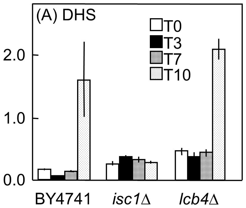
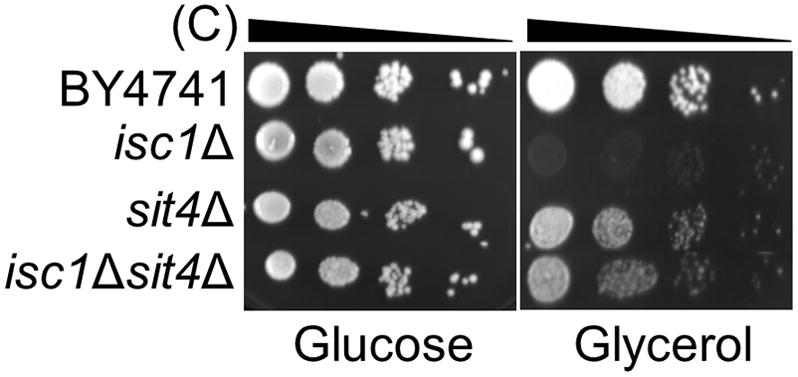
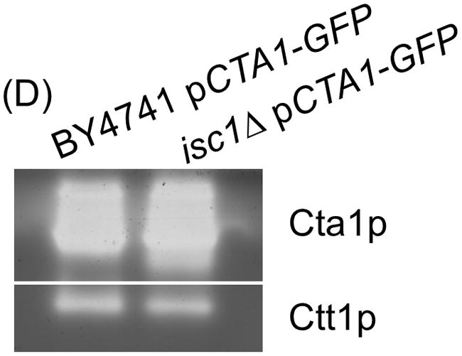
Saccharomyces cerevisiae cells lacking Isc1p, an orthologue of mammalian neutral sphingomyelinase 2, display a shortened lifespan and an increased sensitivity to oxidative stress. A lipidomic analysis revealed specific changes in sphingolipids that accompanied the premature ageing of Isc1p-deficient cells under severe calorie restriction conditions, including a decrease of dihydrosphingosine levels and an increase of dihydro-C(26) -ceramide and phyto-C(26) -ceramide levels, the latter raising the possibility of activation of ceramide-dependent protein phosphatases. Consequently, deletion of the SIT4 gene, which encodes for the catalytic subunit of type 2A ceramide-activated protein phosphatase in yeast, abolished the premature ageing and hydrogen peroxide sensitivity of isc1Δ cells. SIT4 deletion also abolished the respiratory defects and catalase A deficiency exhibited by isc1Δ mutants. These results are consistent with catabolic derepression associated with the loss of Sit4p. The overall results show that Isc1p is an upstream regulator of Sit4p and implicate Sit4p activation in mitochondrial dysfunction leading to the shortened chronological lifespan and oxidative stress sensitivity of isc1Δ mutants.


Figures

Fig. 1
Fig. 1
The de novo synthesis and catabolism of sphingolipids in yeast. LCFA, long chain fatty acid (C16–18); MLCFA, medium long chain fatty acid (C20–22); VLCFA, very long chain fatty acid (C24–26), FFA, free fatty acid.
Fig. 2
Fig. 2
Levels of long chain sphingoid bases during chronological aging. S. cerevisiae BY4741, isc1Δand lcb4Δ cells were grown in YPD medium to the post-diauxic phase, washed twice with H2O and kept in H2O at 26°C. (A) DHS; (B) PHS; (C) DHS-1-P; (D) PHS-1-P. Data are expressed as pmol of lipid/nmol lipid Pi and are means ± SD of three independent experiments.
Fig. 2
Fig. 2
Levels of long chain sphingoid bases during chronological aging. S. cerevisiae BY4741, isc1Δand lcb4Δ cells were grown in YPD medium to the post-diauxic phase, washed twice with H2O and kept in H2O at 26°C. (A) DHS; (B) PHS; (C) DHS-1-P; (D) PHS-1-P. Data are expressed as pmol of lipid/nmol lipid Pi and are means ± SD of three independent experiments.
Fig. 3
Fig. 3
Levels of ceramides during chronological aging. S. cerevisiae BY4741, isc1Δ and lcb4Δ cells were grown in YPD medium to the post-diauxic phase, washed twice with H2O and kept in H2O at 26°C. (A) total dihydroceramide (dh-Cer); (B) dihydro-C26-ceramide (dh-C26-Cer); (C) total phytoceramide (phyto-Cer); (D) phyto-C26-ceramide (phyto-C26-Cer); (E) total α–hydroxylated-phytoceramide (αHO-phyto-Cer). Data are expressed as pmol of lipid/nmol lipid Pi and are means ± SD of three independent experiments. See Tables S1–S3 for levels of individual ceramide species.
Fig. 3
Fig. 3
Levels of ceramides during chronological aging. S. cerevisiae BY4741, isc1Δ and lcb4Δ cells were grown in YPD medium to the post-diauxic phase, washed twice with H2O and kept in H2O at 26°C. (A) total dihydroceramide (dh-Cer); (B) dihydro-C26-ceramide (dh-C26-Cer); (C) total phytoceramide (phyto-Cer); (D) phyto-C26-ceramide (phyto-C26-Cer); (E) total α–hydroxylated-phytoceramide (αHO-phyto-Cer). Data are expressed as pmol of lipid/nmol lipid Pi and are means ± SD of three independent experiments. See Tables S1–S3 for levels of individual ceramide species.
Fig. 4
Fig. 4
SIT4 disruption suppresses the shortened chronological lifespan of isc1Δ cells. S. cerevisiae BY4741, iscΔ, sit4Δ and sit4Δisc1Δ mutant cells were grown in YPD medium to post-diauxic phase. (A) Cells were washed twice with H2O, and kept in H2O at 26°C. The viability was determined by standard dilution plate counts and expressed as the percentage of the colony-forming units at time 0h. (B) Protein carbonylation. Protein extracts were derivatized with DNPH and slot-blotted into a PVDF membrane. Immunodetection was performed using an anti-DNP antibody, as described in Materials and Methods. Quantitative analysis of total protein carbonyl content was performed by densitometry using data taken from the same membrane. Data are expressed as the carbonyl content ratio between cells of day 14 and cells of day 0. Values are means ± SD of three independent experiments. *p<0.05.
Fig. 4
Fig. 4
SIT4 disruption suppresses the shortened chronological lifespan of isc1Δ cells. S. cerevisiae BY4741, iscΔ, sit4Δ and sit4Δisc1Δ mutant cells were grown in YPD medium to post-diauxic phase. (A) Cells were washed twice with H2O, and kept in H2O at 26°C. The viability was determined by standard dilution plate counts and expressed as the percentage of the colony-forming units at time 0h. (B) Protein carbonylation. Protein extracts were derivatized with DNPH and slot-blotted into a PVDF membrane. Immunodetection was performed using an anti-DNP antibody, as described in Materials and Methods. Quantitative analysis of total protein carbonyl content was performed by densitometry using data taken from the same membrane. Data are expressed as the carbonyl content ratio between cells of day 14 and cells of day 0. Values are means ± SD of three independent experiments. *p<0.05.
Fig. 5
Fig. 5
SIT4 deletion suppresses hydrogen peroxide sensitivity of isc1Δ cells. S. cerevisiae BY4741, isc1Δ, sit4Δ and sit4Δisc1Δ mutant cells were grown in YPD medium to the exponential phase (OD600=0.6) (white bars) and exposed to 1.5 mM H2O2 for 30 min (black bars). (A) Cell viability was determined by standard dilution plate counts and expressed as the percentage of the colony-forming units of non-stressed cells. (B) Intracellular oxidation. Cells were labeled with the molecular probe H2DCFDA and lysed as described in Materials and Methods. Data was normalized for protein content. (C) Protein carbonylation. Proteins were derivatized with DNPH and slot-blotted into a PVDF membrane. Immunodetection was performed using an anti-DNP antibody, as described in Materials and Methods. Quantitative analysis of total protein carbonyl content was performed by densitometry using data taken from the same membrane. Values are means ± SD of three independent experiments. *p<0.05, **p<0.01.
Fig. 5
Fig. 5
SIT4 deletion suppresses hydrogen peroxide sensitivity of isc1Δ cells. S. cerevisiae BY4741, isc1Δ, sit4Δ and sit4Δisc1Δ mutant cells were grown in YPD medium to the exponential phase (OD600=0.6) (white bars) and exposed to 1.5 mM H2O2 for 30 min (black bars). (A) Cell viability was determined by standard dilution plate counts and expressed as the percentage of the colony-forming units of non-stressed cells. (B) Intracellular oxidation. Cells were labeled with the molecular probe H2DCFDA and lysed as described in Materials and Methods. Data was normalized for protein content. (C) Protein carbonylation. Proteins were derivatized with DNPH and slot-blotted into a PVDF membrane. Immunodetection was performed using an anti-DNP antibody, as described in Materials and Methods. Quantitative analysis of total protein carbonyl content was performed by densitometry using data taken from the same membrane. Values are means ± SD of three independent experiments. *p<0.05, **p<0.01.
Fig. 6
Fig. 6
SIT4 disruption abolishes mitochondrial dysfunctions seen in isc1Δ cells. S. cerevisiae BY4741, isc1Δ, sit4Δ and sit4Δisc1Δ mutant cells were grown in YPD medium to exponential (log) or post-diauxic phase (PDS). (A) Oxygen consumption rates were measured as described in Materials and Methods. (B) Cytochrome C Oxidase (COX) specific activity. Cells were lysed, and enzyme activity was measured as described in Materials and Methods. (C) Cells were plated in 5-fold dilution series on glucose or glycerol as carbon source. Values are means ± SD of three independent experiments. **p<0.01.
Fig. 6
Fig. 6
SIT4 disruption abolishes mitochondrial dysfunctions seen in isc1Δ cells. S. cerevisiae BY4741, isc1Δ, sit4Δ and sit4Δisc1Δ mutant cells were grown in YPD medium to exponential (log) or post-diauxic phase (PDS). (A) Oxygen consumption rates were measured as described in Materials and Methods. (B) Cytochrome C Oxidase (COX) specific activity. Cells were lysed, and enzyme activity was measured as described in Materials and Methods. (C) Cells were plated in 5-fold dilution series on glucose or glycerol as carbon source. Values are means ± SD of three independent experiments. **p<0.01.
Fig. 7
Fig. 7
SIT4 disruption suppresses catalase A deficiency seen in isc1Δ cells and CTA1 overexpression partially suppresses the shortened chronological lifespan of isc1Δ cells. S. cerevisiae BY4741, isc1Δ, sit4Δ and sit4Δisc1Δ mutant cells were grown in YPD medium to post-diauxic phase (PDS). Catalaseactivity was determined (A) spectrophotometrically or (B) detected in situ after non-denaturing polyacrylamide gel electrophoresis, using the H2O2/peroxidase system. (C) S. cerevisiae BY4741 and isc1Δ mutant cells transformed with pPGK-M28-I (empty vector) or pCTA1-GFP were grown in SC medium lacking uracil to post-diauxic phase. Cells were washed twice with H2O, and kept in H2O at 26°C. The viability was determined by standard dilution plate counts and expressed as the percentage of the colony-forming units at time 0h. (D) CTA1 overexpression increases Cta1p activity in both S. cerevisiae BY4741 and isc1Δ cells. Enzyme activity was detected as in (b). Values are means ± SD of three independent experiments. **p<0.01.
Fig. 7
Fig. 7
SIT4 disruption suppresses catalase A deficiency seen in isc1Δ cells and CTA1 overexpression partially suppresses the shortened chronological lifespan of isc1Δ cells. S. cerevisiae BY4741, isc1Δ, sit4Δ and sit4Δisc1Δ mutant cells were grown in YPD medium to post-diauxic phase (PDS). Catalaseactivity was determined (A) spectrophotometrically or (B) detected in situ after non-denaturing polyacrylamide gel electrophoresis, using the H2O2/peroxidase system. (C) S. cerevisiae BY4741 and isc1Δ mutant cells transformed with pPGK-M28-I (empty vector) or pCTA1-GFP were grown in SC medium lacking uracil to post-diauxic phase. Cells were washed twice with H2O, and kept in H2O at 26°C. The viability was determined by standard dilution plate counts and expressed as the percentage of the colony-forming units at time 0h. (D) CTA1 overexpression increases Cta1p activity in both S. cerevisiae BY4741 and isc1Δ cells. Enzyme activity was detected as in (b). Values are means ± SD of three independent experiments. **p<0.01.
Fig. 8
Fig. 8
Proposed model for Sit4p-dependent oxidative stress sensitivity and shortened chronological lifespan of isc1Δ cells. Cells lacking Isc1p display an increase in dh-C26-Cer and phyto-C26-Cer species, probably due to de novo biosynthesis, that activate Sit4p. Dephosphorylation of Sit4p target proteins causes mitochondrial dysfunction, leading to oxidative stress sensitivity and shortened chronological lifespan in isc1Δ cells. SIT4 deletion restores mitochondrial function of isc1Δ cells, increasing oxidative stress resistance and chronological lifespan.

Similar articles

Cited by

References

    1. Aebi H. Catalase in vitro. Methods Enzymol. 1984;105:121–126. - PubMed
    1. Aerts AM, Francois IE, Bammens L, Cammue BP, Smets B, Winderickx J, Accardo S, De Vos DE, Thevissen K. Level of M(IP)2C sphingolipid affects plant defensin sensitivity, oxidative stress resistance and chronological life-span in yeast. FEBS Lett. 2006;580:1903–1907. - PubMed
    1. Aerts AM, Zabrocki P, Francois IE, Carmona-Gutierrez D, Govaert G, Mao C, Smets B, Madeo F, Winderickx J, Cammue BP, Thevissen K. Ydc1p ceramidase triggers organelle fragmentation, apoptosis and accelerated ageing in yeast. Cell Mol Life Sci. 2008;65:1933–1942. - PMC - PubMed
    1. Almeida T, Marques M, Mojzita D, Amorim MA, Silva RD, Almeida B, Rodrigues P, Ludovico P, Hohmann S, Moradas-Ferreira P, Corte-Real M, Costa V. Isc1p plays a key role in hydrogen peroxide resistance and chronological lifespan through modulation of iron levels and apoptosis. Mol Biol Cell. 2008;19:865–876. - PMC - PubMed
    1. Angeles de la Torre-Ruiz M, Torres J, Arino J, Herrero E. Sit4 is required for proper modulation of the biological functions mediated by Pkc1 and the cell integrity pathway in Saccharomyces cerevisiae. J Biol Chem. 2002;277:33468–33476. - PubMed

Publication types

MeSH terms