. 2011 Jun 27:5:239.
doi: 10.1186/1752-1947-5-239.

Fibrous dysplasia of bone associated with soft-tissue myxomas as well as an intra-osseous myxoma in a woman with Mazabraud's syndrome: a case report

Affiliations

Fibrous dysplasia of bone associated with soft-tissue myxomas as well as an intra-osseous myxoma in a woman with Mazabraud's syndrome: a case report

Wybren A van der Wal et al. J Med Case Rep. .

Abstract

Introduction: Mazabraud's syndrome is a rare but well-described disorder characterized by fibrous dysplasia in single or multiple bones associated with one or more soft-tissue myxomas. In this report, we describe what is, to the best of our knowledge, the first case involving an intra-osseous myxoma. This finding supports, and could provide new insight into, the pathological association between fibrous dysplasia and myxomas.

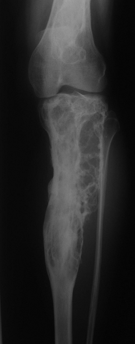
Case presentation: In this report, we describe the case of a 49-year-old Caucasian woman known for years to have fibrous dysplasia in the left femur and tibia who presented with progressive pain in her left leg and soft swelling in the left quadriceps region. After surgical intervention with excision of the soft-tissue mass, the diagnosis of Mazabraud's syndrome was confirmed. During follow-up, our patient presented with a painless mass located on the lateral side of the left knee, next to a second, intra-osseous lesion with the same characteristics in the left lateral tibial plateau. The histopathological examination was consistent with a soft-tissue intra-osseous myxoma.

Conclusion: In the international literature, 67 cases of Mazabraud's syndrome have been described so far.To our knowledge, the present case report is the first to describe the combination of polyostotic fibrous dysplasia and intra-muscular as well as intra-osseous myxoma.


Figures

Figure 1
Figure 1
X-ray of the left femur. Anteroposterior plain film of the upper left leg with typical osseous changes consistent with fibrous dysplasia and shepherd's crook deformity.
Figure 2
Figure 2
X-ray of the left tibia. Anteroposterior plain film of the left tibia showing fibrous dysplasia.
Figure 3
Figure 3
Histology of fibrous dysplasia of our patient. Photomicrograph showing curvilinear, slender trabeculae of woven bone surrounded by cellular fibroblastic tissue. There is no osteoblastic rim at the bone-stromal interface.
Figure 4
Figure 4
Histology of myxoma of our patient. Photomicrograph showing a paucicellular myxomatous lesion with dispersed, inconspicuous spindle cells.
Figure 5
Figure 5
Anteroposterior X-ray of the whole left leg. Situation after fibrous dysplasia excision of the left proximal femur and left tibia with correctional osteotomy of the left hip and fixation with a proximal femoral nail, as well as allograft fixation with AO screws of the tibia.
Figure 6
Figure 6
Lateral X-ray of the whole left leg. This X-ray shows the situation after fibrous dysplasia excision of the left proximal femur and left tibia.
Figure 7
Figure 7
T1-weighted magnetic resonance imaging scan of our patient. Axial spin echo T1-weighted pre-contrast magnetic resonance imaging (MRI) scan of the left knee reveals two well-delineated masses (arrows) with a homogeneous low signal intensity in the lateral retinaculum and in the lateral tibia plateau.
Figure 8
Figure 8
T2-weighted MRI scan of our patient. This T2-weighted image shows identical thin rim enhancement and heterogeneous intra-lesional enhancement in both masses. These findings may suggest myxomatous masses. In contrast, fibrous dysplasia can be noticed in the anterior tibial plateau (arrowhead).
Figure 9
Figure 9
Histology of intra-osseous myxoma of our patient. Bone within a paucicellular myxoid lesion with small, bland spindle cells.

References

    1. Henschen F. Fall von ostitis fibrosa mit multipelen tumoren in den umgebenden muskulatur. Verh Dtsch Ges Pathol. 1926;21:93–97.
    1. Mazabraud A, Semat P, Roze R. Apropos of the association of fibromyxomas of the soft tissues with fibrous dysplasia of the bones in French. Presse Med. 1967;75:2223–2228. - PubMed
    1. Campanacci M. Bone and Soft Tissue Tumors: Clinical Features, Imaging, Pathology and Treatment. 2. New York: Springer; 1999.
    1. DiCaprio MR, Enneking WF. Fibrous dysplasia: pathophysiology, evaluation, and treatment. J Bone Joint Surg Am. 2005;87:1848–1864. doi: 10.2106/JBJS.D.02942. - DOI - PubMed
    1. Marie PJ, de Pollak C, Chanson P, Lomri A. Increased proliferation of osteoblastic cells expressing the activating Gsα mutation in monostotic and polyostotic fibrous dysplasia. Am J Pathol. 1997;150:1059–1069. - PMC - PubMed

LinkOut - more resources