

Successful management of aggressive fibromatosis of the neck using wide surgical excision: a case report
- PMID: 21707981
- PMCID: PMC3141701
- DOI: 10.1186/1752-1947-5-244
Successful management of aggressive fibromatosis of the neck using wide surgical excision: a case report
Abstract
Introduction: Aggressive fibromatosis is a benign tumor, thought to arise from deep musculoaponeurotic structures, rarely found in the head or neck. However, when it does occur in the head and neck region, it tends to be more aggressive and associated with significant morbidity, which may be attributed to the vital vascular, neurological or anatomical structures in close proximity.
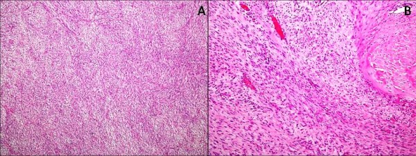
Case presentation: We report the case of a 39-year-old Pakistani man who presented with a two-month history of a lump on the right side of his neck. The mass was excised and histopathological analysis revealed a case of aggressive fibromatosis.
Conclusion: Due to the rarity of the condition no guidelines are available on the indications and extent of each modality. Due to its aggressive behavior and tendency to invade local structures and recur, a multi-modality management strategy is usually employed. On the basis of this case, we suggest that aggressive surgery is a viable management option and may be successfully used as a single modality treatment.
Figures


Similar articles
-
Successful Management of Aggressive Fibromatosis of the Neck: A Case Report.Balkan Med J. 2018 May 29;35(3):278-281. doi: 10.4274/balkanmedj.2017.0509. Balkan Med J. 2018. PMID: 29843498 Free PMC article.
-
Desmoid tumours of the head and neck: A case report.Eur Ann Otorhinolaryngol Head Neck Dis. 2019 Jun;136(3):207-209. doi: 10.1016/j.anorl.2019.02.008. Epub 2019 Mar 14. Eur Ann Otorhinolaryngol Head Neck Dis. 2019. PMID: 30880032
-
Pediatric aggressive fibromatosis of the head and neck: a 20-year retrospective review.J Pediatr Surg. 2008 Sep;43(9):1596-604. doi: 10.1016/j.jpedsurg.2008.02.001. J Pediatr Surg. 2008. PMID: 18778992 Review.
-
Desmoid tumor (aggressive fibromatosis) of the neck.Indian J Otolaryngol Head Neck Surg. 2000 Apr;52(2):182-4. doi: 10.1007/BF03000347. Indian J Otolaryngol Head Neck Surg. 2000. PMID: 23119668 Free PMC article.
-
Desmoid-type fibromatosis of the head and neck in children: a case report and review of the literature.J Med Case Rep. 2016 Jun 10;10:173. doi: 10.1186/s13256-016-0949-9. J Med Case Rep. 2016. PMID: 27286970 Free PMC article. Review.
Cited by
-
Desmoid tumor of trapezius muscle: A case report.Ann Med Surg (Lond). 2021 Dec 1;72:103127. doi: 10.1016/j.amsu.2021.103127. eCollection 2021 Dec. Ann Med Surg (Lond). 2021. PMID: 34925822 Free PMC article.
-
Desmoid tumor (fibromatosis) of the head and neck.Saudi Med J. 2015 Jan;36(1):101-3. doi: 10.15537/smj.2015.1.10504. Saudi Med J. 2015. PMID: 25630012 Free PMC article.
-
Recurrent Desmoid Tumor of the Neck: A Case Report of a Benign Disease with Aggressive Behavior.Case Rep Otolaryngol. 2018 Nov 28;2018:6573587. doi: 10.1155/2018/6573587. eCollection 2018. Case Rep Otolaryngol. 2018. PMID: 30622828 Free PMC article.
-
Desmoid fibromatosis of submandibular region.J Surg Tech Case Rep. 2014 Jan;6(1):21-5. doi: 10.4103/2006-8808.135144. J Surg Tech Case Rep. 2014. PMID: 25013548 Free PMC article.
References
-
- Sze H, Yeung MW. Fibromatosis of the neck causing airway obstruction managed effectively with weekly low-dose methotrexate and vinblastine. Hong Kong Med J. 2009;15:221–223. - PubMed
-
- El-Haddad M, El-Sebaie M, Ahmad R, Khalil E, Shahin M, Pant R, Memon M, Al-Hebshi A, Khafaga Y, Al-Shabanah M, Allam A. Treatment of aggressive fibromatosis: the experience of a single institution. Clin Oncol (R Coll Radiol) 2009;21:775–780. - PubMed
LinkOut - more resources
Full Text Sources
