

Low expression of aldehyde dehydrogenase 1A1 (ALDH1A1) is a prognostic marker for poor survival in pancreatic cancer
- PMID: 21708005
- PMCID: PMC3135572
- DOI: 10.1186/1471-2407-11-275
Low expression of aldehyde dehydrogenase 1A1 (ALDH1A1) is a prognostic marker for poor survival in pancreatic cancer
Abstract
Background: Aldehyde dehydrogenase 1 (ALDH1) has been characterised as a cancer stem cell marker in different types of tumours. Additionally, it plays a pivotal role in gene regulation and endows tumour cells with augmented chemoresistance. Recently, ALDH1A1 has been described as a prognostic marker in a pancreatic cancer tissue microarray. The aim of this study was to reevaluate the expression of ALDH1A1 as a prognostic marker on whole-mount tissue sections.
Methods: Real-time-quantitative-PCR (qRT-PCR) and Western blotting were used to evaluate the expression profile of ALDH1A1 in seven pancreatic cancer cell lines and one non-malignant pancreatic cell line. Immunostaining against ALDH1A1 and Ki-67 was performed on paraffin-embedded samples from 97 patients with pancreatic cancer. The immunohistochemical results were correlated to histopathological and clinical data.
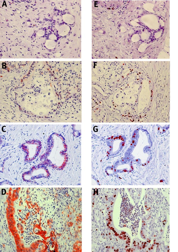
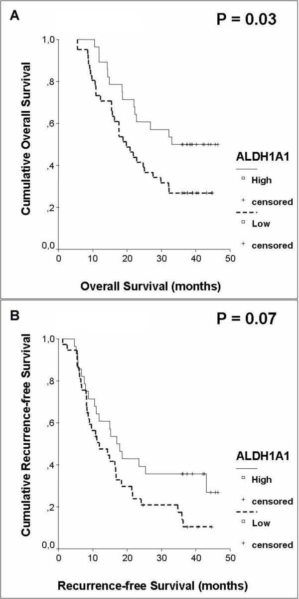
Results: qRT-PCR and Western blotting revealed a different expression pattern of ALDH1A1 in different malignant and non-malignant pancreatic cell lines. Immunohistochemical analysis demonstrated that ALDH1A1 was confined to the cellular cytoplasm and occurred in 72 cases (74%), whereas it was negative in 25 cases (26%). High expression of ALDH1A1 was significantly correlated to an increased proliferation rate (Spearman correlation, p = 0.01). Univariate and multivariate analyses showed that decreased expression of ALDH1A1 is an independent adverse prognostic factor for overall survival.
Conclusions: Immunohistochemical analysis on whole-mount tissue slides revealed that ALDH1A1 is more abundantly expressed in pancreatic cancer than initially reported by a tissue microarray analysis. Moreover, high expression of ALDH1A1 correlated significantly with the proliferation of tumour cells. Intriguingly, this study is the first which identifies low expression of ALDH1A1 as an independent adverse prognostic marker for overall survival in pancreatic cancer.
Figures


Similar articles
-
Expression analysis of aldehyde dehydrogenase 1A1 (ALDH1A1) in colon and rectal cancer in association with prognosis and response to chemotherapy.Ann Surg Oncol. 2012 Dec;19(13):4193-201. doi: 10.1245/s10434-012-2518-9. Epub 2012 Aug 10. Ann Surg Oncol. 2012. PMID: 22878609
-
Increased expression of ALDH1A1 protein is associated with poor prognosis in clear cell renal cell carcinoma.Med Oncol. 2013;30(2):574. doi: 10.1007/s12032-013-0574-z. Epub 2013 Apr 13. Med Oncol. 2013. PMID: 23585015
-
Linc00675 is a novel marker of short survival and recurrence in patients with pancreatic ductal adenocarcinoma.World J Gastroenterol. 2015 Aug 21;21(31):9348-57. doi: 10.3748/wjg.v21.i31.9348. World J Gastroenterol. 2015. PMID: 26309360 Free PMC article.
-
Aldehyde Dehydrogenase-1A1 (ALDH1A1): The Novel Regulator of Chemoresistance in Pancreatic Cancer Cells.Cancer Control. 2024 Jan-Dec;31:10732748241305835. doi: 10.1177/10732748241305835. Cancer Control. 2024. PMID: 39611960 Free PMC article. Review.
-
Aldehyde dehydrogenase 1A1 and 1A3 isoforms - mechanism of activation and regulation in cancer.Cell Signal. 2021 Nov;87:110120. doi: 10.1016/j.cellsig.2021.110120. Epub 2021 Aug 21. Cell Signal. 2021. PMID: 34428540 Free PMC article. Review.
Cited by
-
Aldehyde dehydrogenase 1A1 in stem cells and cancer.Oncotarget. 2016 Mar 8;7(10):11018-32. doi: 10.18632/oncotarget.6920. Oncotarget. 2016. PMID: 26783961 Free PMC article. Review.
-
Biomarkers for predicting future metastasis of human gastrointestinal tumors.Cell Mol Life Sci. 2013 Oct;70(19):3631-56. doi: 10.1007/s00018-013-1266-8. Epub 2013 Jan 31. Cell Mol Life Sci. 2013. PMID: 23370778 Free PMC article. Review.
-
Cancer stem cell-related marker expression in lung adenocarcinoma and relevance of histologic subtypes based on IASLC/ATS/ERS classification.Onco Targets Ther. 2013 Nov 8;6:1597-604. doi: 10.2147/OTT.S52353. eCollection 2013. Onco Targets Ther. 2013. PMID: 24235845 Free PMC article.
-
Proteomics Profiling of Pancreatic Cancer and Pancreatitis for Biomarkers Discovery.J Cell Sci Ther. 2018;9(4):287. doi: 10.4172/2157-7013.1000287. Epub 2018 Nov 28. J Cell Sci Ther. 2018. PMID: 31032145 Free PMC article.
-
Cancer Stem-Like Cells in a Case of an Inflammatory Myofibroblastic Tumor of the Lung.Front Oncol. 2020 May 15;10:673. doi: 10.3389/fonc.2020.00673. eCollection 2020. Front Oncol. 2020. PMID: 32500024 Free PMC article.
References
MeSH terms
Substances
LinkOut - more resources
Full Text Sources
Medical
Research Materials
Miscellaneous
