. 2011 Sep 1;357(1):83-95.
doi: 10.1016/j.ydbio.2011.06.014. Epub 2011 Jun 25.

Evolution of nubbin function in hemimetabolous and holometabolous insect appendages

Affiliations

Evolution of nubbin function in hemimetabolous and holometabolous insect appendages

Nataliya Turchyn et al. Dev Biol. .

Abstract

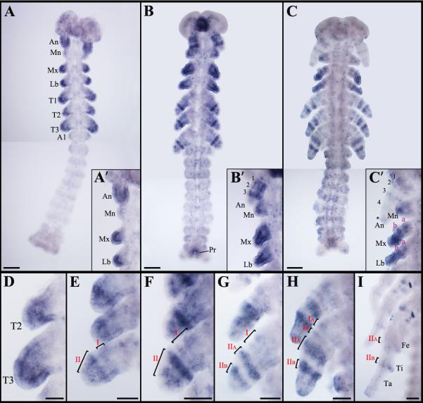
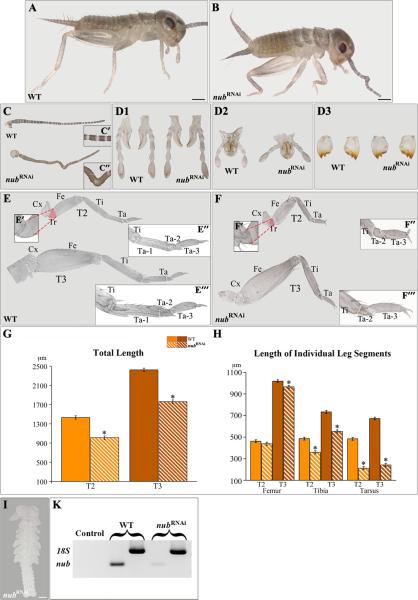
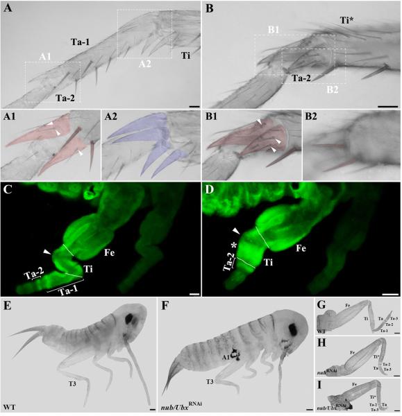
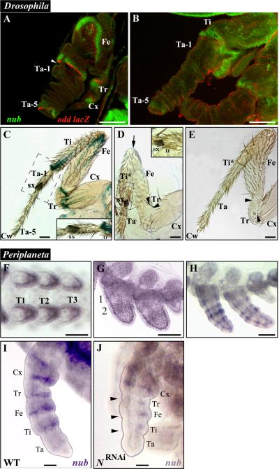
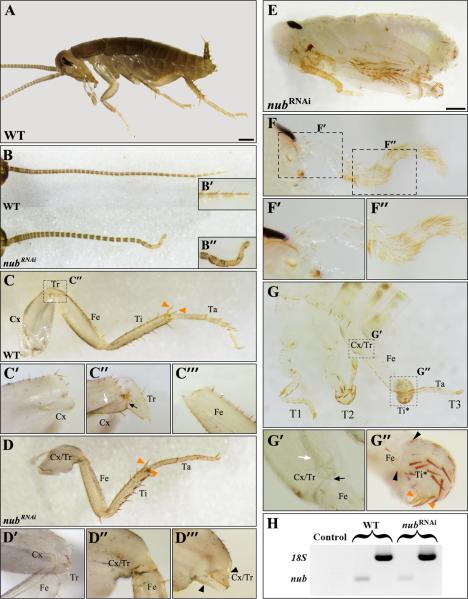
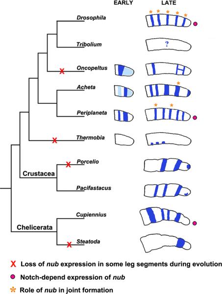
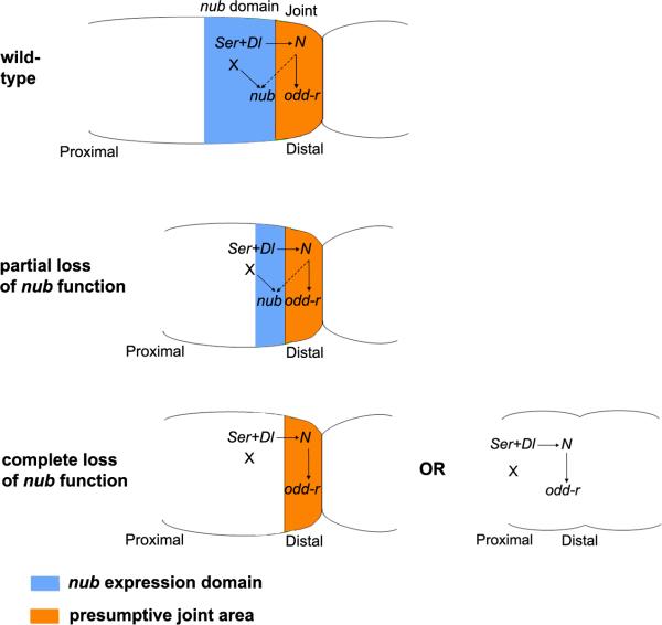
Insects display a whole spectrum of morphological diversity, which is especially noticeable in the organization of their appendages. A recent study in a hemipteran, Oncopeltus fasciatus (milkweed bug), showed that nubbin (nub) affects antenna morphogenesis, labial patterning, the length of the femoral segment in legs, and the formation of a limbless abdomen. To further determine the role of this gene in the evolution of insect morphology, we analyzed its functions in two additional hemimetabolous species, Acheta domesticus (house cricket) and Periplaneta americana (cockroach), and re-examined its role in Drosophila melanogaster (fruit fly). While both Acheta and Periplaneta nub-RNAi first nymphs develop crooked antennae, no visible changes are observed in the morphologies of their mouthparts and abdomen. Instead, the main effect is seen in legs. The joint between the tibia and first tarsomere (Ta-1) is lost in Acheta, which in turn, causes a fusion of these two segments and creates a chimeric nub-RNAi tibia-tarsus that retains a tibial identity in its proximal half and acquires a Ta-1 identity in its distal half. Similarly, our re-analysis of nub function in Drosophila reveals that legs lack all true joints and the fly tibia also exhibits a fused tibia and tarsus. Finally, we observe a similar phenotype in Periplaneta except that it encompasses different joints (coxa-trochanter and femur-tibia), and in this species we also show that nub expression in the legs is regulated by Notch signaling, as had previously been reported in flies and spiders. Overall, we propose that nub acts downstream of Notch on the distal part of insect leg segments to promote their development and growth, which in turn is required for joint formation. Our data represent the first functional evidence defining a role for nub in leg segmentation and highlight the varying degrees of its involvement in this process across insects.


Figures

Fig. 1
Fig. 1
Expression patterns of nub mRNA in wild type Acheta embryos. (A) At 22% development, nub transcript accumulates in all head appendages and thoracic limb buds with varying levels of intensity and in the posterior-most edge of the abdomen. (A’, inset) Close-up view of the head region at this stage shows that while antennae and distal ends of the maxillary and labial palps exhibit a strong signal, only low level of nub is present in the mandibles. (B) nub mRNA expression at 30% development. The signal resolves into several rings in the antennae and in thoracic legs. Expression is also observed in the posterior-most abdominal segment that corresponds to the presumptive proctodeum. (B’, inset) Close-up view of the head appendages at this stage shows the presence of three bands of nub in the antennae, a diffuse signal in the mandibles, and strong expression in the distal parts of the maxillary and labial palps. (C) At 50% development, new rings of nub appear in the antennae, maxillary and labial palps, and legs. Expression in the developing proctodeum remains. (C’, inset) At this stage, another band and a diffuse patch (asterisk) of nub emerge in the distal-most part of the antennae. In the maxillary and labial palps, two rings and a “sock” are observed in the proximal-mid region and at the distal-most end, respectively. (D-I) Magnified images of thoracic legs at 22%, 27%, 30%, 40%, 50%, and 60% development, respectively. (D) nub mRNA accumulates in the mid-distal region of each leg. (E) The expression pattern resolves into a series of rings, labeled I (proximal) and II (distal). (F-H) These rings expand in size (F) and eventually resolve into two additional rings. (G) First, the distal ring II splits into IIA and IIB, then the proximal ring I splits into IA and IB (H). The observed rings have varying levels of intensity with IA, IB, and the distal tip of the leg being the weakest. (I) As leg segments become apparent, there are several clusters of cells in the coxa and in the distal femur and tibia that continue to express nub. Scale bars: 100 μm. Abbreviations: An, antenna; Mn, mandible; Mx, maxilla; Lb, labium; T1-T3, thoracic legs 1-3; A1, first abdominal segment; Pr, proctodeum. Small Roman letters (a-b) denote nub expression in the maxillary and labial palps, whereas the Arabic (1-4) mark the rings of nub expression in the antennae. Roman numerals (I-IIB) represent expression domains of nub in the thoracic legs.
Fig. 2
Fig. 2
nub RNAi phenotypes in Acheta. (A-B) Compared to wild type, class I hatchlingsexhibit morphological changes in the head and thorax. (C-D3) Dissected antenna (C) and gnathal appendages: labium (D1), maxilla (D2), and mandible (D3) of wild type and nub RNAi first nymphs. The mid-distal region of the antenna becomes crooked in nub-depleted individuals (C’-C”), whereas the morphologies of their mouthparts remain unaltered. (E and F) Dissected T2 and T3 legs of wild type and nub RNAi hatchlings, respectively. (E’-E’”, F’-F’”, insets) The corresponding magnified images of the trochanter, distal tibia, and tarsus of wild type and nub-depleted individuals. Compared to wild type, nub RNAi legs have visibly reduced trochanter and no distinct first tarsomere. (G-H) Total leg length and the length of mid-distal segments in wild type and nub-depleted first nymphs, respectively. In the latter, the legs are significantly undersized due to the shortening of mainly the tibia and tarsus. . (I) The accumulation of nub transcript is effectively abolished in Acheta nub RNAiembryos. (K) Compared to wild type, nub-depleted embryos show greatly reduced levels of nub mRNA. White solid lines mark the boundaries between the femur and tibia, tibia and tarsus, and each tarsomere, while broken lines separate the T3 coxa from the body wall. Error bars represent the 95% confidence intervals, n=16. *p < 0.05 as determined by ANOVA. Scale bars: 200 μm (A and B); 100 μm (I). Abbreviations: WT, wild type; T2-T3, thoracic legs 2-3; Cx, coxa; Tr, trochanter; Fe, femur; Ti, tibia; Ta, tarsus; Ta 1-3, tarsomeres 1-3.
Fig. 3
Fig. 3
nub and Ubx are required for the proper leg formation in Acheta. (A-B) Magnified images of the metathoracic tibia and tarsus of wild type and class I first nymphs, respectively. (A1-A2) Close-up views of the boundaries between the first and second tarsomeres (A1) and between the tibia and first tarsomere (A2) in wild type. In order to better visualize their morphologies, spines and spurs were artificially colored. Ta-1/Ta-2 boundary bears a pair of spines and two spurs covered with bristles (arrowheads), while four spurs free of bristles surround Ti/Ta-1 boundary. (B1-B2) Close-up views of the corresponding structures in nub-depleted individuals. The distal region of the tibia now bears one pair of spurs covered with bristles (B1, arrowheads) and one pair of spines (B2), both cuticular features that are normally found on the first tarsomere. (C, D) Ubx expression patterns in the hind legs of wild type and nub-depleted embryos, respectively. (C) The T3 legs of wild type Acheta embryos exhibit an ectodermal Ubx expression in the tibia and mesodermal signal in the femur, first and second tarsomeres. (D) In contrast, nub RNAi embryos have a novel mesodermal expression of Ubx in the mid-distal region of the tibia (asterisk) and a single ring in the tarsus that corresponds to the second tarsomere. In both panels, an arrowhead indicates the absence of signal in the small area of the proximal tibia. (E, F) Lateral views of wild type and nub/Ubx RNAi embryos, respectively. The latter grows ectopic appendages on the first abdominal segment and has severely shortened thoracic legs. (G-I) Dissected hind legs of wild type (G), nub RNAi (H), and nub/Ubx RNAi embryos (I). Compared to wild type and nub RNAi alone, the T3 legs of double nub/Ubx RNAi embryos are more drastically reduced in size due to a significant shortening of the femur, tibia, and tarsus. White solid lines mark the boundaries between individual segments. Scale bars: 50 μm (A, B, C, and D); 100 μm (E-I). Abbreviations: WT, wild type; T3, hind leg; Fe, femur; Ti, tibia; Ta, tarsus; Ta 1-3, tarsomeres 1-3; Ti*, fused tibia and first tarsomere; A1, first abdominal segment.
Fig. 4
Fig. 4
nub phenotypes in Drosophila mutants and expression patterns of Periplaneta nub mRNA in wild type and Notch RNAi embryos. (A) Developing Drosophila leg at 4-8 hours after puparium formation (APF) when eversion of the leg from the leg disc is underway, stained for nub (green) and odd-skipped lacZ (odd-lacZ, red) expressions. nub expression is adjacent and proximal to that of odd-lacZ. odd-lacZ expression labels the proximal side of the joints between the pretarsus and fifth tarsomere, between the first tarsomere and tibia, between the tibia and femur, between the femur and trochanter, and between the trochanter and coxa. At this stage, the joints appear as constrictions in the tissue (white arrowhead). (B) Developing Drosophila leg at 8-12 hours APF, showing nub (green) and odd-lacZ (red) expressions. The leg segments are fully everted and continue to elongate to their final sizes, while the leg joints still appear as constrictions between segments. (C) Adult prothoratic male leg stained for odd-lacZ expression. Notice the expression of odd-lacZ at the proximal side of Ti/Ta-1 joint and at the fifth tarsomere just proximal to the claw. (C, inset) Close-up view of the Ti/Ta-1 boundary showing the presence of the transverse rows of bristles (tr) and sex combs (sx) at the distal ends of the tibia and first tarsomere, respectively. (D) Adult prothoratic leg from a nubE37/Df(2L)GR4 male. All structures exhibiting nub expression in A and B are defective: Ta-5 and the claws are lost, the tibia and first tarsomere are fused together, and the joints between the tibia and femur (arrow), the femur and trochanter (black arrowhead), and the trochanter and coxa (black arrowhead) have not formed. (D, inset) Close-up view of the Ta/Ta-1 boundary showing sex combs and transverse row of bristles joined together due to the loss of the tissue in between. (E) Rare mild phenotype displayed by a nubE37/Df(2L)prd1.7 leg. While Ta-5 has not formed properly, the claws are still present at the tip of the leg. In addition, the first tarsomere is fused to the tibia and the joints between the tibia and femur, the femur and trochanter (black arrowhead), and the trochanter and coxa (black arrowhead) have not formed completely. (F-I) nub expression in developing legs of Periplaneta. (F) In the early limb bud stage, nub is expressed from the middle to distal tip of each leg. (G) As the legs develop further, two broad rings of nub expression appear in their proximal (1) and medial (2) portions. (H) These rings eventually split and a new ring appears at the distal tip of each leg, giving a total of five rings. (I) As leg segments become visible, nub expression is localized proximal to each joint, with an exception in tarsus. (J) While nub signal is reduced in N-RNAi treated embryos of comparable age, it can still be observed in Tr, Fe, and Ti (black arrowheads). Scale bars: 50 μm. Abbreviations: WT, wild type; Cx, coxa; Tr, trochanter; Fe, femur; Ti, tibia; Ti*, fused tibia and first tarsomere; Ta, tarsus; Ta 1-5, tarsomeres 1-5; Cw, claws; sx, sex combs; tr, transverse row of bristles. T1-T3, thoracic legs 1-3.
Fig. 5
Fig. 5
nub RNAi phenotypes in Periplaneta americana. (A) Lateral view of wild type first nymph. (B) Dissected antennae of wild type (top) and moderately affected nub-depleted individuals (bottom). The latter is visibly shortened and abnormally bent at its distal-most end. (B’-B”, insets) Close-up views of the corresponding antennal regions showing loss of segmentation in nub RNAi first nymphs (B”) compared to wild type (B’). (C) Dissected T3 leg of wild type. (C’-C’”) Close-up views of the distal coxa (C’), the boundary between the coxa and trochanter (C”), and of the proximal femur (C’”). The distal coxa is characterized by the presence of suture (white arrow) and a “bulge” (black arrow). (D) Dissected T3 leg of moderately affected nub-depleted individual. (D’-D’”) Close-up views of the greatly reduced Tr (D’), Cx/Tr-Fe joint (D”), and fused coxa-trochanter (D’”). Compared to wild type (C”), the coxa and trochanter become fused together as a result of the joint loss between these two segments (black arrowheads). (E) Lateral view of strongly affected nub RNAi embryo. (F) Dissected antenna of nub RNAi individual showing severe reduction in size and bending along its entire length. (F’-F”) Close-up views of its proximal and mid-distal regions showing loss of segmentation. (G) T1, T2, and T3 legs of strong phenotype. (G’-G”) Strongly affected nub RNAi individuals do not only lose a joint between Cx and Tr (G’) but also between Fe and Ti (black arrowheads in G”). The black arrow points to the “bulge” of coxa, which marks the distal-most portion of this segment. Note that while the T3 tibia is deformed, it still retains a pair of spurs (orange arrowheads) at its distal end that are also found in a moderate nub RNAi phenotype (panel D) and in wild type (panel C). (H) RT-PCR analysis of nub mRNA in late embryos showing only trace levels in nub-depleted individuals compared to wild type. Scale bars: 500 μm. Abbreviations: WT, wild type; T3, hind leg; Cx, coxa; Tr, trochanter; Fe, femur; Ti, tibia; T*, deformed tibia; Ta, tarsus; Cx/Tr, fused coxa and trochanter.
Fig. 6
Fig. 6
Summary of changes in nub expression and function during arthropod evolution. In the primitively wingless insect Thermobia, the expression of nub is confined to three patches of cells in the proximal half of the leg. In winged insects, starting with Acheta, multiple rings of expression are observed that correspond to individual leg segments. In Oncopeltus, several rings are absent and the two distal-most rings are linked forming a letter H-like pattern. Functional analysis from this work and other (see text) indicates that nub is important for leg segment growth and joint formation in winged insects. The requirements for the latter function (orange stars) differ among species studied and may encompass all leg joints, such as in Drosophila, or just few (Acheta and Periplaneta). The role of nub has not been studied outside of the insects, but its expression patterns have been examined in other arthropod species, including crustaceans and chelicerates. In both group, nub is expressed in all or most leg segments, with the exception of Steatoda. These data indicate repeated instances of loss (red X) and/or acquisition of nub expression during arthropod evolution. Several lines of work in Drosophila, Periplaneta, and Cupiennius (see text for details) show that nub expression in these organisms is dependent on Notch signaling (pink circles).
Fig. 7
Fig. 7
Model for the regulation and function of nub in insect legs. Solid arrows denote direct gene and protein interactions, while broken arrows represent indirect ones. The expression of nub (blue) is activated proximal to the presumptive joint area (orange). Notch signalling sets up both of these territories. Cells expressing the Notch ligands Serrate (Ser) and Delta (Dl) appear between presumptive leg segments. These Ser and Dl expressing cells do not signal to each other but to the adjacent distal cells that will form the presumptive joint itself. The activation of the Notch signalling pathway in these distal cells leads to the activation of direct Notch targets, such as the odd-r, E(spl), and bib genes (Bishop et al., 1999; de Celis et al., 1998; Rauskolb and Irvine, 1999). Note that although Notch signalling is required for nub expression, nub is not activated in the presumptive joint cells, but proximal to them, essentially coinciding with Notch ligand expression (Fig. 4 C) (Campbell, 2005; Greenberg and Hatini, 2009; Mirth and Akam, 2002). Hence, the expression of nub must be indirectly activated by a secondary, Notch-dependent signal (broken arrow). In addition to this Notch-dependent signal, the evidence suggests that nub expression may require another input from a Notch-independent local factor (X) (see text). The function of nub is to promote the growth and development of the cells proximal to the joint, which in turn, are required for a correct morphogenesis and differentiation of the joint. In legs suffering a partial lack of nub function, the joint proximal territory (orange) is reduced; the leg segment appears smaller but the joint still forms. In legs with a total loss of nub (or beyond a certain threshold), the whole nub-expressing territory is lost resulting in two alternate outcomes. First, the segment may be reduced, although the joint remains unaffected (e.g. the femoral segment in Oncopeltus or the trochanter in Acheta). Second, the segment shortens and the joint fails to form. This, in turn, causes segments to fuse (e.g. the tibia and tarsus in Acheta or coxa and trochanter in Periplaneta) or disappear altogether as seems to be the case for small subsegments (the fifth tarsomere in Drosophila).

Similar articles

Cited by

References

    1. Abu-Shaar M, Mann RS. Generation of multiple antagonistic domains along the proximodistal axis during Drosophila leg development. Development. 1998;125:3821–30. - PubMed
    1. Abzhanov A, Kaufman TC. Homologs of Drosophila appendage genes in the patterning of arthropod limbs. Dev Biol. 2000;227:673–89. - PubMed
    1. Angelini DR, Kaufman TC. Functional analyses in the hemipteran Oncopeltus fasciatus reveal conserved and derived aspects of appendage patterning in insects. Dev Biol. 2004;271:306–21. - PubMed
    1. Averof M, Patel NH. Crustacean appendage evolution associated with changes in Hox gene expression. Nature. 1997;388:682–6. - PubMed
    1. Beermann A, Jay DG, Beeman RW, Hulskamp M, Tautz D, Jurgens G. The Short antennae gene of Tribolium is required for limb development and encodes the orthologue of the Drosophila Distal-less protein. Development. 2001;128:287–97. - PubMed

Publication types

LinkOut - more resources